Chinese Journal of Tissue Engineering Research ›› 2020, Vol. 24 ›› Issue (23): 3702-3707.doi: 10.3969/j.issn.2095-4344.2685
Previous Articles Next Articles
Establishment of a VX2 bone tumor model in rabbit tibia
Zhao Jingxin1, Zhang Meng2
- 1Department of Traumatology, 2Department of Emergency, Affiliated Hospital of Chengde Medical College, Chengde 067000, Hebei Province, China
-
Received:2019-08-28Revised:2019-08-30Accepted:2019-10-09Online:2020-08-18Published:2020-07-30 -
Contact:Zhang Meng, Master, Physician, Department of Emergency, Affiliated Hospital of Chengde Medical College, Chengde 067000, Hebei Province, China -
About author:Zhao Jingxin, Associate chief physician, Department of Traumatology, Affiliated Hospital of Chengde Medical College, Chengde 067000, Hebei Province, China -
Supported by:the Instructional Project of Hebei Province Science and Technology Department in 2014, No. 14277790D; 2013 Major Medical Research Project in Hebei Province, No. zd2013048
CLC Number:
Cite this article
Zhao Jingxin, Zhang Meng. Establishment of a VX2 bone tumor model in rabbit tibia[J]. Chinese Journal of Tissue Engineering Research, 2020, 24(23): 3702-3707.
share this article
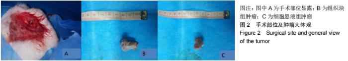
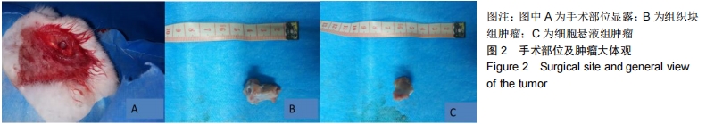
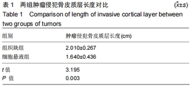
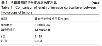
2.1 实验动物数量分析 组织块组有1只实验动物于术后死亡,脱组,细胞悬液组所有实验动物均存活。 2.2 X射线检查及肿瘤大体观察结果 所有实验动物进行X射线检查均发现肿瘤组织,组织块组肿瘤组织及细胞悬液组肿瘤组织均侵犯皮质层,切除肿瘤组织后,组织块组肿瘤侵犯骨皮质长度为(2.010±0.267) cm,细胞悬液组肿瘤侵犯骨皮质长度为(1.640±0.436) cm,见图2和表1。 "
"
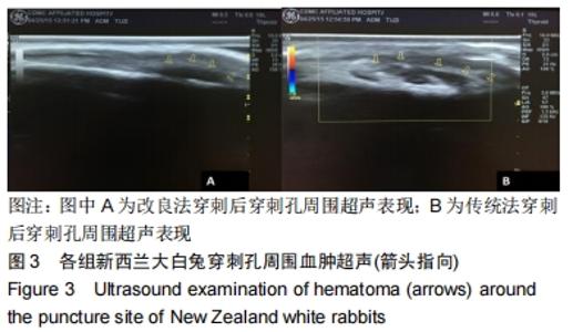
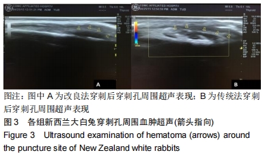
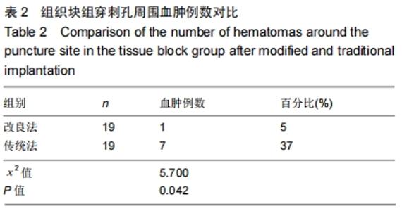
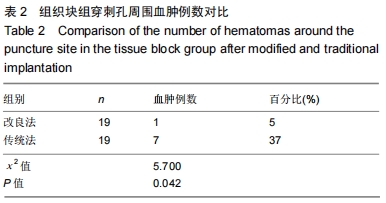
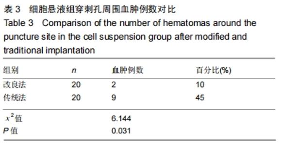
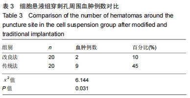
2.3 穿刺部位超声检查结果 组织块组改良法植入1 h后发现有1例穿刺孔周围血肿,传统法植入1 h后有7例穿刺孔周围血肿;细胞悬液组改良法植入1 h后发现2例穿刺孔周围血肿,传统法植入1 h后有9例穿刺孔周围血肿,见图3,表2,3。 "
"
"
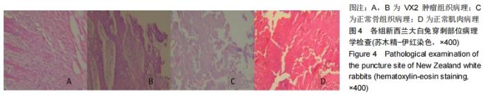
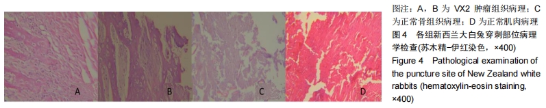
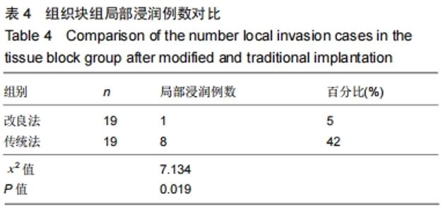
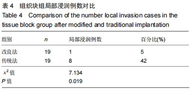
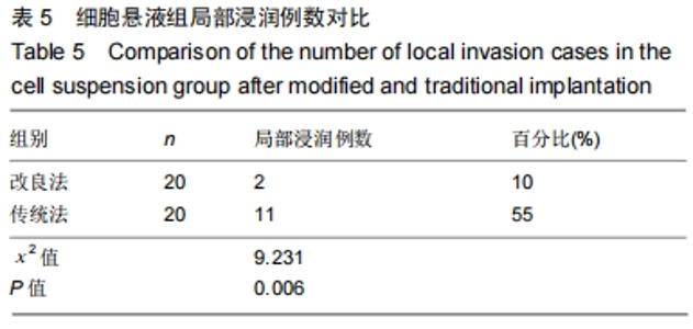
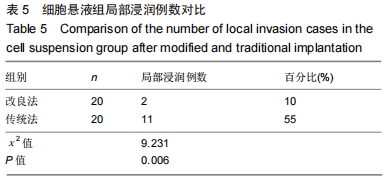
2.4 病理学检查结果 组织块组改良法植入后3周有1例软组织肿瘤细胞,即出现肿瘤局部浸润,传统法植入后3周有8例出现肿瘤局部浸润;细胞悬液组改良法植入后有2例出现肿瘤局部浸润,传统法植入后有11例出现肿瘤局部浸润,见图4,表4,5。 "
"
"
| [1]SMITH-GUZMÁN NE, TORETSKY JA, TSAI J, et al. A probable primary malignant bone tumor in a pre-Columbian human humerus from Cerro Brujo, Bocas del Toro, Panamá. Int J Paleopathol. 2018; 21:138-146. [2]WANG L, LONG NJ, LI L, et al. Multi-functional bismuth-doped bioglasses: combining bioactivity and photothermal response for bone tumor treatment and tissue repair. Light Sci Appl. 2018;7:1. [3]OGURA K, HOSODA F, NAKAMURA H, et al. Highly recurrent H3F3A mutations with additional epigenetic regulator alterations in giant cell tumor of bone. Genes Chromosomes Cancer. 2017;56(10):711-718. [4]OU JY, SPRAKER-PERLMAN H, DIETZ AC, et al. Conditional survival of pediatric, adolescent, and young adult soft tissue sarcoma and bone tumor patients. Cancer Epidemiol. 2017; 50(Pt A):150-157. [5]WANG LJ, WU HB, ZHOU WL, et al. Gummatous Syphilis Mimicking Malignant Bone Tumor on FDG PET/CT. Clin Nucl Med. 2019;44(4): 313-316. [6]HIGUCHI T, YAMAMOTO N, HAYASHI K, et al. Calcium Phosphate Cement in the Surgical Management of Benign Bone Tumors. Anticancer Res. 2018;38(5):3031-3035. [7]VAN BREUGEL JMM, GESCHWIND JF, MIRPOUR S, et al. Theranostic application of lipiodol for transarterial chemoembolization in a VX2 rabbit liver tumor model. Theranostics. 2019;9(13): 3674-3686. [8]SUN Y, XIONG X, PANDYA D, et al. Enhancing tissue permeability with MRI guided preclinical focused ultrasound system in rabbit muscle: From normal tissue to VX2 tumor. J Control Release. 2017;256:1-8. [9]DESCHAMPS F, FAROUIL G, GONZALEZ W, et al. Stabilization Improves Theranostic Properties of Lipiodol®-Based Emulsion During Liver Trans-arterial Chemo-embolization in a VX2 Rabbit Model. Cardiovasc Intervent Radiol. 2017;40(6):907-913. [10]BING C, PATEL P, STARUCH RM, et al. Longer heating duration increases localized doxorubicin deposition and therapeutic index in Vx2 tumors using MR-HIFU mild hyperthermia and thermosensitive liposomal doxorubicin. Int J Hyperthermia. 2019;36(1):196-203. [11]HUNG SH, JAO JC, TZENG JS, et al. Evaluation of Rabbit VX2 Tumor Model Using Magnetic Resonance T1-Mapping and T2-Mapping Techniques at 1.5 T. Journal of Medical and Biological Engineering. 2018; 38(4): 607-617. [12]PELLERIN O, AMARA I, SAPOVAL M, et al. Hepatic Intra-arterial Delivery of a "Trojan-horses" Gene Therapy: A Pilot Study on Rabbit VX2 Hepatic Tumor Model. Cardiovasc Intervent Radiol. 2018;41(1): 153-162. [13]曹海营,金宇,冯震,等.改良穿刺法改善针道肿瘤细胞转移情况的研究[J].中国临床研究,2016,29(1):40-42,45. [14]曹海营,金宇,赵景新,等.改良穿刺活检技术用于降低 VX2 肌肉肿瘤穿刺并发症的研究[J].中国全科医学,2016,19(5): 560-564. [15]张猛,魏俊强,段建伟,等.股骨骨折模型兔外固定架稳定性与股四头肌肌腱的关系[J].中国组织工程研究,2016,20(39): 5834-5839. [16]张猛,魏俊强,段建伟,等.外固定架制作兔股骨骨折模型[J].中国临床研究, 2016,29(5):685-686. [17]段建伟,魏俊强,张猛,等.外固定架加压-牵开-再加压治疗非感染性骨折不愈合实验研究[J].新医学,2016,47(5):318-323. [18]魏然,赵景新,孔庆柱,等.一种新的骨肿瘤穿刺活检器械的研究[J].中国医疗设备,2015,30(11):98-99. [19]魏然,孔庆柱,赵景新,等.自创封闭穿刺技术应用于骨肿瘤诊断的动物实验研究[J].新医学,2016,47(2):92-96.
[20]LI C, ZHANG Y, CHEN G, et al. Engineered Multifunctional Nanomedicine for Simultaneous Stereotactic Chemotherapy and Inhibited Osteolysis in an Orthotopic Model of Bone Metastasis. Adv Mater. 2017;29(13): 1605754.
[30]LI SY, HUANG PT, FANG Y, et al. Ultrasonic Cavitation Ameliorates Antitumor Efficacy of Residual Cancer After Incomplete Radiofrequency Ablation in Rabbit VX2 Liver Tumor Model. Transl Oncol. 2019;12(8):1113-1121. |
| [1] | Tan Xinfang, Guo Yanxing, Qin Xiaofei, Zhang Binqing, Zhao Dongliang, Pan Kunkun, Li Yuzhuo, Chen Haoyu. Effect of uniaxial fatigue exercise on patellofemoral cartilage injury in a rabbit [J]. Chinese Journal of Tissue Engineering Research, 2022, 26(在线): 1-6. |
| [2] | Wang Baojuan, Zheng Shuguang, Zhang Qi, Li Tianyang. Miao medicine fumigation can delay extracellular matrix destruction in a rabbit model of knee osteoarthritis [J]. Chinese Journal of Tissue Engineering Research, 2022, 26(8): 1180-1186. |
| [3] | Lü Yiyan, Li Hanbing, Ma Xiaoqing, Zhang Han, Zhang Yuhang, Li Genlin. Establishment and characteristic analysis of interior heat and diabetes mouse model using compound factors [J]. Chinese Journal of Tissue Engineering Research, 2022, 26(8): 1187-1193. |
| [4] | Zhu Chan, Han Xuke, Yao Chengjiao, Zhang Qiang, Liu Jing, Shao Ming. Acupuncture for Parkinson’s disease: an insight into the action mechanism in animal experiments [J]. Chinese Journal of Tissue Engineering Research, 2022, 26(8): 1272-1277. |
| [5] | Wang Xinmin, Liu Fei, Xu Jie, Bai Yuxi, Lü Jian. Core decompression combined with dental pulp stem cells in the treatment of steroid-associated femoral head necrosis in rabbits [J]. Chinese Journal of Tissue Engineering Research, 2022, 26(7): 1074-1079. |
| [6] | Feng Jianbo, Li Chencheng, Liu Jinyue, Wang Xiaomin, Peng Jiachen. Implantation of Kirschner wire with Staphylococcus aureus biofilm establishes a traumatic osteomyelitis model in rats [J]. Chinese Journal of Tissue Engineering Research, 2022, 26(5): 700-705. |
| [7] | Wang Shihui, Cheng Yang, Zhu Yunjie, Cheng Shaodan, Mao Jianying. Effect of arc edge needle-scalpel therapy on inflammatory factors and histomorphology of the frozen shoulder in rabbit models [J]. Chinese Journal of Tissue Engineering Research, 2022, 26(5): 706-711. |
| [8] | Yang Qian, Zhang Yiou, Jia Lili, Xie Jun, Feng Mali, Li Tingkai. Establishment and disease progression in a rat myocardial infarction model [J]. Chinese Journal of Tissue Engineering Research, 2022, 26(23): 3733-3737. |
| [9] | Su Jianqing, Sun Bo, Ding Yunrong, Liu Guangming, Ji Wei, Jiang Enyu, Yang Jiayu. Preparing a rabbit model of synovitis in knee osteoarthritis based on the “injury-repair-reinjury” method [J]. Chinese Journal of Tissue Engineering Research, 2022, 26(23): 3738-3743. |
| [10] | Wang Jiajia, Liu Jie, Wang Min. Establishing a murine model of experimental apical periodontitis induced by Fusobacterium nucleatum [J]. Chinese Journal of Tissue Engineering Research, 2022, 26(2): 176-181. |
| [11] | Li Shulun, Hao Peng, Hao Fei, Duan Hongmei, Zhao Wen, Gao Yudan, Yang Chaoyang, Li Xiaoguang. Pathological changes in rats with ischemic stroke induced by improved photochemical embolization [J]. Chinese Journal of Tissue Engineering Research, 2022, 26(2): 218-224. |
| [12] | Jia Qiyu, Guo Jian, Wei Qin, Guo Xiaobin, Chen Dongsheng, Feng Dongwei, Liu Yanshi, Ma Chuang. Establishment and evaluation of a validation model for the efficacy of external femoral fixator screws in rats [J]. Chinese Journal of Tissue Engineering Research, 2022, 26(18): 2854-2861. |
| [13] | Chen Xianxiang, Liao Shijie, Li Boxiang, Li Chong, Huang Qian, Lin Chengsen, Liu Yun, Lu Rongbin, Ding Xiaofei. Characteristics and application on animal models of different species of Perthes disease [J]. Chinese Journal of Tissue Engineering Research, 2022, 26(18): 2922-2929. |
| [14] | Wan Zhe, Du Jun, He Jing, Hu Yang. Effect of Gushukang on osteogenic differentiation in a Beagle dog model during orthodontic root resorption and its mechanism [J]. Chinese Journal of Tissue Engineering Research, 2022, 26(17): 2654-2659. |
| [15] | Yang Qin, Wang Min. Constructing a mouse model of periodontitis with occlusal trauma [J]. Chinese Journal of Tissue Engineering Research, 2022, 26(17): 2667-2672. |
| Viewed | ||||||
|
Full text |
|
|||||
|
Abstract |
|
|||||