Chinese Journal of Tissue Engineering Research ›› 2017, Vol. 21 ›› Issue (26): 4160-4166.doi: 10.3969/j.issn.2095-4344.2017.26.011
Previous Articles Next Articles
Silk fibroin/nano-hydroxyapatite composite material as a lumbar interbody implant: a study on initial segmental stability
- Department of Spine Surgery, the Central Hospital of Wuhan, Tongji Medical College, Huazhong University of Science and Technology, Wuhan 430000, Hubei Province, China
-
Received:2017-04-12Online:2017-09-18Published:2017-09-28 -
About author:Deng Bi-quan, M.D., Attending physician, Department of Spine Surgery, the Central Hospital of Wuhan, Tongji Medical College, Huazhong University of Science and Technology, Wuhan 430000, Hubei Province, China -
Supported by:the Natural Science Foundation of Hubei Province in 2014, No. 2014CFB459
CLC Number:
Cite this article
Deng Bi-quan, Teng Yu, Hu Hua, Li Jian, Jiang Hong-hui, Zhang Wei-guo.
share this article
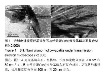
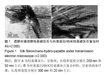
2.1 复合支架材料的表征 图1是在投射电镜下拍摄的典型羟基磷灰石和丝素蛋白/纳米轻基磷灰石复合材料照片,两者之间在形状上差别很大,前者呈棒状,长度和宽度分别在200 nm和50 nm左右;复合材料呈束状纳米纤维,长度和宽度分别在300 nm和20 nm左右。"
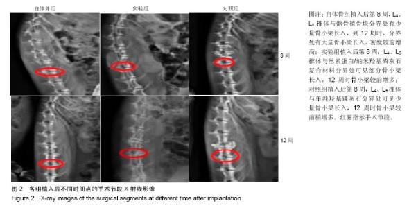
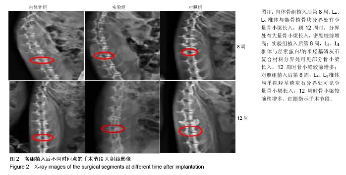
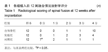
2.2 术后动物一般情况及大体观察 实验动物当日清醒,术后正常进食进水,精神状态良好。1只因术中麻醉药过敏致死,1只因失血过多死亡,均于术后补充完整。 植入后观察12周,所有存活动物伤口恢复良好,未出现神经损伤、出血、红肿、渗出及感染等术后并发症。3组标本重点观察融合部位,质地均较硬,其中自体骨组可见大量骨小梁长入,有骨连接;实验组材料与周围肌肉组织结合紧密,有融合迹象,可见部分骨小梁长入,已形成骨连接;对照组有少量骨小梁长入,骨性连接不坚固。 2.3 动物实验影像学检查结果 如图2所示:①自体骨组:植入后第8周,L4、L5椎体与髂骨植骨块分界处有少量骨小梁长入,到12周时,分界处有大量骨小梁长入,密度较前增高,X射线评估有11只符合融合标准;②实验组:植入后第8周,L4、L5椎体与丝素蛋白/纳米羟基磷灰石复合材料分界处可见部分骨小梁长入,12周时骨小梁较前增多,X射线评估有7只符合融合标准;③对照组:植入后第8周,L4、L5椎体与单纯羟基磷灰石分界处可见少量骨小梁长入,12周时骨小梁较前稍增多,X射线检测有2只评定为融合,见表1。"

"
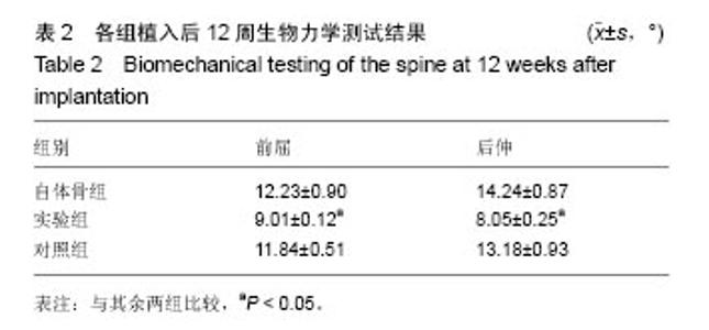
2.4 动物实验生物力学测试结果 植入后12周,通过对两组手术节段的脊柱三维非破坏性生物力学试验,结果显示,3组总体比较差异有显著性意义,其中,实验组、对照组前屈、后伸的脊柱活动度值均小于自体骨组,但仅实验组与自体骨组比较差异有显著性意义(P < 0.05);实验组脊柱活动度值低于比对照组(P < 0.05),见表2。"

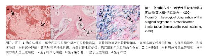
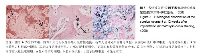
2.5 动物实验组织学观察结果 图3为植入后12周时,各组在光学显微镜下组织学观察到的典型图片代表(由于在第8周时,各个组别的病理图片区分度不多,所以组织学展示的是12周各组病理图片),自体骨组髂骨和周边组织分界处可见骨性连接,新骨周边可见大量骨母细胞,表面亦可见纤维母细胞,内部见编织骨;实验组材料部分降解,其周边可见纤维组织,内部有新生编织骨,陷窝细胞和骨细胞散在分布;对照组材料周边可见血管增生,材料内部有大量巨噬细胞;其中自体骨组中接近图3A病理类型改变的有10只,实验组中接近图3B病理类型改变的有8只,对照组中接近图3C病理改变的有9只。 "
| [1]Chen Y,Wang X,Chen D,et al.Posterior hybrid technique for ossification of the posterior longitudinal ligament associated with segmental instability in the cervical spine.J Spinal Disord Tech.2014;27(4):240-244.[2]Barz T,Melloh M,Lord SJ,et al.A Conceptual Model of Compensation/ Decompensation in Lumbar Segmental Instability. Med Hypotheses.2014;83(3):312-316.[3]费琦,赵凡,杨雍,等.腰椎后路融合手术对失稳模型节段稳定性及相邻节段力学的影响[J].中华医学杂志, 2015,95(45): 3681-3686.[4]Li H,Sun S,Liu H,et al.Use of a biological reactor and platelet-rich plasma for the construction of tissue-engineered boneto repair articular cartilage defects.Exp Ther Med. 2016; 12(2):711-719.[5]Janson IA,Putnam A J.Extracellular matrix elasticity and topography: material-based cues that affect cell function via conserved mechanisms.J Biomed Mater Res A.2015;103(3): 1246-1258.[6]Bianchi M,Urquia Edreira ER,Wolke JG,et al.Substrate geometry directs the in vitro mineralization of calcium phosphate ceramics.Acta Biomater.2014;10(2):661-669.[7]张琦,浦益琼,王冰,等.羟基磷灰石复合材料制备技术的研究进展[J].中成药,2015,37(12):2722-2725.[8]李浩,曹呈斌,杨惠林,等.丝素蛋白/羟基磷灰石复合物对磷酸钙骨水泥性能的影响[J].现代生物医学进展, 2013,13(33): 6435-6439.[9]Shokrollahi P,Mehmanchi M,Atai M,et al.Effect of interface on mechanical properties and biodegradation of PCL HAp supramolecular nano-composites.J Mater Sci Mater Med. 2014;25(1):23-35.[10]唐俊杰,李文杰,李根,等.骨组织工程诱导性支架材料修复骨缺损[J].中国组织工程研究,2015,19(3):340-346.[11]Tian X,Zhu G,Wang J,et al.Study on the relation between tissues pathologies and traditional chinese medicine syndromes in knee osteoarthritis:Medical image diagnostics by preoperative X-ray and surgical arthroscopy.J Xray Sci Technol.2016;24(4):509-519.[12]Byvaltsev VA,Kalinin AA,Belykh EG,et al.Optimization of segmental lumbar spine instability treatment using minimally invasive spinal fusion technique.Zh Vopr Neirokhir Im N N Burdenko.2015;79(3):45-54.[13]Ploumis A,Christodoulou P,Kapoutsis D,et al.Surgical treatment of lumbar spinal stenosis with microdecompression and interspinous distraction device insertion.A case series.J Orthop Surg Res.2012;(7):35.[14]Samadian M,Hosseinzadeh Bakhtevari M,Jahangiri Babadi A,et al.Congenital Posterior Spinal Agenesis Leads to L2-L3 Instability: a Case Report and Review of the Literature.Arch Iran Med.2015;8(12):861-864.[15]刘晓光.不稳定型脊柱骨折脱位的内固定技术及进展[J].中国骨伤,2014,27(2):89-91.[16]嘉士健,黄翠华,雷行华,等.针刀配合脊椎平衡手法康复训练治疗下颈椎不稳症临床观察[J].中国中医急症, 2015,24(10): 1829-1831.[17]封亚平,章翔,封雨.加强脊髓脊柱术后不稳与重建研究[J].中华神经外科疾病研究杂志,2013,12(2):97-100.[18]Pound BG.Passive films on metallic biomaterials under simulated physiological conditions.J Biomed Mater Res A.2014;102(5):1595-1604.[19]Bing W,Dong J,Li Q,et al.Mechanism of inhibition on L929 rat fibroblasts proliferation induced by potential adhesion barrier material poly(p-dioxanone-co-l-phenylalanine) electrospun membranes].J Biomed Mater Res A. 2014; 102(11):4062-4070.[20]Pa?cu EI,Stokes J,McGuinness GB.McGuinness.Electrospun composites of PHBV,silk fibroin and nano-hydroxyapatite for bone tissue engineering.Mater Sci Eng C Mater Biol Appl. 2013;33(8):4905-4916.[21]侯立刚,杨建义.骨修复中应用的生物降解可吸收材料[J].中国组织工程研究,2016,20(3):441-445.[22]贾世双,张堃,李双.股骨颈并股骨干骨折的修复:骨移植与组织及细胞移植效果评价[J].中国组织工程研究, 2015,19(21): 3424-3428.[23]邢麟子,桂鉴超,徐燕,等.微骨折技术结合自体骨软骨碎屑样移植修复兔膝关节软骨缺损的实验研究[J].中华创伤骨科杂志, 2012, 14(5):424-428.[24]王凤玲.SPIO标记细胞磁靶向移植修复关节软骨缺损可行性的初步研究[D].第三军医大学,2013.[25]李练兵,张其清,侯志伟,等.复合人工骨材料对骨形成影响的实验研究[J].北京生物医学工程,2014,33(3):240-246.[26]马立坤,叶鹏,邓江,等.丝素蛋白/壳聚糖/纳米羟基磷灰石骨组织工程支架材料的体外细胞毒性评价[J].西部医学, 2014,26(8): 975-977,980.[27]帅亚俊,张璨,邓连霞,等.矿化柞蚕丝胶膜表面粗糙度的调控及其对骨髓间充质干细胞生长行为的影响[J].复合材料学报,2 015, 32(5):1527-1535.[28]Zhu M,Kai W,Mei J,et al.Fabrication of highly interconnected porous silk fibroin scaffolds for potential use as vascular grafts.Acta Biomater.2014;10(5):2014-2023.[29]Hofmann S,Stok K S,Kohler T,et al.Effect of sterilization on structural and material properties of 3-D silk fibroin scaffolds. Acta Biomaterialia.2014;10(1):308-317.[30]Lai GJ,Shalumon KT,Chen SH,et al.Composite chitosan/silk fibroin nanofibers for modulation of osteogenic differentiation and proliferation of human mesenchymal stem cells. Carbohydr Polym.2014;111(111):288-297.[31]Niu B,Li B,Gu Y,et al.In vitro evaluation of electrospun silk fibroin/nano-hydroxyapatite/BMP-2 scaffolds for bone regeneration.J Biomater Sci Polym Ed.2017;28(3):257-270. [32]Le TD,Liaudanskaya V,Bonani W,et al.Enhancing bioactive properties of silk fibroin with diatom particles for bone tissue engineering applications.J Tissue Eng Regen Med.2016.doi: 10.1002/term.2373. [Epub ahead of print][33]Yao MZ,Huang-Fu MY,Liu HN,et al.Fabrication and characterization of drug-loaded nano-hydroxyapatite/polyamide 66 scaffolds modified with carbon nanotubes and silk fibroin.Int J Nanomedicine.2016;11:6181-6194.[34]Ran J,Hu J,Sun G,et al.A novel chitosan-tussah silk fibroin/nano-hydroxyapatite composite bone scaffold platform with tunable mechanical strength in a wide range.Int J Biol Macromol.2016;93(Pt A):87-97. [35]Ding Z,Fan Z,Huang X,et al.Silk-Hydroxyapatite Nanoscale Scaffolds with Programmable Growth Factor Delivery for Bone Repair.ACS Appl Mater Interfaces.2016;8(37): 24463-24470.[36]Hu JX,Ran JB,Chen S,et al.Carboxylated Agarose (CA)-Silk Fibroin (SF) Dual Confluent Matrices Containing Oriented Hydroxyapatite (HA) Crystals: Biomimetic Organic/Inorganic Composites for Tibia Repair.Biomacromolecules. 2016; 17(7):2437-2447. [37]Wu J,Liu J,Shi Y,et al.Rheological, mechanical and degradable properties of injectable chitosan/silk fibroin/hydroxyapatite/glycerophosphate hydrogels.J Mech Behav Biomed Mater.2016;64:161-172.[38]Bhattacharjee P,Naskar D,Maiti TK,et al.Non-mulberry silk fibroin grafted poly (?-caprolactone)/nano hydroxyapatite nanofibrous scaffold for dual growth factor delivery to promote bone regeneration.J Colloid Interface Sci.2016;472:16-33. [39]Sinlapabodin S,Amornsudthiwat P,Damrongsakkul S,et al.An axial distribution of seeding, proliferation, and osteogenic differentiation of MC3T3-E1 cells and rat bone marrow-derived mesenchymal stem cells across a 3D Thai silk fibroin/gelatin/hydroxyapatite scaffold in a perfusion bioreactor.Mater Sci Eng C Mater Biol Appl.2016;58:960-970. [40]王海莹,张旭,丁文元,等.椎间盘退变动物模型的研究进展[J].中国脊柱脊髓杂志,2015,25(3):279-282.[41]钟锐,刘少喻.腰椎间盘退变动物模型构建的研究进展[J].中华实验外科杂志,2015,32(7):1760-1762.[42]王鹏.骨质疏松大鼠脊柱融合后邻近节段椎间盘退变的实验性研究[D].河北联合大学,2015.[43]杨泽雨,杨欣建,陈扬,等.新型3D打印多孔钛人工椎体在猪脊柱模型置换前后的生物力学测试研究[J].生物骨科材料与临床研究,2016,13(1):7-9.[44]牛伟民,于德水,李名武,等.椎体间纤维性融合重建兔脊柱稳定性的初步探索[J].中国脊柱脊髓杂志,2012,22(12):1108-1112.[45]杨洋,黎庆初,朱召银,等.双节段前路颈椎自锁式融合器融合术后矢状位影像学参数的变化[J].中国脊柱脊髓杂志,2016,26(2): 116-123.[46]谭海涛,孟志斌,黄涛,等.三点稳定式脊柱融合技术效果的动物实验研究[J].临床医学工程,2014,21(3):278-280.[47]张春霖,王若愚,李莹,等.腰椎失稳症患者腰椎间隙X线解剖分型及其临床意义[J].中华解剖与临床杂志,2015,20(6):488-493.[48]吕厚辰,唐佩福.骨组织形态及微观结构的影像学评价及进展[J].中华骨质疏松和骨矿盐疾病杂志,2016,9(1):68-74.[49]江祖炘,谭波,代辉,等.骨髓间充质干细胞和软骨细胞共培养在组织工程化软骨中的研究进展[J].实用医院临床杂志,2014,11(1): 188-190.[50]孙庆治.纳米羟基磷灰石/丝素蛋白人工骨修复骨缺损[J].中国组织工程研究,2015,19(8):1190-1194. |
| [1] | Yao Xiaoling, Peng Jiancheng, Xu Yuerong, Yang Zhidong, Zhang Shuncong. Variable-angle zero-notch anterior interbody fusion system in the treatment of cervical spondylotic myelopathy: 30-month follow-up [J]. Chinese Journal of Tissue Engineering Research, 2022, 26(9): 1377-1382. |
| [2] | An Weizheng, He Xiao, Ren Shuai, Liu Jianyu. Potential of muscle-derived stem cells in peripheral nerve regeneration [J]. Chinese Journal of Tissue Engineering Research, 2022, 26(7): 1130-1136. |
| [3] | Zhang Jinglin, Leng Min, Zhu Boheng, Wang Hong. Mechanism and application of stem cell-derived exosomes in promoting diabetic wound healing [J]. Chinese Journal of Tissue Engineering Research, 2022, 26(7): 1113-1118. |
| [4] | Yang Jun, Yang Qun, Zhang Rui, Jiang Chang. A novel slidable pedicle screw-rod system for lumbar tuberculosis: promoting bone graft fusion by producing stress stimulation to fused segment [J]. Chinese Journal of Tissue Engineering Research, 2022, 26(6): 914-918. |
| [5] | Li Jian, Bao Zhengqi, Zhou Pinghui, Zhu Ruizhi, Li Zhixiang, Wang Jinzi. Effects of posterior single open-door laminoplasty and anterior cervical corpectomy fusion on cervical sagittal balance parameters in the treatment of multilevel cervical spondylotic myelopathy [J]. Chinese Journal of Tissue Engineering Research, 2022, 26(6): 949-953. |
| [6] | He Yunying, Li Lingjie, Zhang Shuqi, Li Yuzhou, Yang Sheng, Ji Ping. Method of constructing cell spheroids based on agarose and polyacrylic molds [J]. Chinese Journal of Tissue Engineering Research, 2022, 26(4): 553-559. |
| [7] | He Guanyu, Xu Baoshan, Du Lilong, Zhang Tongxing, Huo Zhenxin, Shen Li. Biomimetic orientated microchannel annulus fibrosus scaffold constructed by silk fibroin [J]. Chinese Journal of Tissue Engineering Research, 2022, 26(4): 560-566. |
| [8] | Chen Xiaoxu, Luo Yaxin, Bi Haoran, Yang Kun. Preparation and application of acellular scaffold in tissue engineering and regenerative medicine [J]. Chinese Journal of Tissue Engineering Research, 2022, 26(4): 591-596. |
| [9] | Kang Kunlong, Wang Xintao. Research hotspot of biological scaffold materials promoting osteogenic differentiation of bone marrow mesenchymal stem cells [J]. Chinese Journal of Tissue Engineering Research, 2022, 26(4): 597-603. |
| [10] | Shen Jiahua, Fu Yong. Application of graphene-based nanomaterials in stem cells [J]. Chinese Journal of Tissue Engineering Research, 2022, 26(4): 604-609. |
| [11] | Zhang Tong, Cai Jinchi, Yuan Zhifa, Zhao Haiyan, Han Xingwen, Wang Wenji. Hyaluronic acid-based composite hydrogel in cartilage injury caused by osteoarthritis: application and mechanism [J]. Chinese Journal of Tissue Engineering Research, 2022, 26(4): 617-625. |
| [12] | Li Hui, Chen Lianglong. Application and characteristics of bone graft materials in the treatment of spinal tuberculosis [J]. Chinese Journal of Tissue Engineering Research, 2022, 26(4): 626-630. |
| [13] | Gao Cangjian, Yang Zhen, Liu Shuyun, Li Hao, Fu Liwei, Zhao Tianyuan, Chen Wei, Liao Zhiyao, Li Pinxue, Sui Xiang, Guo Quanyi. Electrospinning for rotator cuff repair [J]. Chinese Journal of Tissue Engineering Research, 2022, 26(4): 637-642. |
| [14] | Guan Jian, Jia Yanfei, Zhang Baoxin , Zhao Guozhong. Application of 4D bioprinting in tissue engineering [J]. Chinese Journal of Tissue Engineering Research, 2022, 26(3): 446-455. |
| [15] | Huang Bo, Chen Mingxue, Peng Liqing, Luo Xujiang, Li Huo, Wang Hao, Tian Qinyu, Lu Xiaobo, Liu Shuyun, Guo Quanyi . Fabrication and biocompatibility of injectable gelatin-methacryloyl/cartilage-derived matrix particles composite hydrogel scaffold [J]. Chinese Journal of Tissue Engineering Research, 2022, 10(16): 2600-2606. |
| Viewed | ||||||
|
Full text |
|
|||||
|
Abstract |
|
|||||