Chinese Journal of Tissue Engineering Research ›› 2017, Vol. 21 ›› Issue (22): 3541-3546.doi: 10.3969/j.issn.2095-4344.2017.22.017
Previous Articles Next Articles
Biocompatibility of modified nano-hydroxyapatite/polyvinyl alcohol porous composite hydrogel as an artificial corneal material
- 1Department of Ophtalmology, Nanyang Centre Hospital, Nanyang 473000, Henan Province, China; 2Public Health Division, Tanghe Suburban Hospital, Nanyang 473000, Henan Province, China
-
Received:2017-06-13Online:2017-08-08Published:2017-09-01 -
About author:Du Qian, Master, Attending physician, Department of Ophtalmology, Nanyang Centre Hospital, Nanyang 473000, Henan Province, China
CLC Number:
Cite this article
Du Qian, Du Chen, Jin Gui-yu, Tian Hua.
share this article
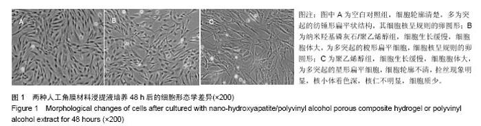
2.1 人工角膜材料浸提液培养兔角膜基质成纤维细胞的形态学差异 与空白对照组相比,加入2种材料提取液培养细胞48 h后,细胞均稀疏,生长速度缓慢,其中纳米羟基 磷灰石/聚乙烯醇组细胞拉丝现象不明显(图1)。"

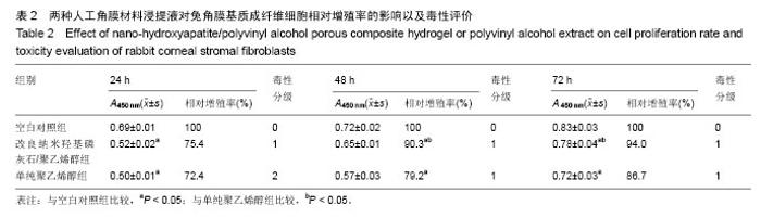
2.2 人工角膜材料浸提液对兔角膜基质成纤维细胞的毒性评价 与空白对照组相比,加入2种材料提取液培养细胞不同时间后,细胞相对增殖率均明显降低(P < 0.05),改良纳米羟基磷灰石/聚乙烯醇组的细胞增殖率高于聚乙烯醇组(P < 0.05);除了单纯聚乙烯醇组培养24 h后的毒性属待定,其余均属合格,见表2。"

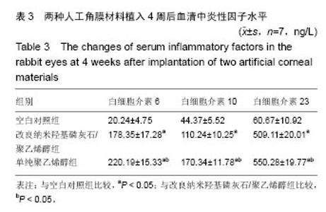
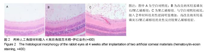
2.3 两种材料植入后实验兔过敏、热原、死亡发生情况 植入2种材料后,纳米羟基磷灰石/聚乙烯醇组仅有2只兔子发生过敏反应,无热原反应和死亡,而聚乙烯醇组有2只兔子发生过敏反应,2只发生热原反应,无死亡。 2.4 两种材料植入后角膜苏木精-伊红染色以及血清中炎性因子水平 植入4周后,与空白对照组比较,两组实验兔血清炎性因子水平明显增加(P < 0.05),纳米羟基磷灰石/聚乙烯醇组炎性因子少于聚乙烯醇组(P < 0.05);苏木精-伊红染色显示,与空白对照组相比,植入2种材料组炎性浸润明显增加,而改良纳米羟基磷灰石/聚乙烯醇组的炎性反应较聚乙烯醇组轻,见表3,图2。"
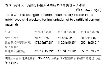
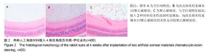
"

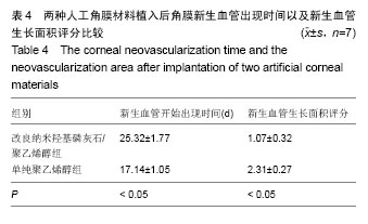
2.5 两种人工角膜材料植入后角膜新生血管出现的时间以及新生血管生长面积评分比较 植入人工角膜材料术后第1周,纳米羟基磷灰石/聚乙烯醇组和聚乙烯醇组均有球结膜充血水肿、房水闪辉及前房炎性渗出。术后第2周,两组球结膜充血减轻,房水闪辉减少。术后第3周,聚乙烯醇组出现点状炎症浸润,角膜上皮出现缺损,且4只出现角膜血管新生情况。术后4周,纳米羟基磷灰石/聚乙烯醇组有2只出现血管新生情况,且发生角膜新生血管生长面积小于聚乙烯醇组(图3)。纳米羟基磷灰石/聚乙烯醇组的兔新生血管出现时间晚于聚乙烯醇组(P < 0.05),新生血管生长面积评分小于聚乙烯醇组(P < 0.05),见表4。"
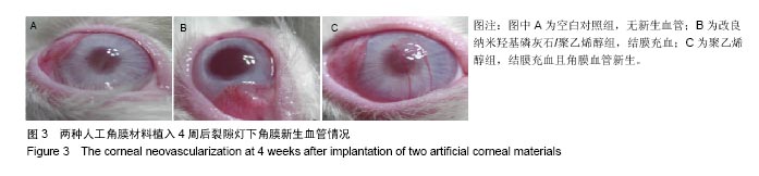
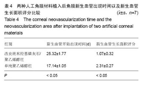
"
| [1]Zhang B, Gu J, Zhang X, et al. Biomechanical Measurement of Rabbit Cornea by a Modified Scheimpflug Device. J Ophthalmol. 2016;2016:8271762.[2]Zhang F, Wu J, Kang D, et al. Development of a complex hydrogel of hyaluronan and PVA embedded with silver nanoparticles and its facile studies on Escherichia coli. J Biomater Sci Polym Ed. 2013;24(12):1410-1425.[3]Yusong P, Qianqian S, Chengling P, et al. Prediction of mechanical properties of multilayer gradient hydroxyapatite reinforced poly(vinyl alcohol) gel biomaterial. J Biomed Mater Res B Appl Biomater. 2013;101(5):729-735.[4]D'Amico DJ. New methods for retinal examination in eyes with a Boston keratoprosthesis. Retina. 2013;33(6): 1097-1098.[5]Du LQ, Chen HM, Yan Y, et al. In vivo biological stability of chemically pretreated silicone gel inserts intended for use in keratoprostheses. Chin Med J (Engl). 2012;125(23): 4239-4244.[6]Jiang H, Zuo Y, Zhang L, et al. Property-based design: optimization and characterization of polyvinyl alcohol (PVA) hydrogel and PVA-matrix composite for artificial cornea. J Mater Sci Mater Med. 2014;25(3):941-952.[7]Bakhshandeh H, Soleimani M, Hosseini SS, et al. Poly (epsilon-caprolactone) nanofibrous ring surrounding a polyvinyl alcohol hydrogel for the development of a biocompatible two-part artificial cornea. Int J Nanomedicine. 2011;6:1509-1515.[8]Wang L, Ma R, Du G, et al. Biocompatibility of helicoidal multilamellar arginine-glycine-aspartic acid-functionalized silk biomaterials in a rabbit corneal model. J Biomed Mater Res B Appl Biomater. 2015;103(1):204-211.[9]Liu Y, Ren L, Wang Y. Crosslinked collagen-gelatin-hyaluronic acid biomimetic film for cornea tissue engineering applications. Mater Sci Eng C Mater Biol Appl. 2013;33(1): 196-201. [10]Hicks CR, Chirila TV, Vijayasekaran S, et al. PHEMA as a keratoprosthesis material. Br J Ophthalmol. 2006;90(1):124.[11]Carreira AS, Ferreira P, Ribeiro MP, et al. New drug-eluting lenses to be applied as bandages after keratoprosthesis implantation. Int J Pharm. 2014;477(1-2):218-226.[12]Hwang Y, Kim G. Evaluation of stability and biocompatibility of PHEMA-PMMA keratoprosthesis by penetrating keratoplasty in rabbits. Lab Anim Res. 2016;32(4):181-186.[13]Vajaranant TS, Liu J, Wilensky J, et al. Innovative approaches to glaucoma management of Boston keratoprosthesis type 1. Curr Ophthalmol Rep. 2016;4(3):147-153.[14]Huhtinen R, Sandeman S, Rose S, et al. Examining porous bio-active glass as a potential osteo-odonto-keratoprosthetic skirt material. J Mater Sci Mater Med. 2013;24(5):1217-1227.[15]Viitala R, Franklin V, Green D, et al. Towards a synthetic osteo-odonto-keratoprosthesis. Acta Biomater. 2009;5(1): 438-452.[16]Lee JH, Wee WR, Chung ES, et al. Development of a newly designed double-fixed Seoul-type keratoprosthesis. Arch Ophthalmol. 2000;118(12):1673-1678.[17]Wang L, Jeong KJ, Chiang HH, et al. Hydroxyapatite for keratoprosthesis biointegration. Invest Ophthalmol Vis Sci. 2011;52(10):7392-7399.[18]Mehta JS, Futter CE, Sandeman SR, et al. Hydroxyapatite promotes superior keratocyte adhesion and proliferation in comparison with current keratoprosthesis skirt materials. Br J Ophthalmol. 2005;89(10):1356-1362.[19]Mittal A, Garg S, Kohli D, et al. Effect of cross linking of PVA/starch and reinforcement of modified barley husk on the properties of composite films. Carbohydr Polym. 2016;151: 926-938.[20]Luo L, Cai W, Zhou J, et al. Facile synthesis of boehmite/PVA composite membrane with enhanced adsorption performance towards Cr(VI). J Hazard Mater. 2016;318:452-459.[21]Ehrenberg M, Zolotariov E, Loeb E, et al. Combining Sodium Hyaluronate and Polyvinylpyrrolidone Therapies for the Rabbit Cornea: A New Approach to Relief of the Human Dry Eye Syndrome. Curr Eye Res. 2015;40(9):913-922.[22]Li L, Zhou D, Wang XM, et al. Expression of matrix metalloproteinases and inhibitor on the cornea tissue in rabbit after implantation of modified titanium skirt for keratoprosthesis. Zhonghua Yan Ke Za Zhi. 2012;48(1): 20-26.[23]Kornilovskiy IM, Sultanova AI, Burtsev AA. Riboflavin photoprotection with cross-linking effect in photorefractive ablation of the cornea. Vestn Oftalmol. 2016;132(3):37-41.[24]Su P, Yang Y, Zhang L, et al. Biomechanical simulation of needle insertion into cornea based on distortion energy failure criterion. Acta Bioeng Biomech. 2016;18(1):65-75.[25]Su C, Su Y, Li Z, et al. In situ synthesis of bilayered gradient poly(vinyl alcohol)/hydroxyapatite composite hydrogel by directional freezing-thawing and electrophoresis method. Mater Sci Eng C Mater Biol Appl. 2017;77:76-83.[26]Iwatsubo T, Kishi R, Miura T, et al. Formation of Hydroxyapatite Skeletal Materials from Hydrogel Matrices via Artificial Biomineralization. J Phys Chem B. 2015;119(28): 8793-8799.[27]Jiang H, Zuo Y, Zhang L, et al. Property-based design: optimization and characterization of polyvinyl alcohol (PVA) hydrogel and PVA-matrix composite for artificial cornea. J Mater Sci Mater Med. 2014;25(3):941-952. [28]Xiao Q, Zhou K, Chen C, et al. Hollow and porous hydroxyapatite microspheres prepared with an O/W emulsion by spray freezing method. Mater Sci Eng C Mater Biol Appl. 2016;69:1068-1074.[29]Sturm D, Schmidt-Wilcke T, Greiner T, et al. Confocal Cornea Microscopy Detects Involvement of Corneal Nerve Fibers in a Patient with Light-Chain Amyloid Neuropathy Caused by Multiple Myeloma: A Case Report.Case Rep Neurol. 2016; 8(2):134-139. |
| [1] | Yao Xiaoling, Peng Jiancheng, Xu Yuerong, Yang Zhidong, Zhang Shuncong. Variable-angle zero-notch anterior interbody fusion system in the treatment of cervical spondylotic myelopathy: 30-month follow-up [J]. Chinese Journal of Tissue Engineering Research, 2022, 26(9): 1377-1382. |
| [2] | Zhang Jinglin, Leng Min, Zhu Boheng, Wang Hong. Mechanism and application of stem cell-derived exosomes in promoting diabetic wound healing [J]. Chinese Journal of Tissue Engineering Research, 2022, 26(7): 1113-1118. |
| [3] | An Weizheng, He Xiao, Ren Shuai, Liu Jianyu. Potential of muscle-derived stem cells in peripheral nerve regeneration [J]. Chinese Journal of Tissue Engineering Research, 2022, 26(7): 1130-1136. |
| [4] | Chen Xiaoxu, Luo Yaxin, Bi Haoran, Yang Kun. Preparation and application of acellular scaffold in tissue engineering and regenerative medicine [J]. Chinese Journal of Tissue Engineering Research, 2022, 26(4): 591-596. |
| [5] | Kang Kunlong, Wang Xintao. Research hotspot of biological scaffold materials promoting osteogenic differentiation of bone marrow mesenchymal stem cells [J]. Chinese Journal of Tissue Engineering Research, 2022, 26(4): 597-603. |
| [6] | Shen Jiahua, Fu Yong. Application of graphene-based nanomaterials in stem cells [J]. Chinese Journal of Tissue Engineering Research, 2022, 26(4): 604-609. |
| [7] | Zhang Tong, Cai Jinchi, Yuan Zhifa, Zhao Haiyan, Han Xingwen, Wang Wenji. Hyaluronic acid-based composite hydrogel in cartilage injury caused by osteoarthritis: application and mechanism [J]. Chinese Journal of Tissue Engineering Research, 2022, 26(4): 617-625. |
| [8] | Li Hui, Chen Lianglong. Application and characteristics of bone graft materials in the treatment of spinal tuberculosis [J]. Chinese Journal of Tissue Engineering Research, 2022, 26(4): 626-630. |
| [9] | Gao Cangjian, Yang Zhen, Liu Shuyun, Li Hao, Fu Liwei, Zhao Tianyuan, Chen Wei, Liao Zhiyao, Li Pinxue, Sui Xiang, Guo Quanyi. Electrospinning for rotator cuff repair [J]. Chinese Journal of Tissue Engineering Research, 2022, 26(4): 637-642. |
| [10] | Huang Chuanjun, Zou Yu, Zhou Xiaoting, Zhu Yangqing, Qian Wei, Zhang Wei, Liu Xing. Transplantation of umbilical cord mesenchymal stem cells encapsulated in RADA16-BDNF hydrogel promotes neurological recovery in an intracerebral hemorrhage rat model [J]. Chinese Journal of Tissue Engineering Research, 2022, 26(4): 510-515. |
| [11] | He Yunying, Li Lingjie, Zhang Shuqi, Li Yuzhou, Yang Sheng, Ji Ping. Method of constructing cell spheroids based on agarose and polyacrylic molds [J]. Chinese Journal of Tissue Engineering Research, 2022, 26(4): 553-559. |
| [12] | He Guanyu, Xu Baoshan, Du Lilong, Zhang Tongxing, Huo Zhenxin, Shen Li. Biomimetic orientated microchannel annulus fibrosus scaffold constructed by silk fibroin [J]. Chinese Journal of Tissue Engineering Research, 2022, 26(4): 560-566. |
| [13] | Guan Jian, Jia Yanfei, Zhang Baoxin , Zhao Guozhong. Application of 4D bioprinting in tissue engineering [J]. Chinese Journal of Tissue Engineering Research, 2022, 26(3): 446-455. |
| [14] | Liu Jiali, Suo Hairui, Yang Han, Wang Ling, Xu Mingen. Influence of lay-down angles on mechanical properties of three-dimensional printed polycaprolactone scaffolds [J]. Chinese Journal of Tissue Engineering Research, 2022, 10(16): 2612-2617. |
| [15] | Huang Bo, Chen Mingxue, Peng Liqing, Luo Xujiang, Li Huo, Wang Hao, Tian Qinyu, Lu Xiaobo, Liu Shuyun, Guo Quanyi . Fabrication and biocompatibility of injectable gelatin-methacryloyl/cartilage-derived matrix particles composite hydrogel scaffold [J]. Chinese Journal of Tissue Engineering Research, 2022, 10(16): 2600-2606. |
| Viewed | ||||||
|
Full text |
|
|||||
|
Abstract |
|
|||||