Chinese Journal of Tissue Engineering Research ›› 2017, Vol. 21 ›› Issue (2): 165-170.doi: 10.3969/j.issn.2095-4344.2017.02.001
Growth factor composite scaffolds for bone defect repair via immediate implantation of bone defects
- 1Department of Stomatology, 2Medical Insurance Office, Hebei General Hospital, Shijiazhuang 050051, Heibei Province, China
-
Received:2016-10-23Online:2017-01-18Published:2017-02-27 -
Contact:Wu Yong-sheng, Master, Professor, Chief physician, Master’s supervisor, Department of Stomatology, Hebei General Hospital, Shijiazhuang 050051, Heibei Province, China -
About author:Yang Yu-peng, Master, Attending physician, Department of Stomatology, Hebei General Hospital, Shijiazhuang 050051, Heibei Province, China -
Supported by:the Medical Science Research Project of Hebei Province in 2014, No. ZL20140216
CLC Number:
Cite this article
Yang Yu-peng1, Yang Sheng-jun2, Cheng Feng-xia1, Gu Jian-qi1, Zheng Yao1, Li Juan1, Hao Wei1, Wu Yong-sheng1 .
share this article
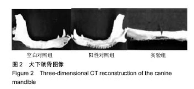
2.1 实验动物数量分析 实验选用犬5只,实验过程无脱失,全部进入结果分析。 2.2 犬下颌骨CT三维图像重建结果 空白对照组犬骨缺损中央没有新骨生成,周围有少量新生骨生成;阳性对照组和实验组犬骨缺损处骨小梁从中间向四周扩散形成骨修复,其中实验组和阳性对照组比较,骨小梁比较粗,数量多,骨质比较之谜,骨小梁之间分离度比较小,新骨形成比较多。见图2。"

2.3 犬术后下颌骨种植体周围骨缺损的愈合情况比较 由表1看出:3组犬术后10周时下颌骨种植体周围骨骨小梁的骨密度、分离度、数量和厚度比较差异有显著性意义(P < 0.05)。 其中实验组犬下颌骨种植体周围骨小梁的骨密度高于阳性对照组和空白对照组(P < 0.05),阳性对照组犬下颌骨种植体周围骨小梁的骨密度高于空白对照组,差异均有显著性意义(P < 0.05)。 实验组犬下颌骨种植体周围骨小梁分离度低于阳性对照组和空白对照组(P < 0.05),阳性对照组犬下颌骨种植体周围骨小梁分离度低于空白对照组(P < 0.05)。 "
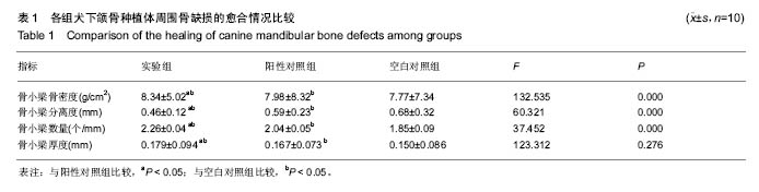
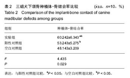
实验组犬下颌骨种植体周围骨小梁数量高于阳性对照组和空白对照组(P < 0.05),阳性对照组犬下颌骨种植体周围骨小梁数量高于空白对照组,差异均有显著性意义(P < 0.05)。 实验组犬下颌骨种植体周围骨小梁厚度高于阳性对照组和空白对照组(P < 0.05),阳性对照组犬下颌骨种植体周围骨小梁厚度高于空白对照组(P < 0.05)。 2.4 犬术后下颌骨种植体-骨结合率比较 由表2看出:3组犬术后10周时下颌骨种植体-骨结合率比较差异有显著性意义(P < 0.05),其中实验组犬下颌骨种植体-骨结合率高于阳性对照组和空白对照组(P < 0.05),阳性对照组犬下颌骨种植体-骨结合率高于空白对照组,差异均有显著性意义(P < 0.05)。"

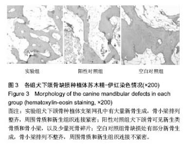
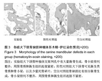
2.5 犬术后10周苏木精-伊红染色情况 在术后10周时,实验组犬的下颌骨种植体支架网孔中出现钙化的新生骨,形成成熟的骨小梁,复合支架材料上长满成熟的骨小梁,支架材料之间的骨小梁互相连接成板状和网状,有纤维骨痂组织穿破之间间隙;阳性对照组犬的下颌骨可见新生类骨质和骨小梁,以及少量死骨碎片;空白对照组犬的骨缺损处有部分新骨生成,肉芽组织减少,骨小梁排列不整齐,周围骨质和新生组织连接不紧密,其间有大量炎性细胞、新生血管、间充质细胞、成纤维细胞以及软骨细胞等,见图3。"
| [1]邸萍,林野,罗佳,等. 上颌前牙单牙种植修复中过渡义齿对软组织成型作用的临床研究[J]. 北京大学学报:医学版, 2012,44(1): 59-64.[2]Happe A, Kunz A.Complex fixed implant-supported restoration in a site compromised by periodontitis: a case report.Int J Esthet Dent.2016;11(2):186-202.[3]邱立新,胡秀莲,陈波,等.上颌窦底冲压提升法种植修复122例缺牙的临床观察[J]. 中华口腔医学杂志,2006,41(3):136-139.[4]吴展,李婧,陈卓凡. 上颌前牙即刻种植即刻修复的临床应用研究[J]. 中国口腔种植学杂志,2012,17(2):67-71+82.[5]Saito H, Chu SJ, Reynolds MA, et al.Provisional Restorations Used in Immediate Implant Placement Provide a Platform to Promote Peri-implant Soft Tissue Healing: A Pilot Study.Int J Periodontics Restorative Dent.2016;36(1):47-52. [6]李伟. 美学区单牙即刻种植即刻修复与延期种植修复临床效果评价[D].安徽医科大学,2015.[7]Levin BP, Wilk BL.The teamwork approach to esthetic tooth replacement with immediate implant placement andimmediate temporization.Compend Contin Educ Dent. 2015;36(9):682-688.[8]Keady F, Murphy BP.Investigating the feasibility of using a grit blasting process to coat nitinol stents withhydroxyapatite.J Mater Sci Mater Med.2013;24(1):97-103. [9]Bornapour M, Muja N,Shum-Tim D,et al.Biocompatibility and biodegradability of Mg-Sr alloys: the formation of Sr-substitutedhydroxyapatite.Acta Biomater.2013;9(2): 5319-5330. [10]王冠聪. 具有二级三维网络结构的壳聚糖/羟基磷灰石骨组织工程复合支架材料的构建及其生物性能研究[D].山东大学,2012.[11]Almeida JC, Wacha A, Gomes PS,et al.A biocompatible hybrid material with simultaneous calcium and strontium release capability forbone tissue repair.Mater Sci Eng C Mater Biol Appl.2016;1(62):429-438. [12]Raucci MG, Alvarez-Perez M, Giugliano D,et al.Properties of carbon nanotube-dispersed Sr-hydroxyapatite injectable material for bone defects.Regen Biomater.2016;3(1):13-23. [13]Sariibrahimoglu K, Yang W, Leeuwenburgh SC, et al. Development of porous polyurethane/strontium-substituted hydroxyapatite composites for boneregeneration.J Biomed Mater Res A.2015;103(6):1930-1939. [14]Neves N, Campos BB, Almeida IF, et al.Strontium-rich injectable hybrid system for bone regeneration.Mater Sci Eng C Mater Biol Appl.2016;1(59):818-827. [15]Meloni M, Cesselli D, Caporali A,et al.Cardiac Nerve Growth Factor Overexpression Induces Bone Marrow-derived Progenitor Cells Mobilization and Homing to the Infarcted Heart.Mol Ther.2015;23(12):1854-1866. [16]Chen WH, Mao CQ, Zhuo LL, et al.Beta-nerve growth factor promotes neurogenesis and angiogenesis during the repair of bonedefects.Neural Regen Res.2015;10(7):1159-1165. [17]King P, Maiorana C, Luthardt RG,et al.Clinical and Radiographic Evaluation of a Small-Diameter Dental Implant Used for the Restorationof Patients with Permanent Tooth Agenesis (Hypodontia) in the Maxillary Lateral Incisor and Mandibular Incisor Regions: A 36-Month Follow-Up.Int J Prosthodont.2016;29(2):147-153. [18]胡秀莲,李健慧,邱立新,等. 先天缺牙患者种植修复[J]. 北京大学学报:医学版,2011,43(1):62-66.[19]Stevens CJ.Technology to Control Excessive Occlusal Contact Force: Enhancing Implant RestorationLongevity.Dent Today.2016;35(1):112, 114, 116-117. [20]徐欣.当代口腔种植修复技术新进展[J].口腔医学, 2015,35(4): 241-244.[21]Khzam N, Arora H, Kim P, et al.Systematic Review of Soft Tissue Alterations and Esthetic Outcomes Following Immediate ImplantPlacement and Restoration of Single Implants in the Anterior Maxilla.J Periodontol.2015;86(12): 1321-1330. [22]贺刚,陈治清,王培志,等. 改良式引导骨再生术+即刻修复技术在上前牙即刻种植中的应用评价[J]. 口腔医学, 2013,33(11): 739-742.[23]苏媛,吕影涛,刘卫平,等. 美学区即刻种植即刻修复的临床应用[J].实用医学杂志,2014,30(11):1797-1799.[24]许勇.可注射性纳米羟基磷灰石、壳聚糖复合材料与骨髓基质细胞生物相容性及其修复骨缺损的实验研究[D].南方医科大学, 2010.[25]Alviar CL, Tellez A, Wang M,et al. Low-dose sirolimus-eluting hydroxyapatite coating on stents does not increase platelet activation and adhesion ex vivo.J Thromb Thrombolysis. 2012;34(1):91-98. [26]Costa JR Jr, Abizaid A, Costa R,et al.1-year results of the hydroxyapatite polymer-free sirolimus-eluting stent for the treatment of single de novo coronary lesions: the VESTASYNC I trial.JACC Cardiovasc Interv.2009;2(5): 422-427. [27]van der Giessen WJ, Sorop O, Serruys PW, et al.Lowering the dose of sirolimus, released from a nonpolymeric hydroxyapatite coated coronarystent, reduces signs of delayed healing.JACC Cardiovasc Interv.2009;2(4):284-290. [28]Han J, Wan P, Ge Y, et al.Tailoring the degradation and biological response of a magnesium-strontium alloy for potentialbone substitute application. Mater Sci Eng C Mater Biol Appl.2016;1(58):799-811. [29]Park JW, Kang DG, Hanawa T.New bone formation induced by surface strontium-modified ceramic bone graft substitute. Oral Dis.2016;22(1):53-61. [30]Li Y,Shui X,Zhang L,et al.Cancellous bone healing around strontium-doped hydroxyapatite in osteoporotic rats previously treated with zoledronic acid.J Biomed Mater Res B Appl Biomater.2016;104(3):476-481. [31]Boanini E, Torricelli P, Sima F,et al.Strontium and zoledronate hydroxyapatites graded composite coatings for bone prostheses.J Colloid Interface Sci.2015;15(448):1-7. [32]Monish B,Anthony L,Shilpa K.Immediate Implant Placement: Clinical Decisions, Advantages,and Disadvantages .J Prosthodontics.2008;17(7): 576-581. [33]Faleh A,Stefan R,Noel C,et al.Effect of grafting materials on osseointegration of dental implants surrounded by circumferential bone defects. An experimental study in the dog.Clin Oral Implants Res.2008;19(4):329-344. [34]Christophe DE,marie-Therese C,Christele C,et al.Surface enrichment of biomimetic apatites with biologically-active ions Mg2+ and Sr2+: A preamble to the activation of bone repair materials. Mater Sci and Eng C.2008;28(8):1544-1550. [35]Gentleman E,Fredholm Y,Jell G,et al.The effects of strontiumsubstituted bioactive glasses on osteoblasts and osteoclasts in vitro.Biomaterials.2010;31(14):3949-3956.[36]Goldenberg-Cohen N,Avraham-Lubin BC,Sadikov T,et al. Effect of coadministration of neuronal growth factors on neuroglial differentiation of bone marrow-derived stem cells in the ischemic retina.Invest Ophthalmol Vis Sci. 2014;55(1): 502-512. [37]Wang L,Zhou S,Liu B,et al.Locally applied nerve growth factor enhances bone consolidation in a rabbit model of mandibular distraction osteogenesis.Orthop Res.2006;24(12): 2238-2245. [38]王亮,曾剑玉,谭包生,等.神经生长因子在种植体周围早期骨愈合中的表达[J].北京口腔医学,2010,18(2):69-72. [39]梅玲,汪琼,王恩群,等.神经生长因子对兔下颌骨缺损愈合影响的实验研究.口腔医学研究[J].2012,28(8)775-779. [40]李俊杰,梅玲,王恩群,等.神经生长因子复合支架材料修复兔下颌骨缺损的实验研究. 现代口腔医学[J].2013,27(1)42-46. |
| [1] | Yao Xiaoling, Peng Jiancheng, Xu Yuerong, Yang Zhidong, Zhang Shuncong. Variable-angle zero-notch anterior interbody fusion system in the treatment of cervical spondylotic myelopathy: 30-month follow-up [J]. Chinese Journal of Tissue Engineering Research, 2022, 26(9): 1377-1382. |
| [2] | Xiao Yang, Gong Liqiong, Fei Jing, Li Leiji. Effect of electroacupuncture on nerve growth factor and its receptor expression in facial nerve nucleus after facial nerve injury in rabbits [J]. Chinese Journal of Tissue Engineering Research, 2022, 26(8): 1253-1259. |
| [3] | An Weizheng, He Xiao, Ren Shuai, Liu Jianyu. Potential of muscle-derived stem cells in peripheral nerve regeneration [J]. Chinese Journal of Tissue Engineering Research, 2022, 26(7): 1130-1136. |
| [4] | Zhang Jinglin, Leng Min, Zhu Boheng, Wang Hong. Mechanism and application of stem cell-derived exosomes in promoting diabetic wound healing [J]. Chinese Journal of Tissue Engineering Research, 2022, 26(7): 1113-1118. |
| [5] | He Yunying, Li Lingjie, Zhang Shuqi, Li Yuzhou, Yang Sheng, Ji Ping. Method of constructing cell spheroids based on agarose and polyacrylic molds [J]. Chinese Journal of Tissue Engineering Research, 2022, 26(4): 553-559. |
| [6] | He Guanyu, Xu Baoshan, Du Lilong, Zhang Tongxing, Huo Zhenxin, Shen Li. Biomimetic orientated microchannel annulus fibrosus scaffold constructed by silk fibroin [J]. Chinese Journal of Tissue Engineering Research, 2022, 26(4): 560-566. |
| [7] | Yang Feng, Zhao Qian, Zhang Shixuan, Zhao Tienan, Feng Bo. Effectiveness and safety of rapamycin combined with CD133 antibody stent in preventing vascular restenosis [J]. Chinese Journal of Tissue Engineering Research, 2022, 26(4): 579-584. |
| [8] | Chen Xiaoxu, Luo Yaxin, Bi Haoran, Yang Kun. Preparation and application of acellular scaffold in tissue engineering and regenerative medicine [J]. Chinese Journal of Tissue Engineering Research, 2022, 26(4): 591-596. |
| [9] | Kang Kunlong, Wang Xintao. Research hotspot of biological scaffold materials promoting osteogenic differentiation of bone marrow mesenchymal stem cells [J]. Chinese Journal of Tissue Engineering Research, 2022, 26(4): 597-603. |
| [10] | Shen Jiahua, Fu Yong. Application of graphene-based nanomaterials in stem cells [J]. Chinese Journal of Tissue Engineering Research, 2022, 26(4): 604-609. |
| [11] | Zhang Tong, Cai Jinchi, Yuan Zhifa, Zhao Haiyan, Han Xingwen, Wang Wenji. Hyaluronic acid-based composite hydrogel in cartilage injury caused by osteoarthritis: application and mechanism [J]. Chinese Journal of Tissue Engineering Research, 2022, 26(4): 617-625. |
| [12] | Li Hui, Chen Lianglong. Application and characteristics of bone graft materials in the treatment of spinal tuberculosis [J]. Chinese Journal of Tissue Engineering Research, 2022, 26(4): 626-630. |
| [13] | Gao Cangjian, Yang Zhen, Liu Shuyun, Li Hao, Fu Liwei, Zhao Tianyuan, Chen Wei, Liao Zhiyao, Li Pinxue, Sui Xiang, Guo Quanyi. Electrospinning for rotator cuff repair [J]. Chinese Journal of Tissue Engineering Research, 2022, 26(4): 637-642. |
| [14] | Guan Jian, Jia Yanfei, Zhang Baoxin , Zhao Guozhong. Application of 4D bioprinting in tissue engineering [J]. Chinese Journal of Tissue Engineering Research, 2022, 26(3): 446-455. |
| [15] | Huang Bo, Chen Mingxue, Peng Liqing, Luo Xujiang, Li Huo, Wang Hao, Tian Qinyu, Lu Xiaobo, Liu Shuyun, Guo Quanyi . Fabrication and biocompatibility of injectable gelatin-methacryloyl/cartilage-derived matrix particles composite hydrogel scaffold [J]. Chinese Journal of Tissue Engineering Research, 2022, 10(16): 2600-2606. |
| Viewed | ||||||
|
Full text |
|
|||||
|
Abstract |
|
|||||