Chinese Journal of Tissue Engineering Research ›› 2016, Vol. 20 ›› Issue (19): 2756-2762.doi: 10.3969/j.issn.2095-4344.2016.19.003
Previous Articles Next Articles
Bone marrow mesenchymal stem cells co-cultured with allogenic bone in the articular cavity
Gao Feng-guang1, Zhou Bai-sui1, Mu Hai-bo2
- 1Department of Orthopedics and Joints, 2Department of Traumatology, Affiliated Yantai Hospital of Binzhou Medical University, Yantai 264100, Shandong Province, China
-
Received:2016-03-02Online:2016-05-06Published:2016-05-06 -
Contact:Mu Hai-bo, Department of Traumatology, Affiliated Yantai Hospital of Binzhou Medical University, Yantai 264100, Shandong Province, China -
About author:Gao Feng-guang, Associate chief physician, Department of Orthopedics and Joints, Affiliated Yantai Hospital of Binzhou Medical University, Yantai 264100, Shandong Province, China
CLC Number:
Cite this article
Gao Feng-guang, Zhou Bai-sui, Mu Hai-bo. Bone marrow mesenchymal stem cells co-cultured with allogenic bone in the articular cavity[J]. Chinese Journal of Tissue Engineering Research, 2016, 20(19): 2756-2762.
share this article
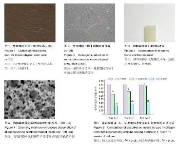
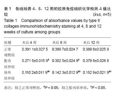
2.1 实验动物数量分析 关节腔内观察实验纳入15只成年新西兰白兔,全部进入结果分析,无脱失。 2.2 兔骨髓间充质干细胞培养结果 对兔骨髓间充质干细胞进行原代培养,培养后1 d后,出现少量细胞贴壁生长。接种3 d,贴壁生长细胞数量增加。经传代培养,细胞呈旋涡状或螺旋状,传至第3代未出现明显的改变(见图1)。 2.3 兔骨髓间充质干细胞成骨诱导结果 对第3代骨髓间充质干细胞进行软骨定向诱导之后,连续培养2周,可观察到细胞周围出现基质分泌(见图2)。 2.4 同种异体骨形态观察 制备得到的同种异体骨支架材料触之具有一定的轻度和韧性,可剪裁为不同的大小(见图3)。经电镜扫描,材料呈疏松多孔状结构(见图4)。 2.5 术后动物情况观察 对新西兰白兔进行同种异体骨-骨髓间充质干细胞复合物膝关节腔内植入之后,未出现明显的排异反应。 术后切口干燥,未出现渗出、红肿,动物活动正常,未出现明显的关节活动受限。术后动物均正常饮食,未出现动物死亡。 2.6 苏木精-伊红染色结果 培养12周后进行苏木精-伊红染色和观察,可见:①正常对照组:细胞质和软骨基质出现红染,细胞核出现蓝染,软骨细胞按照一定的方向呈紧密状有序排列。②腔内培养组:细胞质和软骨基质出现红染,细胞核出现蓝染,支架材料基本吸收。软骨细胞长入支架之中,细胞按照一定应力方向排列,且形态变小。③体外培养组:软骨细胞出现大量增殖,但呈无序状排列。 2.7 免疫组织化学染色结果 经免疫组织化学染色和观察,正常对照软骨细胞外基质中可发现大量棕黄色颗粒,经Ⅱ型胶原染色呈现出强阳性。腔内培养组经Ⅱ型胶原染色呈现出阳性,胞浆和基质均为棕黄色,且随着时间的不断推移,棕黄色颗粒数量越来越多。4周体外培养组可观察到出现少量Ⅱ型胶原表达的情况;8周时胞周存在棕黄色颗粒;12周经免疫组织化学反应Ⅱ型胶原呈强阳性,软骨细胞胞浆及胞外基质可观察到少量的黄色颗粒。 经计数可得,随着培养时间的推移,腔内培养组的A值呈现出不断上升的情况,体外培养组和正常对照组均未出现明显的改变。且培养4,8,12周,正常对照组和腔内培养组的A值均显著高于体外培养组(P < 0.05)。培养4,8周,腔内培养组的A值均显著低于正常对照组(P < 0.05)。但培养12周,两组之间A值经比较差异无显著性意义(P > 0.05)。具体结果见表1及 图5。"

"

| [1] 肖丰伟.骨髓间充质干细胞联合异体骨治疗骨缺损的实验研究[D].泰山医学院,2010. [2] 王斌,吕浩然,王伟雄,等.异体皮质人工骨复合物治疗骨折不愈合及骨缺损[J].中国组织工程研究,2013,17(51): 8821-8826.[3] Han Y,Lan N,Pang C, et al.Bone marrow-derived mesenchymal stem cells enhance cryopreserved trachea allograft epithelium regeneration and vascular endothelial growth factor expression. Transplantation: Official Journal of the Transplantation Society. 2011, 92(6):620-626.[4] Lee JY,Choi MH,Shin EY, et al. Autologous mesenchymal stem cells loaded in Gelfoam for structural bone allograft healing in rabbits. Cell Tissue Bank. 2011;12(4):299-309.[5] Dong JL, Li LX, Mu WD, et al. Bone Regeneration with BMP-2 Gene-modified Mesenchymal Stem Cells Seeded on Nano-hydroxyapatite/Collagen/ Poly(L-Lactic Acid) Scaffolds.J Bioact Compat Polymer. 2010;25(6):547-566.[6] 朱肖奇,贺用礼,马雪峰,等.生物衍生骨与骨髓间充质干细胞复合修复免桡骨大段缺损[J].中国组织工程研究与临床康复,2009,13(3):417-422.[7] Tran CT,Gargiulo C,Thao HD, et al.Culture and differentiation of osteoblasts on coral scaffold from human bone marrow mesenchymal stem cells. Cell Tissue Bank.2011;12(4):247-261.[8] Reinders ME, de Fijter JW, Roelofs H, et al. Autologous bone marrow-derived mesenchymal stromal cells for the treatment of allograft rejection after renal transplantation:Results of a phase I study. Stem Cells Trans Med. 2013;2(2):107-111.[9] 江标,李明.骨组织工程中骨髓间充质干细胞与细胞因子的研究进展[J].重庆医学,2006,35(24):2290-2293.[10] 黄相杰,姜红江,孟鹏,等.自体间充质干细胞移植异体韧带重建治疗膝交叉韧带损伤[J].中国实用医药,2011,6(36): 92-95.[11] Itakura S,Asari S,Rawson J, et al.Mesenchymal stem cells facilitate the induction of mixed hematopoietic chimerism and islet allograft tolerance without GVHD in the rat. Am J Trans. 2007;7(2):336-346.[12] Tang TT, Lu H, Dai KR, et al.Osteogenesis of freeze-dried cancellous bone allograft loaded with autologous marrow-derived mesenchymal cells. Mat Sci Eng. 2002;20(1/2):57-61.[13] 刘彩云.骨髓间充质干细胞联合异体骨治疗蒙古羊胫骨缺损的实验研究[D].内蒙古农业大学,2014.[14] 刘阳.富集骨髓间充质干细胞复合nHA/CS材料修复兔骨缺损的实验研究[D].南方医科大学,2011.[15] 王宏彬.同种异体骨髓间充质干细胞的研究现状[J].中国组织工程研究,2012,16(23):4306-4309.[16] 杨楠,何惠宇,胡杨,等.复合骨髓间充质干细胞同种异体支架骨修复羊髂骨极限缺损[J].中国组织工程研究,2013, 17(16):2859-2868.[17] 刘晓阳,李广润,刘洪涛,等.硫酸钙人工骨/骨髓间充质干细胞构建组织工程化骨诱导脊柱融合[J].中国组织工程研究,2014,18(21):3281-3286.[18] Zhang Q, Yang H, Wei YG, et al. Selection of Destination Ports of Inland-Port-Transferring RHCTS Based on Sea-Rail Combined Container Transportation.// Innovation and sustainability of modern railway: Third international symposium on Innovation and sustainability of modern railway (ISMR 2012), September 20-21, 2012, Nanchang, China, 2012:675-680.[19] Khojasteh A, Eslaminejad MB, Nazarian H, et al. Vertical bone augmentation with simultaneous implant placement using particulate mineralized bone and mesenchymal stem cells: a preliminary study in rabbit. J Oral Implantol. 2013;39(1):3-13.[20] Wei X,Mao Z,Hou Y, et al. Local administration of TGFbeta-1/VEGF165 gene-transduced bone mesenchymal stem cells for Achilles allograft replacement of the anterior cruciate ligament in rabbits. Biochem Biophys Res Comm. 2011;406(2):204-210.[21] 谭新颖.同种异体骨髓间充质干细胞复合同种异体冻干骨修复比格犬半侧下颌骨缺损的实验研究[D].中国人民解放军医学院,2014.[22] 邢志远,张继波,孔令菊,等.深低温冻存羟基磷灰石/骨髓间充质干细胞修复兔桡骨缺损[J].中国组织工程研究, 2013,17(25):4629-4636.[23] 刘昌奎,谭新颖,徐娟,等.骨髓间充质干细胞复合同种异体骨支架体内构建组织工程骨修复髁突缺损的实验研究[C].//2013年全国第五届口腔颌面外科修复重建学术会议暨国际研讨会论文集.2013:54-55.[24] Vulcano E, Murena L, Falvo DA, et al. Bone marrow aspirate and bone allograft to treat acetabular bone defects in revision total hip arthroplasty: Preliminary report. Eur Rev Med Pharmacol Sci. 2013;17(16): 2240-2249.[25] Kuo YR,Chen CC,Shih HS, et al.Prolongation of composite tissue allotransplant survival by treatment with bone marrow mesenchymal stem cells is correlated with T-cell regulation in a Swine hind-limb model. Plastic Reconstruct Surg. 2011;127(2):569- 579.[26] Nather A,David V,Teng JW, et al.Effect of autologous mesenchymal stem cells on biological healing of allografts in critical-sized tibial defects simulated in adult rabbits. Ann Acad Med. 2010;39(8):599-606.[27] 刘昌奎,谭新颖,徐娟,等.骨髓间充质干细胞复合同种异体骨支架体内构建组织工程骨修复髁突缺损的实验研究[C].//第五届全国口腔颅颌面修复重建外科学术会议暨国际研讨会论文集.2013:54-55.[28] 李涛,宋国栋,包崇云,等.骨髓间充质干细胞作为干细胞参与生物材料异位诱导成骨[J].中国组织工程研究与临床康复,2010,14(21):3819-3822.[29] 费志强.BMP-2基因修饰的骨髓间充质干细胞复合PLLA/n-HA人工骨修复大鼠股骨缺损的实验研究[D].中山大学,2013.[30] 李光辉.外周血CD34+细胞与骨髓间充质干细胞共培养修复兔颅骨缺损的实验研究[D].第四军医大学,2013.[31] 王小志.基因转染骨髓间充质干细胞复合同种异体骨修复羊髂骨极限缺损[D].新疆医科大学,2014.[32] Hara Y,Stolk M,Ringe J,et al.In vivo effect of bone marrow-derived mesenchymal stem cells in a rat kidney transplantation model with prolonged cold ischemia. Transpl Int. 2011;24(11):1112-1123.[33] Aksu AE,Horibe E,Sacks J,et al.Co-infusion of donor bone marrow with host mesenchymal stem cells treats GVHD and promotes vascularized skin allograft survival in rats. Clin Immunol. 2008;127(3):348-358.[34] 钱三祥.兔同种异体脱钙骨复合骨髓间充质干细胞体外培养的成软骨观察[D].安徽医科大学,2011.[35] 王小志,何惠宇,杨楠,等.基因转染骨髓间充质干细胞复合同种异体骨修复绵羊极限骨缺损[J].中国组织工程研究, 2013,17(47):8141-8148.[36] 刘杰,许建中,王序全,等.脱钙骨基质支架构建组织工程骨的实验研究[J].第三军医大学学报,2005,27(9):888-891.[37] 景彩霞,刘昌奎,谭新颖,等.骨髓间充质干细胞与同种异体骨复合修复犬下颌骨缺损:成骨能力检测[J].中国组织工程研究,2015,19(14):2138-2143.[38] 王刚,刘一,单玉兴,等.不同应力环境对兔骨髓间充质干细胞修复关节软骨缺损的影响[J].中国修复重建外科杂志, 2004,18(2):96-99[39] Smith RL,Lin J,Trindade MC,et al.Time-dependent effects of intermittent hydrostatic pressure on articular chondrocyte type II collagen and aggrecan mRNA expression.J Rehabil Res Dev.2000;37:153-161.[40] 赵子义,杨雷,徐鹏,等.同种异体骨髓间充质干细胞关节腔内成软骨分化的实验研究[J].中华外科杂志,2005,43(20): 1340-1343.[41] 徐斌,宣涛,徐洪港,等.骨髓间充质干细胞与同种异体脱钙骨基质组合生长因子诱导向软骨细胞分化[J].中国组织工程研究与临床康复,2011,15(21):3822-3828. |
| [1] | Jiang Tao, Ma Lei, Li Zhiqiang, Shou Xi, Duan Mingjun, Wu Shuo, Ma Chuang, Wei Qin. Platelet-derived growth factor BB induces bone marrow mesenchymal stem cells to differentiate into vascular endothelial cells [J]. Chinese Journal of Tissue Engineering Research, 2021, 25(25): 3937-3942. |
| [2] | Chen Yang, Huang Denggao, Gao Yuanhui, Wang Shunlan, Cao Hui, Zheng Linlin, He Haowei, Luo Siqin, Xiao Jingchuan, Zhang Yingai, Zhang Shufang. Low-intensity pulsed ultrasound promotes the proliferation and adhesion of human adipose-derived mesenchymal stem cells [J]. Chinese Journal of Tissue Engineering Research, 2021, 25(25): 3949-3955. |
| [3] | Zhang Lishu, Liu Anqi, He Xiaoning, Jin Yan, Li Bei, Jin Fang. Alpl gene affects the therapeutic effect of bone marrow mesenchymal stem cells on ulcerative colitis [J]. Chinese Journal of Tissue Engineering Research, 2021, 25(25): 3970-3975. |
| [4] | Ruan Guangping, Yao Xiang, Liu-Gao Miyang, Cai Xuemin, Li Zian, Pang Rongqing, Wang Jinxiang, Pan Xinghua. Umbilical cord mesenchymal stem cell transplantation for traumatic systemic inflammatory response syndrome in tree shrews [J]. Chinese Journal of Tissue Engineering Research, 2021, 25(25): 3994-4000. |
| [5] | Mo Jianling, He Shaoru, Feng Bowen, Jian Minqiao, Zhang Xiaohui, Liu Caisheng, Liang Yijing, Liu Yumei, Chen Liang, Zhou Haiyu, Liu Yanhui. Forming prevascularized cell sheets and the expression of angiogenesis-related factors [J]. Chinese Journal of Tissue Engineering Research, 2021, 25(22): 3479-3486. |
| [6] | Chen Lei, Zheng Rui, Jie Yongsheng, Qi Hui, Sun Lei, Shu Xiong. In vitro evaluation of adipose-derived stromal vascular fraction combined with osteochondral integrated scaffold [J]. Chinese Journal of Tissue Engineering Research, 2021, 25(22): 3487-3492. |
| [7] | Wei Qin, Zhang Xue, Ma Lei, Li Zhiqiang, Shou Xi, Duan Mingjun, Wu Shuo, Jia Qiyu, Ma Chuang. Platelet-derived growth factor-BB induces the differentiation of rat bone marrow mesenchymal stem cells into osteoblasts [J]. Chinese Journal of Tissue Engineering Research, 2021, 25(19): 2953-2957. |
| [8] | Chen Xiao, Guo Zhi, Chen Lina, Liu Xuanyong, Zhang Yihuizhi, Li Xumian, Wang Yueqiao, Wei Liya, Xie Jing, Lin Li. Factors affecting the mobilization and collection of autologous peripheral blood hematopoietic stem cells [J]. Chinese Journal of Tissue Engineering Research, 2021, 25(19): 2958-2962. |
| [9] | Guo Zhibin, Wu Chunfang, Liu Zihong, Zhang Yuying, Chi Bojing, Wang Bao, Ma Chao, Zhang Guobin, Tian Faming. Simvastatin stimulates osteogenic differentiation of bone marrow mesenchymal stem cells [J]. Chinese Journal of Tissue Engineering Research, 2021, 25(19): 2963-2968. |
| [10] | Li Congcong, Yao Nan, Huang Dane, Song Min, Peng Sha, Li Anan, Lu Chao, Liu Wengang. Identification and chondrogenic differentiation of human infrapatellar fat pad derived stem cells [J]. Chinese Journal of Tissue Engineering Research, 2021, 25(19): 2976-2981. |
| [11] | Gao Yuanhui, Xiang Yang, Cao Hui, Wang Shunlan, Zheng Linlin, He Haowei, Zhang Yingai, Zhang Shufang, Huang Denggao. Comparison of biological characteristics of adipose derived mesenchymal stem cells in Wuzhishan inbreed miniature pigs aged two different months [J]. Chinese Journal of Tissue Engineering Research, 2021, 25(19): 2988-2993. |
| [12] | Cao Yang, Zhang Junping, Peng Li, Ding Yi, Li Guanghui. Isolation and culture of rabbit aortic endothelial cells and biological characteristics [J]. Chinese Journal of Tissue Engineering Research, 2021, 25(19): 3000-3003. |
| [13] | Dai Min, Wang Shuai, Zhang Nini, Huang Guilin, Yu Limei, Hu Xiaohua, Yi Jie, Yao Li, Zhang Ligang. Biological characteristics of hypoxic preconditioned human amniotic mesenchymal stem cells [J]. Chinese Journal of Tissue Engineering Research, 2021, 25(19): 3004-3008. |
| [14] | Qin Yanchun, Rong Zhen, Jiang Ruiyuan, Fu Bin, Hong Xiaohua, Mo Chunmei. Chinese medicine compound preparation inhibits proliferation of CD133+ liver cancer stem cells and the expression of stemness transcription factors [J]. Chinese Journal of Tissue Engineering Research, 2021, 25(19): 3016-3023. |
| [15] | Dai Yaling, Chen Lewen, He Xiaojun, Lin Huawei, Jia Weiwei, Chen Lidian, Tao Jing, Liu Weilin. Construction of miR-146b overexpression lentiviral vector and the effect on the proliferation of hippocampal neural stem cells [J]. Chinese Journal of Tissue Engineering Research, 2021, 25(19): 3024-3030. |
| Viewed | ||||||
|
Full text |
|
|||||
|
Abstract |
|
|||||