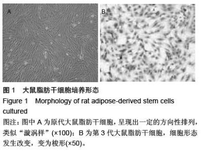
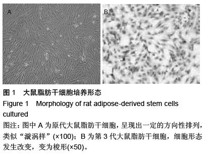
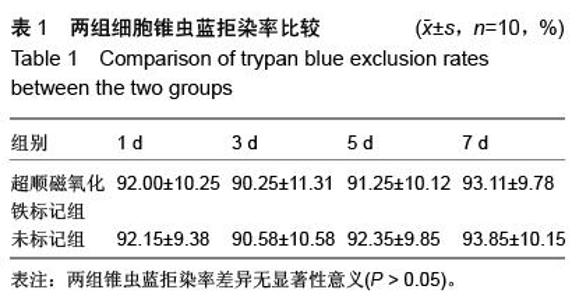
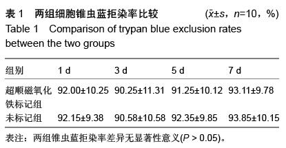
| [1] 陈长青,王小宜,陈晨,等.SPIO标记脂肪干细胞移植治疗大鼠脑梗死的磁共振示踪成像研究[J].磁共振成像, 2010, 1(1):50-54.
[2] 薛静,高培毅,李晋,等.胎鼠神经干细胞超顺磁性氧化铁颗粒标记移植后MRI研究[J].放射学实践,2006,21(2): 110-113.
[3] 晋光荣,徐汉荣,居胜红,等.超顺磁性氧化铁标记神经干细胞移植入局灶性脑缺血大鼠纹状体的MRI示踪及其对学习记忆的影响[J].临床神经病学杂志,2007,20(3): 203-206.
[4] 葛风,朱剑虹,刘军,等.纳米磁化标记神经干细胞的MRI大鼠活体示踪实验研究[J].中国临床神经科学,2005, 13(2): 152-155.
[5] 张鹏.骨髓源神经干细胞联合基质胶修复大鼠脑损伤及动态MEMRI成像大鼠视皮层实验研究[D].广州:南方医科大学,2010.
[6] 谢青松,许信龙,魏晓捷,等.新型超顺磁性氧化铁在干细胞活体示踪中的应用研究[J].中国临床药理学与治疗学, 2010,15(3): 272-276.
[7] 陈长青,陈晨,王小宜,等.体外标记脂肪源性干细胞移植治疗缺血性脑损伤的MRI示踪成像研究[J].临床放射学杂志, 2009, 28(2):266-270.
[8] 颜丽芬.超顺磁氧化铁标记对大鼠脂肪干细胞及其心肌样分化细胞转铁蛋白受体和铁蛋白表达的影响[D]. 广州:南方医科大学,2012.
[9] 陈长青.体外标记脂肪源性干细胞移植治疗缺血性脑损伤的MRI示踪成像研究[C].第二届放射青年医师学术论坛暨第一届全国MR功能成像学术研讨会论文集, 2008: 93-96.
[10] 谢光友.磁标记兔脂肪干细胞与建立兔腰椎间盘退变模型及MR定量的研究[D]. 重庆:重庆医科大学,2014.
[11] 景猛,刘新权,梁鹏,等.应用超顺磁性氧化铁纳米粒子标记神经干细胞及活体磁共振示踪的实验研究[J].中华医学杂志,2004, 84(16):1386-1389.
[12] 李宗芳,刘流,田伟,等.MR示踪观察面神经损伤大鼠脑内移植超顺磁性氧化铁标记神经干细胞[J].中国医学影像技术,2009,25(11):1941-1944.
[13] 薛静,高培毅,李晋,等.脑梗死大鼠脑内移植超顺磁性氧化铁颗粒标记神经干细胞后的MR示踪研究[J].中华放射学杂志,2006, 40(2):122-126.
[14] 张勇,程敬亮,李华丽,等.超顺磁性氧化铁标记骨髓间充质干细胞移植治疗大鼠脑梗死的在体磁敏感加权成像研究[J].中华生物医学工程杂志,2011,17(5):420-423.
[15] 徐汉荣.SPIO-PLL及Brdu标记神经干细胞移植入局灶性脑缺血大鼠脑纹状体的实验研究[D].南京:东南大学, 2006.
[16] 陈双庆,蔡庆,沈玉英,等.磁标记神经干细胞在APP/PS1转基因阿尔茨海默病小鼠脑内迁移的MRI研究[J].中华放射学杂志,2013,47(3):250-254.
[17] Zhang P, Li J, Liu Y, et al. Human embryonic neural stem cell transplantation increases subventricular zone cell proliferation and promotes peri-infarct angiogenesis after focal cerebral ischemia. Neuropathology. 2011;31(4):384-391.
[18] 张勇,程敬亮,王娟,等.磁标记大鼠BMSCs移植治疗脑梗死的在体磁敏感加权成像研究[C].第四届全国磁共振新技术应用与管理研讨会暨河南省第五届磁共振临床应用研讨会论文集,2013:126.
[19] Zhang P, Li J, Liu Y, et al. Transplanted human embryonic neural stem cells survive, migrate, differentiate and increase endogenous nestin expression in adult rat cortical peri-infarction zone. Neuropathology. 2009;29(4):410-421.
[20] 高伟,雷鹏,周杰,等.脂肪源性干细胞移植对颅脑损伤大鼠神经功能评分和神经细胞凋亡的影响[J].中国康复医学杂志,2010,25(11):1040-1043.
[21] 邓兴力,王应莉,杨智勇,等.SPIO、EGFP双标GDNF基因修饰中脑神经干细胞移植治疗帕金森病[J].中风与神经疾病杂志,2010,27(2):109-113.
[22] 黎国雄,王传湄,赵洪洋,等.磁性纳米粒标记的神经干细胞不同移植途径对大鼠脑损伤治疗的实验研究[J].中国现代医学杂志,2009,19(8):1134-1138.
[23] Zuk PA, Zhu M, Mizuno H, et al. Multilineage cells from human adipose tissue: implications for cell-based therapies.Tissue Eng. 2001;7(2):211-228.
[24] 谢光友,杨海涛,吕富荣,等.兔脂肪间充质干细胞的培养鉴定及体外磁标记MR成像[J].第三军医大学学报,2014, 36(15):1567-1571.
[25] 刘斌,董静,刘宁,等.人脂肪组织来源的神经干细胞移植治疗大鼠脑缺血再灌注损伤[J].基础医学与临床,2011, 31(7): 788-792.
[26] 王莹,李文媛,张大伟,等.电针联合脂肪源性干细胞移植对大鼠脑缺血再灌注后血管生成的影响[J].安徽中医学院学报,2012,31(2):29-33.
[27] 褚纯,赵元立.脂肪干细胞及其在脑缺血性疾病治疗中的研究进展[J].中国卒中杂志,2009,4(11):916-919.
[28] 王美芹,沈钧康,王诚,等.帕金森病大鼠尾静脉注射和脑内两点直接注射超顺磁性氧化铁标记的BMSCs后活体MRI示踪观察[J].苏州大学学报:医学版,2008,28(2): 226-228,265,封3.
[29] Ishikane S, Hosoda H, Yamahara K, et al. Allogeneic transplantation of fetal membrane-derived mesenchymal stem cell sheets increases neovascularization and improves cardiac function after myocardial infarction in rats.Transplantation. 2013; 96(8):697-706.
[30] Zimmet H, Porapakkham P, Porapakkham P, et al. Short- and long-term outcomes of intracoronary and endogenously mobilized bone marrow stem cells in the treatment of ST-segment elevation myocardial infarction: a meta-analysis of randomized control trials Eur J Heart Fail. 2012;14(1):91-105.
[31] 陈晓丹,郑洁,李成鹏,等.外磁场作用下SPIO标记脂肪干细胞经尾静脉移植治疗急性脑缺血[J].激光杂志,2013, 34(4):95-97.
[32] 王杰华,刘楠,杜厚伟,等.脂肪来源的干细胞移植对大鼠脑缺血后微血管生成及bFGF和VEGF表达的影响[J].细胞与分子免疫学杂志,2008,24(10):958-961,965.
[33] Liu YP, Seçkin H, Izci Y, et al. Neuroprotective effects of mesenchymal stem cells derived from human embryonic stem cells in transient focal cerebral ischemia in rats. J Cereb Blood Flow Metab. 2009; 29(4):780-791.
[34] Gambarota G, Leenders W. Characterization of tumor vasculature in mouse brain by USPIO contrast- enhanced MRI. Methods Mol Biol. 2011;771:477-487.
[35] Mou Y, Chen B, Zhang Y, et al. Influence of synthetic superparamagnetic iron oxide on dendritic cells. Int J Nanomedicine. 2011;6:1779-1786.
[36] 刘再毅,王瑛,王广谊,等.超顺磁性氧化铁标记脂肪干细胞移植入大鼠心脏后的活体磁共振示踪成像[J].中国医学科学院学报,2009,31(2):187-191,后插10.
[37] 朱文珍,李祥,漆剑频,等.SPIO标记的神经干细胞活体移植后细胞迁徙及功能性分化的MRI研究[C].第一届放射青年医师学术研讨会暨第三届分子影像学术研讨会论文集,2007:3-5.
[38] 白守民,薛卫平,刘宜敏,等.氧化铁标记神经干细胞对大鼠放射性脑损伤的磁共振成像研究[J].中华细胞与干细胞杂志:电子版,2012,2(1):23-26.
[39] 郭静,王兰,张荣俊,等.大鼠脑内移植超顺磁性氧化铁标记骨髓基质干细胞的MRI扫描序列优化研究[J].苏州大学学报:医学版,2010,30(1):31-33,封3.
[40] 彭德新,陈自谦,倪萍,等.大鼠脑创伤SPIO标记骨髓间充质干细胞磁共振成像活体示踪研究[J].功能与分子医学影像学杂志:电子版,2012,1(1):3-8. |