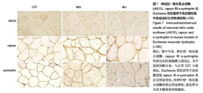
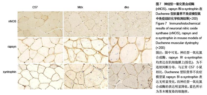
| [1] Emery AE. The muscular dystrophies. Lancet.2002; 359 (9307): 687-95.
[2] 张成,刘焯霖,梁秀龄.关于dystrophin中文译名的商讨[J].中华医学遗传学杂志; 15(2):7-9.
[3] Bulfield G, Siller WG, Wight PA, et al. X-chromosome-linked muscular dystrophy (mdx) in the mouse. Proc Natl Acad Sci. 1984; 81:1189-1192.
[4] Annee D, Jill AR, Judith AS, et al. Utrophin-dystrophin- deficient mice as a model for Duchenne muscular dystrophy. Cell.2002; 90(16):717-722.
[5] Lyons PR, Slater CR. Structure and function of the neuromuscular junction in young adult mdx mice.J Neurocytol.1991; 20: 969-981.
[6] Xu R, Salpeter M. Acetylcholine receptors in innervated muscles of dystrophic mdx mice degrade as after denervation. J Neurosci.1997; 17:8194-8200.
[7] Marques MJ, Pertille A, Carvalho CL, et al.Acetylcholine receptor organization at the dystrophic extraocular muscle neuromuscular junction. Anat Rec.2007; 290:846-854.
[8] Grady RM, Teng H, Nichol MC, et al. Skeletal and cardiac myopathies in mice lacking utrophin and dystrophin: a model for Duchenne muscular dystrophy. Cell. 1997; 90,729-738.
[9] Grady RM, Merlie JP, Sanes JR. Subtle neuromuscular defects in utrophin deficient mice. J Cell Biol.1997; 136: 871-882.
[10] Minatel E, Neto HS, Marques MJ. Acetylcholine receptor distribution and synapse elimination at the developing neuromuscular junction of mdx mice. Muscle Nerve.2001; 28, 561-569.
[11] Gautam M, Noakes PG. Defective neuromuscular synaptogenesis in agrin deficient mutant mice. Cell.1996; 85: 525-535.
[12] Jones G, Moore C, Hashem olhosseini S, et al. Constitutively active MuSK is clustered in the absence of Agrin and induces ectopic postsynaptic like membranes in skeletal muscle fibers. J Neurosc.1999; 19: 3376-3383.
[13] Gautam M, DeChiara TM, Glass DJ, et al. Distinct phenotypes of mutant mice lacking Agrin, MuSk, or Rapsyn. Brain Res Dev Brain Res.1999; 114: 171-178.
[14] Haenggi T, Fritschy JM. Role of dystrophin and utrophin for assembly and function of the dystrophin glycoprotein complex in non-muscle tissue. Cell Mol Life Sci.2006; 63:1614-1631.
[15] Allikian M, McNally EM. Processing and assembly of the dystrophin glycoprotein complex. Traffic.2007; 8:177-183.
[16] Shiao T. Defects in neuromuscular junction structure in dystrophic muscle are corrected by expression of a NOS transgene in dystrophin-deficient muscles, but not in muscles lacking α and β -syntrophins. Hum Mol Genet.2004; 13: 1873-1884.
[17] Lai Y. Dystrophins carrying spectrin-like repeats 16 and 17 anchor nNOS to the sarcolemma and enhance exercise performance in a mouse model of muscular dystrophy. J Clin Invest.2009; 119:624-635. |