| [1] Boffeli TJ,Pfannenstein RR,Thompson JC.Combined medial column primary arthrodesis, middle column open reduction internal fixation, and lateral column pinning for treatment of Lisfranc fracture-dislocation injuries.J Foot Ankle Surg.2014; 53(50):657-663.
[2] Panchbhavi VK,Molina D 4th,Villarreal J,et al.Three- dimensional,digital,and gross anatomy of the Lisfranc ligament. Foot Ankle Int. 2013;34(6): 876-880.
[3] Coss HS,Manos RE,Buoncristiani A,et al.Abduction stress and AP weight bearing radiography of purely ligamentous injury in the tarsometatarsal joint. Foot Ankle Int. 1998;19(8): 537-541.
[4] Castrol M, Melão L, Canella C. Lisfranc Joint Ligamentous Complex: MRI With Anatomic Correlationin Cadavers.AJR Am J Roentgenol.2010;195(6):447-455.
[5] Hirano T, Niki H, Beppu M. Anatomical considerations for reconstruction of the lisfranc ligament. J Orthop Sci. 2013; 18(5):720-726.
[6] Johnson A, Hill K, Ward J, et al. Anatomy of the lisfranc ligament. Foot Ankle Spec.2008;1(1):19-23.
[7] Rettedal DD, Graves NC, Marshall JJ, et al. Reliability of ultrasound imaging in the assessment of the dorsal Lisfranc ligament. J Foot Ankle Res. 2013;6(1):2-7.
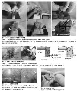
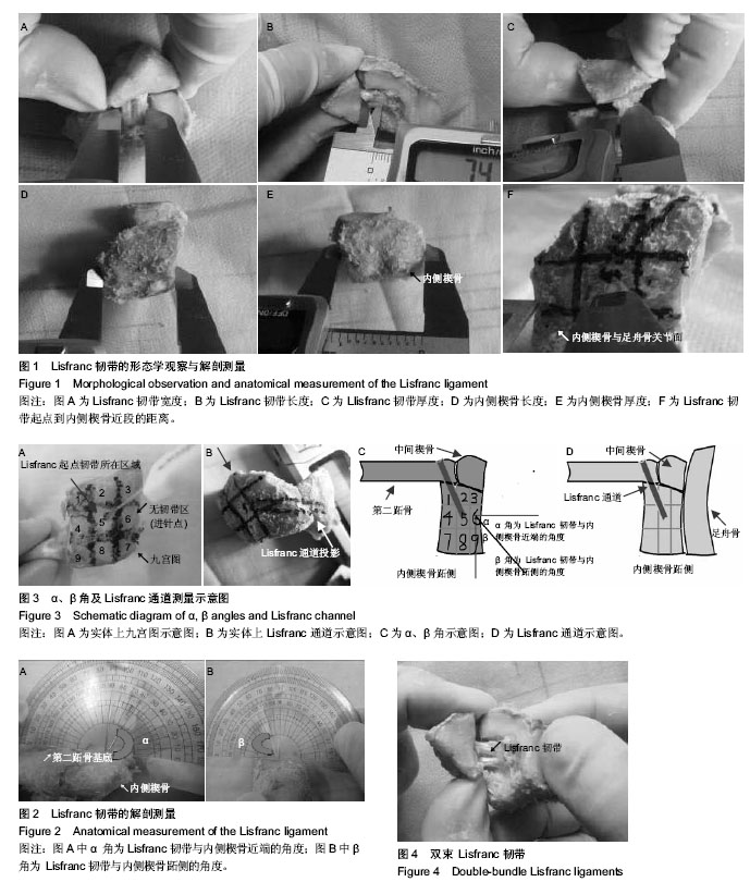
[8] 闫荣亮,曲家富,彭义,等. Lisfranc 韧带解剖与拉伸生物力学研究[J].中华创伤骨科杂志, 2013,15(3):230-234.
[9] Tarczyńska M,Gaweda K,Dajewski Z,et al.Comparison of treatment results of acute and late injuries of the lisfranc joint. Acta Ortop Bras. 2013;21(6):344-346.
[10] Diebal AR,Westrick RB,Alitz C, et al. Lisfranc injury in a west point cadet. Sports Heslth.2013;5(3):282-285.
[11] Sanli J, Hermus J,Poeze M.Primary internal fixation and soft-tissue reconstruction in the treatment for an open Lisfranc fracture-dislocation.Musculoskelet Surg. 2012;96(1):59-62.
[12] Miswan MF, Singh VA, Yasin NF.Outcome of surgically treated Lisfranc injury:A review of 34 cases.Ulus Travma Acil Cerrahi Derg. 2011;17(6):504-508.
[13] Kuo RS, Tejwani NC, Digiovanni CW, et al.Outcome after open reduction and internal fixation of Lisfranc joint injuries. J Bone Joint Surg. 2000;82-A(11):1609-1618.
[14] Lee CA, Birkedal JP, Dickerson EA, et al. Stabilization of Lisfranc joint injuries: a biomechanical study. Foot Ankle Int. 2004;25(5):365-370.
[15] Rammelt S, Schneiders W, Schikore H, et al.Primary open reduction and fixation compared with delayed corrective arthrodesis in the treatment of tarsometatarsal (Lisfranc) fracture dislocation. J Bone Joint Surg. 2008;90(11):499-506.
[16] Thordarson DB. Lisfranc ORIF with absorbable fixation. Foot Ankle Int. 2003;2(1):21-26.
[17] Mittlmeier T, Beck M. Injuries of midfoot. Chirurg. 2011; 82(2): 169-188. |