Chinese Journal of Tissue Engineering Research ›› 2015, Vol. 19 ›› Issue (4): 606-611.doi: 10.3969/j.issn.2095-4344.2015.04.020
Previous Articles Next Articles
Clinical significance of CT image digital screw channel analog measurement of C4-C6 anterior fixed parameters
Fu Yu1, Lin Bing2, Li Xiao-he3
- 1Department of Spine Surgery, Second Affiliated Hospital, Inner Mongolia Medical University, Hohhot 010110, Inner Mongolia Autonomous Region, China; 2Department of Radiology Laboratory, Armed Police Frontier Corps Hospital in Fujian Province, Quanzhou 362000, Fujian Province, China; 3Department of Human Anatomy, Basic Medical College, Inner Mongolia Medical University, Hohhot 010110, Inner Mongolia Autonomous Region, China
-
Revised:2014-11-24Online:2015-01-22Published:2015-01-22 -
Contact:Li Xiao-he, Ph.D., Associate professor, Department of Human Anatomy, Basic Medical College, Inner Mongolia Medical University, Hohhot 010110, Inner Mongolia Autonomous Region, China -
About author:Fu Yu, M.D., Associate chief physician, Department of Spine Surgery, Second Affiliated Hospital, Inner Mongolia Medical University, Hohhot 010110, Inner Mongolia Autonomous Region, China -
Supported by:the Natural Science Foundation of Inner Mongolia Autonomous Region, No. 2012MS1117; the National Natural Science Foundation of China, No. 81460330
CLC Number:
Cite this article
Fu Yu, Lin Bing, Li Xiao-he. Clinical significance of CT image digital screw channel analog measurement of C4-C6 anterior fixed parameters [J]. Chinese Journal of Tissue Engineering Research, 2015, 19(4): 606-611.
share this article
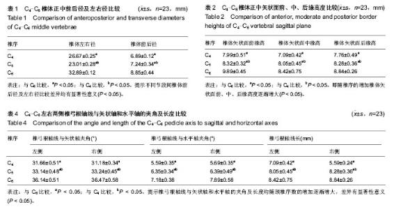
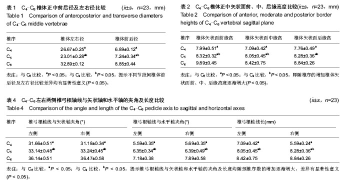
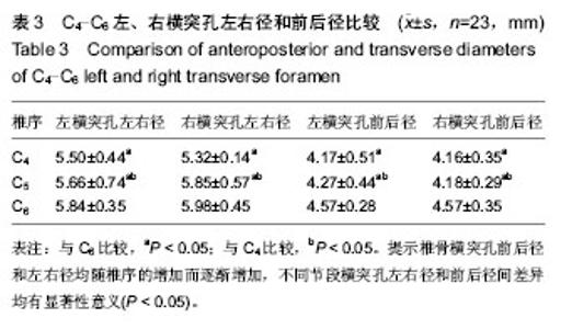
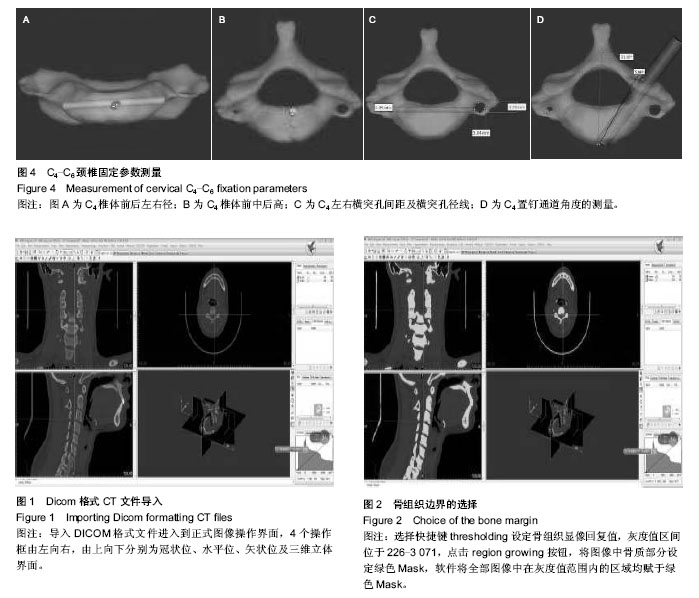
2.1 椎体前后径及左右径 椎体左右径C4-C6由(26.67± 0.25) mm逐渐增长到(32.89±0.12) mm,椎体前后径C4-C6由(6.89±0.12) mm逐渐增长到(8.85±0.44) mm,不同节段间比较差异均有显著性意义(表1)。 2.2 椎体矢状面高度测量结果 测量结果提示,C4-C6椎体正中矢状面前、中、后缘高度从C4到C6,椎体间比较差异有显著性意义(P < 0.05),即随椎序的增加椎体矢状面前、中、后缘高度逐渐增大(表2)。 2.3 椎骨横突孔的前后径及左右径测量结果 椎骨横突孔前后径和左右径均随椎序的增加而逐渐增加,相同椎骨左右横突孔前后及左右径间差异无显著性意义(P > 0.05),不同节段横突孔左右径和前后径间差异均有显著性意义(P < 0.05,表3)。 2.4 左右两侧横突孔内侧缘距离 C4-C6左右两侧横突孔内侧缘距离由(25.10±0.45) mm逐渐增长到(28.89± 0.56) mm,不同节段间比较差异有显著性意义(P < 0.05)。 2.5 左右两侧椎弓根轴线与矢状轴和水平轴的夹角及长度 相同节段椎弓根轴线与矢状轴、水平轴长夹角和椎弓根轴线左右两侧差异无显著性意义(P > 0.05),椎弓根轴线与矢状轴和水平轴的夹角及长度均随颈椎序数的增加逐渐增大,差异有显著性意义(P < 0.05,表4)。"
| [1] Lee A, Sommer D, Reddy K,et al.Endoscopic Transnasal Approach to the Craniocervical Junction. Skull Base. 2010; 20(3):199-205.
[2] Ai F, Yin Q, Xia H, et al. Applied anatomy of tranoral atlanto-axial reduction plate internal fixation. Spine. 2006; 31(2):128-132.
[3] Cloward RB. The anterior surgical approach to the cervical spine: the Cloward Procedure: past, present, and future. The presidential guest lecture, Cervical Spine Research Society. Spine.1988;13(7):823-827.
[4] 张帆,王方方,杨志高,等. 颈前路钩椎关节减压联合改良植骨术治疗颈椎病[J].中国脊柱脊髓杂志,2011,21(7):578-582.
[5] 黄阳亮,刘少喻,赵卫东,等.颈前路钢板置入内固定加椎间植骨治疗Ⅱ型Hangman骨折的生物力学评价术[J].中国组织工程研究与临床康复杂志,2010,14(39):7251-7253.
[6] Müller J, Wissel J, Kemmler G,et al. Craniocervical dystonia questionnaire (CDQ-24):development and validation of a disease-specific quality of life instrument. J Neurol Neurosurg Psychiatry. 2004;75(2):749-753.
[7] Martins C, Cardoso AC, Alencastro LF,et al.Endoscopic- assisted lateral transatlantal approach to craniovertebral junction. World Neurosurg.2010;74(2): 351-358.
[8] Pillai P, Baig MN, Karas CS, et al.Endoscopic image-guided transoral approach to the craniovertebral junction: an anatomic study comparing surgical exposure and surgical freedom obtained with the endoscope and the operating microscope.Neurosurgery. 2009;64(5 Suppl 2):437-442.
[9] 赵建华,金大地,李明.脊柱外科实用技术[M].北京:人民军医出版社,2005:323-335.
[10] 冯虎,马志兵,齐祥如,等.颈前路钢板固定位置与相邻节段退变之间关系的研究[J].中国骨与关节损伤杂志,2011,26 (1):1-4.
[11] 李金泉,徐皓,姚晓东,等.两种不同手术入路治疗不稳定Hangman骨折的比较[J].中国矫形外科杂志,2010,18(4): 280-283.
[12] 罗洪艳,李敏,张绍祥,等.数字人图像的自动分割方法[J].华南理工大学学报(自然科学版),2011,39(7):109-113.
[13] 袁元杏,万磊,尹庆水,等.中国数字人CT数据颈椎运动节段有限元模型的建立[J].中国组织工程研究与临床康复, 2011, 15(26): 4915-4917.
[14] 吕婷.数字人体研究及其应用[J].中国组织工程研究与临床康复,2010,14(48):9041-9045.
[15] 王远政.下颈椎前路经椎弓根螺钉内固定技术的研究与应用[D].重庆医科大学,2013.
[16] Hussain M, Nassr A,Raghu N, et al.Biomechanical effects of anterior, posterior, and combined anterior-posterior instrumentation techniques on the stability of a multilevel cervical corpectomy construct: a finite element model analysis. Spine J. 2011;11(4):324-330.
[17] Okawa A, Sakai K, Hirai T, et al. Risk Factors for Early Reconstruction Failure of Multilevel Cervical Corpectomy With Dynamic Plate Fixation. Spine. 2011;36(9):E582-587.
[18] Koller H, Schmidt R, Mayer M,et al. The stabilizing potential of anterior, posterior and combined techniques for the reconstruction of a 2-level cervical corpectomy model: biomechanical study and first results of ATPS prototyping. Eur Spine J. 2010;19(12):2137-2148.
[19] Robinson RA, Smith GW. Anterolateral cervical disc removal and interbody fusion for cervical disc syndrome. SAS J. 2014;12(1):245-248.
[20] Abuzayed B, Tutunculer B, Kucukyuruk B, et al. Anatomic basis of anterior and posterior instrumentation of the spine: morphometric study. Surg Radiol Anat.2010;32(1):75-85.
[21] Barrenechea IJ.One-stage open reduction of an old cervical subluxation: case report.Global Spine J. 2014;4(4): 263-268.
[22] Tschugg A, Neururer S, Scheufler KM,et al.Comparison of posterior foraminotomy and anterior foraminotomy with fusion for treating spondylotic foraminal stenosis of the cervical spine: study protocol for a randomized controlled trial (ForaC).Trials. 2014;15(1):437.
[23] Floeth FW, Herdmann J, Rhee S, et al.Open microsurgical tumor excavation and vertebroplasty for metastatic destruction of the second cervical vertebra-outcome in 7 cases. Spine J.2014; pii:S1529-9430.
[24] Caruso R, Pesce A, Marrocco L, et al.Anterior approach to the cervical spine for treatment of spondylosis or disc herniation: Long-term results. Comparison between ACD, ACDF, TDR. Clin Ter. 2014;165(4):e263-270.
[25] Ito H, Takai K, Taniguchi M.Cervical duraplasty with tenting sutures via laminoplasty for cervical flexion myelopathy in patients with Hirayama disease: successful decompression of a "tight dural canal in flexion" without spinal fusion.J Neurosurg Spine. 2014;21(5):743-752.
[26] Barrett RJ, Sandquist L, Richards BF, et al.Antibiotic- impregnated polymethylmethacrylate as an anterior biomechanical device for the treatment of cervical discitis and vertebral osteomyelitis: technical report of two cases.Turk Neurosurg. 2014;24(4):613-617.
[27] Landi A, Marotta N, Mancarella C, et al.360° fusion for realignment of high grade cervical kyphosis by one step surgery: Case report.World J Clin Cases. 2014;2(7): 289-292.
[28] Oberkircher L, Born S, Struewer J,et al. Biomechanical evaluation of the impact of various facet joint lesions on the primary stability of anterior plate fixation in cervical dislocation injuries: a cadaver study:Laboratory investigation.J Neurosurg Spine. 2014;21(4):634-639.
[29] Yang JS, Chu L, Chen L, et al. Anterior or posterior approach of full-endoscopic cervical discectomy for cervical intervertebral disc herniation? A comparative cohort study.Spine (Phila Pa 1976). 2014;39(21):1743-1750.
[30] An HS, Al-Shihabi L, Kurd M.Surgical treatment for ossification of the posterior longitudinal ligament in the cervical spine.J Am Acad Orthop Surg. 2014;22(7): 420-429.
[31] Subedi A, Tripathi M, Bhattarai B, et al. Successful Intubation with McCoy Laryngoscope in a Patient with Ankylosing Spondylitis.J Nepal Health Res Counc. 2014;12(26):70-72.
[32] Menzer H, Gill GK, Paterson A.Thoracic spine sports-related injuries.Curr Sports Med Rep. 2015;14(1): 34-40.
[33] Khan A, Dubois S, Khan AA, et al.A randomized, double-blind, placebo-controlled study to evaluate the effects of alendronate on bone mineral density and bone remodelling in perimenopausal women with low bone mineral density. J Obstet Gynaecol Can. 2014;36(11): 976-982.
[34] Wong B, Ong BB, Milne N. The source of haemorrhage in traumatic basal subarachnoid haemorrhage.J Forensic Leg Med. 2015;29(1):18-23.
[35] Hoxha R, Islami H, Qorraj-Bytyqi H, et al.Relationship of weight and body mass index with bone mineral density in adult men from kosovo.Mater Sociomed. 2014;26(5): 306-308.
[36] Kasumovic M, Gorcevic E, Gorcevic S. Cervical syndrome - the effectiveness of physical therapy interventions. Med Arch. 2013;67(6):414-417.
[37] Ando K, Imagama S, Ito Z, et al. Progressive relapse of ligamentum flavum ossification following decompressive surgery.Asian Spine J. 2014;8(6):835-839.
[38] Oh JY, Kapoor S, Koh RK, et al. Spinal cord ischemia secondary to hypovolemic shock.Asian Spine J. 2014;8(6): 831-834.
[39] Ando K, Imagama S, Ito Z, et al. Progressive relapse of ligamentum flavum ossification following decompressiv surgery. Asian Spine J. 2014;8(6):835-839.
[40] Oh JY, Kapoor S, Koh RK, et al. Spinal cord ischemia secondary to hypovolemic shock.Asian Spine J. 2014;8(6): 831-834. |
| [1] | Chen Qun-qun, Qiao Rong-qin, Duan Rui-qi, Hu Nian-hong, Li Zhao, Shao Min. Acu-Loc®2 volar distal radius bone plate system for repairing type C fracture of distal radius [J]. Chinese Journal of Tissue Engineering Research, 2017, 21(7): 1025-1030. |
| [2] | Li Peng, Li Hong-wei, Wang Shuang, Wang Hai-zhou. Digital anatomy of lumbar spinous process tilt angle of adults in northeast China: prodinding reference for pedicle screw insertion [J]. Chinese Journal of Tissue Engineering Research, 2017, 21(7): 1064-1068. |
| [3] | Zou Wei, Xiao Jie, Long Hao, Zhang Yang, Wu Chen, Du Yu-hui, Feng Ming-xing, Zhou Chang-jun. Screw placement selection of minimally invasive percutaneous pedicle screw fixation for thoracolumbar fractures [J]. Chinese Journal of Tissue Engineering Research, 2017, 21(3): 356-361. |
| [4] | Chen Lu-yao, Hu Shi-qiang, Wang Xiao-ping, Wu Wei-wei, Wei Zhan-tu, Huang Jian. Accuracy of digital orthopedic three-dimensional reconstruction for thoracolumbar pedicle screw placement [J]. Chinese Journal of Tissue Engineering Research, 2017, 21(3): 373-377. |
| [5] | Du Shi-yao, Zhou Feng-jin, Ni Bin, Chen Bo, Chen Jin-shui. Finite-element analysis of a novel posterior atlantoaxial restricted non-fusion fixation system [J]. Chinese Journal of Tissue Engineering Research, 2017, 21(3): 383-389. |
| [6] | Liu Jun, Liao Su-ping. Three-dimensional finite element analysis of Kirschner nails and external fixation for Bennett fracture [J]. Chinese Journal of Tissue Engineering Research, 2017, 21(3): 390-395. |
| [7] | Sheng Xiao-lei, Yuan Feng, Li Zhi-duo, Yang Yu-ming, Lu Hai-tao, Zhang Jun-wei. Comparison of the accuracy of lower cervical anterior transpedicular screws between three-dimensional printing assembly navigation template and free hand placement [J]. Chinese Journal of Tissue Engineering Research, 2017, 21(3): 406-411. |
| [8] | Wu Min-hao, Sun Wen-chao, Yan Fei-fei, Xie Yuan-long, Hou Zhi-qiang, Feng Fan, Cai Lin . Treatment research and new progress of early-onset scoliosis [J]. Chinese Journal of Tissue Engineering Research, 2017, 21(3): 433-439. |
| [9] | Wang Lei, Wang Feng-feng, Ma Yan-hui, Zhang Jie, Hu Fang, Ma Gai-ping, Liu Mei-mei, Ma Zhang-wen. Meta analysis of clinical outcome of intramedullary nails versus locking plates for two-part proximal humerus fracture [J]. Chinese Journal of Tissue Engineering Research, 2017, 21(3): 478-484. |
| [10] | Hu Jun, Zhang De-qiang, Tang Xin. Postoperative quality of life of internal fixation versus hemiarthroplasty for femoral neck fractures in the elderly [J]. Chinese Journal of Tissue Engineering Research, 2017, 21(19): 2953-2960. |
| [11] | Jiang Wei, Yuan Feng. Unilateral pedicle screw fixation combined with translaminar facet screw fixation versus bilateral pedicle screw fixation for lower lumbar degenerative diseases: a 2-year follow-up [J]. Chinese Journal of Tissue Engineering Research, 2017, 21(19): 2973-2979. |
| [12] | Wang Xiu-ping, Sun Rui-bo, Liu You-wen, Zhang Ying, Jia Yu-dong, Yang Yu-xia, Wang Hui-chao. Femoral neck fractures fixed with intramedullary cannulated screws: factors for postoperative functional recovery [J]. Chinese Journal of Tissue Engineering Research, 2017, 21(19): 2999-3004. |
| [13] | Liu Gang, Zhang Lei, Wang Guo-you, Zhou Xin, Zhang Tao, Guan Tai-yuan, Guo Xiao-guang, Fu Shi-jie. Double-row suture anchors under arthroscopy for avulsion-type greater tuberosity fractures (Mutch type I) [J]. Chinese Journal of Tissue Engineering Research, 2017, 21(19): 3005-3010. |
| [14] | Yang Min, Ma Xiang-yang, Yang Jin-cheng, Chen Shu-jin, Zou Xiao-bao. Biomechanical properties of a novel automatic anti-rotation posterior atlantoaxial internal fixation system: a finite element analysis [J]. Chinese Journal of Tissue Engineering Research, 2017, 21(19): 3031-3037. |
| [15] | Qiu Hao, Lu Min-peng, Dong Jing, Zhang Zhong-zu, Chu Tong-wei, Wang Qun-bo, Quan Zheng-xue, Jiang Dian-ming. Subtotal corpectomy and reconstruction with titanium mesh cage implantation and pedicle screw fixation through posterior approach in treatment of thoracolumbar burst fracture or thoracolumbar fracture dislocation [J]. Chinese Journal of Tissue Engineering Research, 2016, 20(53): 7932-7938. |
| Viewed | ||||||
|
Full text |
|
|||||
|
Abstract |
|
|||||
