Chinese Journal of Tissue Engineering Research ›› 2014, Vol. 18 ›› Issue (52): 8420-8425.doi: 10.3969/j.issn.2095-4344.2014.52.011
Previous Articles Next Articles
Adhesion ability and biofilm formation of Mycobacterium tuberculosis in the interface of polyethylene
Xiong Qing-guang1, Wang Yong-qing1, Zhao Zhi-hui1, Bi Hong-bin2, Sun Jin-bao1, Zhang Qing-jie2, Hao Xiao-hui1, Li Yi1
- 1Department of Orthopedics, the Fourth Central Clinical College, Tianjin Medical University, Tianjin 300140, China; 2Tianjin University of Traditional Chinese Medicine, Tianjin 300193, China
-
Revised:2014-11-12Online:2014-12-17Published:2014-12-17 -
Contact:Wang Yong-qing, Professor, Chief physician, Master’s supervisor, Doctoral supervisor, Department of Orthopedics, the Fourth Central Clinical College, Tianjin Medical University, Tianjin 300140, China -
About author:Xiong Qing-guang, Studying for master’s degree, Department of Orthopedics, the Fourth Central Clinical College, Tianjin Medical University, Tianjin 300140, China -
Supported by:Science and Technology Foundation of Tianjin Bureau of Public Health, No. 2013KY05
CLC Number:
Cite this article
Xiong Qing-guang, Wang Yong-qing, Zhao Zhi-hui, Bi Hong-bin, Sun Jin-bao, Zhang Qing-jie, Hao Xiao-hui, Li Yi. Adhesion ability and biofilm formation of Mycobacterium tuberculosis in the interface of polyethylene[J]. Chinese Journal of Tissue Engineering Research, 2014, 18(52): 8420-8425.
share this article

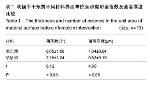
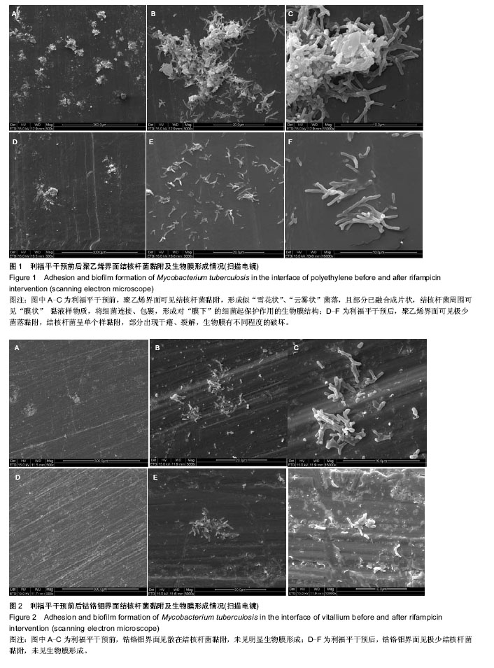
2.1 激光共聚焦显微镜分析结果 利福平干预前,实验组界面单位面积黏附的菌落数与菌落厚度多于对照组(P < 0.05),见表1。利福平干预后,聚乙烯界面单位面积黏附菌落数及菌落厚度分别为(2.24±0.84)个、(0.74±0.22) μm,均较干预前均明显减少(t =8.38,4.45,P < 0.05)。 2.2 扫描电镜观察结果 500倍视野下,聚乙烯界面可见结核杆菌黏附,形成似“雪花状”、“云雾状”,且部分菌落间已融合成片状;放大至5 000,15 000倍,可见大量结核杆菌聚集,成为立体的、复杂的膜样结构,结核杆菌周围可见“膜状” 黏液样物质,将细菌连接、包裹,形成对“膜下”的细菌起保护作用的生物膜结构,见图1A-C。500倍视野下,钴铬钼界面可见散在结核杆菌菌落,但见未见融合;放大至5 000,15 000倍,结核杆菌呈单个样黏附,彼此之间不发生聚集、粘连,未见生物膜结构形成,见图2A-C。利福平干预后,500倍视野下聚乙烯界面黏附菌落数减少,继续放大至5 000,15 000倍,部分结核杆菌出现干瘪、裂解,生物膜有不同程度的破坏,见图1D-F;钴铬钼界面未见菌落形成,偶见单个结核杆菌黏附,未见生物膜形成,见图2D-F。"

| [1] Chocholac D,Kala B,Gallo J,et al.Evaluation of treatment outcomes in tuberculosis of knee and hip joints in 2005-2012. Acta Chir Orthop Traumatol Cech.2013; 80(4): 256-262.
[2] De Haan J,Vreeling AW,Van Hellemondt G.Reactivation of ancient joint tuberculosis of the knee following total knee arthroplasty after 61 years: a case report. Knee.2008;15(4): 336-338.
[3] Furia JP,Box G,Lintner DM.Tuberculous arthritis of the knee presenting as a meniscal tear. Am J Orthop (Belle Mead NJ).1996;25(2):138-142.
[4] Neogi DS,Yadav CS,Ashok K,et al.Total hip arthroplasty in patients with active tuberculosis of the hip with advanced arthritis.Clin Orthop Relat Res.2010; 468(2):605-612.
[5] 王永清,毕红宾,赵志辉,等.晚期活动性全髋关节结核一期病灶清除全髋关节置换术的中远期疗效[J].中华骨科杂志,2013,33(9): 895-900.
[6] Wang Y,Wang J,Xu Z,et al.Total hip arthroplasty for active tuberculosis of the hip.Int Orthop.2010;34(8):1111-1114.
[7] 张晓岗,任姜栋,曹力,等.一期全髋关节置换术治疗晚期活动性髋关节结核[J].中华骨科杂志,2013,33(1):8-13.
[8] Finlay B,Falkow S.Common themes in microbial pathogenicity revisited. Microbiol Mol Biol Rev.1997;61(2):136-169.
[9] 毛彦杰,沈灏,蒋垚.生物膜与人工关节假体感染关系研究进展[J].中国修复重建外科杂志,2010,24(12):1463-1467.
[10] Costerton JW.Biofilm theory can guide the treatment of device-related orthopaedic infections.Clin Orthop Relat Res. 2005;(437):7-11.
[11] 马骏,李国庆,曹力.结核杆菌黏附不同人工关节假体材料的能力研究[J].中国组织工程研究,2012,16(47):8807-8812.
[12] 周劲松,陈建庭,金大地,等.结核分枝杆菌对材料粘附能力的体外实验研究[J].中国脊柱脊髓杂志,2003,13(11):26-29.
[13] 张宏其,郭超峰,唐明星,等.一期后路病灶清除、异形钛网椎间植骨融合治疗胸、腰椎结核[J].中华骨科杂志,2014,34(2): 102-108.
[14] 赵晨,蒲小兵,周强,等.后路病灶清除、椎间植骨融合内固定治疗复杂性胸、腰椎结核[J].中华骨科杂志,2014,34(2):109-115.
[15] 黄迅悟,冯会成,孙继桐,等.活动性髋关节结核一期病灶清除全髋关节置换28例报告[J].中华骨科杂志,2013,33(5):495-500.
[16] 魏召劝,孙俊英,查国春,等.采用高交联聚乙烯与传统聚乙烯髋臼内衬行人工全髋关节置换的比较研究[J].中国修复重建外科杂志, 2013,27(12):1414-1418.
[17] Judd KT,Noiseux N.Concomitant infection and local metal reaction in patients undergoing revision of metal on metal total hip arthroplasty . Iowa Orthop J. 2011; 31:59-63.
[18] Hosman AH,Van Der Mei HC,Bulstra SK,et al.Effects of metal-on-metal wear on the host immune system and infection in hip arthroplasty.Acta Orthop.2010; 81(5):526-534.
[19] Schildhauer TA,Robie B,Muhr G,et al.Bacterial adherence to tantalum versus commonly used orthopedic metallic implant materials.J Orthop Trauma.2006;20(7):476-484.
[20] Finlay B,Falkow S.Common themes in microbial pathogenicity revisited. Microbiol Mol Biol Rev.1997;61(2):136-169.
[21] Arnold WV,Shirtliff ME,Stoodley P.Bacterial biofilms and periprosthetic infections.J Bone Joint Surg Am.2013;95(24): 2223-2229.
[22] Chen WH,Jiang LS,Dai LY.Influence of bacteria on spinal implant centered infection: an in vitro and in vivo experimental comparison between Staphylococcus aureus and mycobacterium tuberculosis. Spine (Phila Pa 1976).2011; 36(2):103-108.
[23] 李传有,端木宏谨.结核分枝杆菌粘附素和结核病[C].中国防痨协会全国学术会议论文集,2005:214-215.
[24] Sanz J,Navarro J,Arbues A,et al.The transcriptional regulatory network of Mycobacterium tuberculosis. PLoS One.2011;6(7):e22178.
[25] 黄良库,徐涛,唐进,等.夫西地酸钠联合利福平对表皮葡萄球菌体外培养生物膜的作用[J].中国组织工程研究与临床康复, 2011, 15(21):3891-3894.
[26] Ojha AK,Baughn AD,Sambandan D,et al.Growth of Mycobacterium tuberculosis biofilms containing free mycolic acids and harbouring drug-tolerant bacteria. Mol Microbiol. 2008;69(1):164-174.
[27] Kulka K,Hatfull G,Ojha AK. Growth of Mycobacterium tuberculosis biofilms. J Vis Exp.2012;(60).pii: 3820.
[28] Grivet M, Morrier JJ, Benay G. Effect of hydrophobicity on in vitro streptococcal adhesion to dental alloys.J Mater Sci Mater Med.2000;11(10):637-642.
[29] Quitynen M,Bollen M.The influence of surface roughness and surface free energy on supra and subgingival plaque formation in man A review of the literature. J Clin Periodontol. 1995;22(1):1-14.
[30] Ramsugit S, Guma S,Pillay B,et al. Pili contributes to biofilm formation in vitro in Mycobacterium tuberculosis. Antonie Van Leeuwenhoek.2013;104(5):725-735.
[31] Alteri CJ,Xicohténcatl-Cortes J,Hess S,et al.Mycobacterium tuberculosis produces pili during human infection.Proc Natl Acad Sci U S A. 2007;104(12):5145-5150.
[32] Gerstel U, Park C, Romling U.Complex regulation of csgD promoter activity by global regulatory proteins. Mol Microbiol. 2003;49:639-654.
[33] Latasa C,Roux A,Toledo-Arana A,et al.BapA, a large secreted protein required for biofilm formation and host colonization of Salmonella enterica serovar Enteritidis. Mol Microbiol. 2005; 58:1322-1339.
[34] 刘霞,郭庆龙,王若珺,等.结核分枝杆菌生物膜形成相关基因的筛选与鉴定[J].中国生物工程杂志,2013,33(4):15-21.
[35] Pang JM.Genetics of biofilm maturation in Mycobacterium tuberculosis. University of Washington,2011.
[36] 王星.表皮葡萄球菌和分枝杆菌生物膜相关基因的鉴定和功能研究[D].复旦大学, 2012.
[37] Pang JM,Layre E,Sweet L,et al.The polyketide Pks1 contributes to biofilm formation in Mycobacterium tuberculosis. J Bacteriol.2012;194(3):715-721.
[38] Fuqua WC,Winans SC,Greenberg EP.Quorum sensing in bacteria: The Lux R-Lux I family of density-responsive transcriptional regulators.J Bacteriol.1994; 176(2):269-275.
[39] Dickschat JS.Quorum sensing and bacterial biofilms. Nat Prod Rep.2010; 27(3):343-369. |
| [1] | Chen Ziyang, Pu Rui, Deng Shuang, Yuan Lingyan. Regulatory effect of exosomes on exercise-mediated insulin resistance diseases [J]. Chinese Journal of Tissue Engineering Research, 2021, 25(25): 4089-4094. |
| [2] | Chen Yang, Huang Denggao, Gao Yuanhui, Wang Shunlan, Cao Hui, Zheng Linlin, He Haowei, Luo Siqin, Xiao Jingchuan, Zhang Yingai, Zhang Shufang. Low-intensity pulsed ultrasound promotes the proliferation and adhesion of human adipose-derived mesenchymal stem cells [J]. Chinese Journal of Tissue Engineering Research, 2021, 25(25): 3949-3955. |
| [3] | Yang Junhui, Luo Jinli, Yuan Xiaoping. Effects of human growth hormone on proliferation and osteogenic differentiation of human periodontal ligament stem cells [J]. Chinese Journal of Tissue Engineering Research, 2021, 25(25): 3956-3961. |
| [4] | Sun Jianwei, Yang Xinming, Zhang Ying. Effect of montelukast combined with bone marrow mesenchymal stem cell transplantation on spinal cord injury in rat models [J]. Chinese Journal of Tissue Engineering Research, 2021, 25(25): 3962-3969. |
| [5] | Gao Shan, Huang Dongjing, Hong Haiman, Jia Jingqiao, Meng Fei. Comparison on the curative effect of human placenta-derived mesenchymal stem cells and induced islet-like cells in gestational diabetes mellitus rats [J]. Chinese Journal of Tissue Engineering Research, 2021, 25(25): 3981-3987. |
| [6] | Hao Xiaona, Zhang Yingjie, Li Yuyun, Xu Tao. Bone marrow mesenchymal stem cells overexpressing prolyl oligopeptidase on the repair of liver fibrosis in rat models [J]. Chinese Journal of Tissue Engineering Research, 2021, 25(25): 3988-3993. |
| [7] | Liu Jianyou, Jia Zhongwei, Niu Jiawei, Cao Xinjie, Zhang Dong, Wei Jie. A new method for measuring the anteversion angle of the femoral neck by constructing the three-dimensional digital model of the femur [J]. Chinese Journal of Tissue Engineering Research, 2021, 25(24): 3779-3783. |
| [8] | Meng Lingjie, Qian Hui, Sheng Xiaolei, Lu Jianfeng, Huang Jianping, Qi Liangang, Liu Zongbao. Application of three-dimensional printing technology combined with bone cement in minimally invasive treatment of the collapsed Sanders III type of calcaneal fractures [J]. Chinese Journal of Tissue Engineering Research, 2021, 25(24): 3784-3789. |
| [9] | Qian Xuankun, Huang Hefei, Wu Chengcong, Liu Keting, Ou Hua, Zhang Jinpeng, Ren Jing, Wan Jianshan. Computer-assisted navigation combined with minimally invasive transforaminal lumbar interbody fusion for lumbar spondylolisthesis [J]. Chinese Journal of Tissue Engineering Research, 2021, 25(24): 3790-3795. |
| [10] | Hu Jing, Xiang Yang, Ye Chuan, Han Ziji. Three-dimensional printing assisted screw placement and freehand pedicle screw fixation in the treatment of thoracolumbar fractures: 1-year follow-up [J]. Chinese Journal of Tissue Engineering Research, 2021, 25(24): 3804-3809. |
| [11] | Shu Qihang, Liao Yijia, Xue Jingbo, Yan Yiguo, Wang Cheng. Three-dimensional finite element analysis of a new three-dimensional printed porous fusion cage for cervical vertebra [J]. Chinese Journal of Tissue Engineering Research, 2021, 25(24): 3810-3815. |
| [12] | Wang Yihan, Li Yang, Zhang Ling, Zhang Rui, Xu Ruida, Han Xiaofeng, Cheng Guangqi, Wang Weil. Application of three-dimensional visualization technology for digital orthopedics in the reduction and fixation of intertrochanteric fracture [J]. Chinese Journal of Tissue Engineering Research, 2021, 25(24): 3816-3820. |
| [13] | Sun Maji, Wang Qiuan, Zhang Xingchen, Guo Chong, Yuan Feng, Guo Kaijin. Development and biomechanical analysis of a new anterior cervical pedicle screw fixation system [J]. Chinese Journal of Tissue Engineering Research, 2021, 25(24): 3821-3825. |
| [14] | Lin Wang, Wang Yingying, Guo Weizhong, Yuan Cuihua, Xu Shenggui, Zhang Shenshen, Lin Chengshou. Adopting expanded lateral approach to enhance the mechanical stability and knee function for treating posterolateral column fracture of tibial plateau [J]. Chinese Journal of Tissue Engineering Research, 2021, 25(24): 3826-3827. |
| [15] | Zhu Yun, Chen Yu, Qiu Hao, Liu Dun, Jin Guorong, Chen Shimou, Weng Zheng. Finite element analysis for treatment of osteoporotic femoral fracture with far cortical locking screw [J]. Chinese Journal of Tissue Engineering Research, 2021, 25(24): 3832-3837. |
| Viewed | ||||||
|
Full text |
|
|||||
|
Abstract |
|
|||||

