Chinese Journal of Tissue Engineering Research ›› 2014, Vol. 18 ›› Issue (48): 7867-7872.doi: 10.3969/j.issn.2095-4344.2014.48.028

Previous Articles    

Prevalence and distribution of Schmorl’s nodes in cervical segment and its relationship with intervertebral disc degeneration

Cao Sheng1, 2, Zhang Xue-li2, Hu Wei2, Zhu Ru-sen2, Wan Jun2, Liu Yan2   

  1. 1Graduate School of Tianjin Medical University, Tianjin 300070, China; 2First Department of Spine, Tianjin People’s Hospital, Tianjin 300121, China
  • Received:2014-09-28 Online:2014-11-26 Published:2014-11-26
  • Contact: Zhang Xue-li, Chief physician, Professor, First Department of Spine, Tianjin People’s Hospital, Tianjin 300121, China
  • About author:Cao Sheng, Studying for master’s degree, Physician, Graduate School of Tianjin Medical University, Tianjin 300070, China

Abstract:

BACKGROUND: The distribution and characteristics of Schmorl’s nodes in lumbar spine have been described, but there are few studies focusing on Schmorl’s nodes in the cervical spine.
OBJECTIVE: To evaluate the distribution of Schmorl’s nodes in patients suffered neck pain and analyze the factors responsible for Schmorl’s nodes. 
METHODS: From January to December in 2012, 820 patients suffered neck pain were examined by magnetic resonance scan and plain film. There were 398 males and 422 females, with an average age of 52.2±11.5 years (range 30-80 years). The distribution features of Schmorl’s nodes in cervical endplate on age, sex, segment, head circumference, cervical curvature, and the type and grade of intervertebral disc degeneration were analyzed retrospectively.  
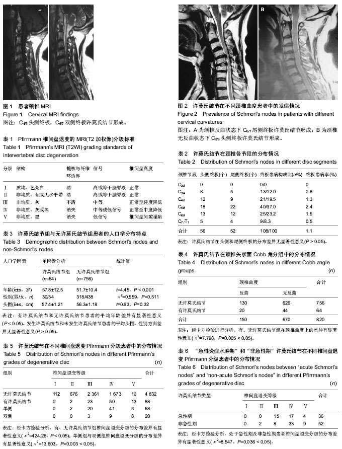
RESULTS AND CONCLUSION: Among 4 920 cervical intervertebral segments of the 820 patients, 64 cases (7.8%) and 108 endplates (1.1%) were involved with Schmorl’s nodes, the incidence of Schmorl’s nodes in the cranial and the caudal endplates was not statistically significant (χ2=1.471, P=0.689 > 0.05). According to the prevalence per cervical segment, C5/6 was the most common level (2.4%), followed by C6/7 (1.5%) and C4/5 (1.3%). The incidence of Schmorl’s nodes was positively correlated with age, cervical curvature, the type and grade of intervertebral disc degeneration    (P < 0.05), but not with sex and head circumference (P > 0.05). The distributions of the grade of intervertebral disc degeneration were significantly different between groups with and without adjacent Schmorl’s nodes (χ2=424.26,      P < 0.05). Intervertebral discs with adjacent Schmorl’s nodes were more degenerated than those without adjacent Schmorl’s nodes (Z=58.80, P < 0.05). The distributions of the grade of intervertebral disc degeneration were significantly different between groups with both sides Schmorl’s nodes and one side Schmorl’s nodes (χ2=13.603, P=0.003 < 0.05). The both sides Schmorl’s nodes were more degenerated (Z=3.44, P < 0.05). The distributions of the grade of intervertebral disc degeneration were significantly different between groups with “acute Schmorl’s nodes” and “non-acute Schmorl’s nodes” (χ2=8.547, P=0.036 < 0.05). The “non-acute Schmorl’s nodes” were more degenerated (Z=2.40,      P < 0.05). Schmorl’s nodes distribution was correlated with disc degeneration, then age, and cervical curvature. Schmorl’s nodes occurred often in C5/6 segment in patients with neck pain. The incidence of Schmorl’s nodes in the cranial and the caudal endplates was not statistically significant. Schmorl’s nodes were correlated with age, cervical curvature, the type and grade of intervertebral disc degeneration, disc degeneration played the most important role.


中国组织工程研究杂志出版内容重点:人工关节;骨植入物;脊柱骨折;内固定;数字化骨科;组织工程


全文链接:

Key words: cervical vertebrae, intervertebral disc degeneration, magnetic resonance imaging, factor analysis, statistics

CLC Number: