Chinese Journal of Tissue Engineering Research ›› 2014, Vol. 18 ›› Issue (33): 5299-5304.doi: 10.3969/j.issn.2095-4344.2014.33.009
Previous Articles Next Articles
Development of acetabulum and the content of bone morphogenetic protein 7 after local lesion of cartilago acetabularis in a rabbit model
Gu Yang-lin, Zhu Guo-xing
- Department of Orthopedics, Wuxi Second Hospital Affiliated to Nanjing Medical University, Wuxi 214002, Jiangsu Province, China
-
Online:2014-08-13Published:2014-08-13 -
Contact:Zhu Guo-xing, Chief physician, Associate professor, Department of Orthopedics, Wuxi Second Hospital Affiliated to Nanjing Medical University, Wuxi 214002, Jiangsu Province, China -
About author:Gu Yang-lin, M.D., Attending physician, Department of Orthopedics, Wuxi Second Hospital Affiliated to Nanjing Medical University, Wuxi 214002, Jiangsu Province, China
CLC Number:
Cite this article
Gu Yang-lin, Zhu Guo-xing. Development of acetabulum and the content of bone morphogenetic protein 7 after local lesion of cartilago acetabularis in a rabbit model[J]. Chinese Journal of Tissue Engineering Research, 2014, 18(33): 5299-5304.
share this article
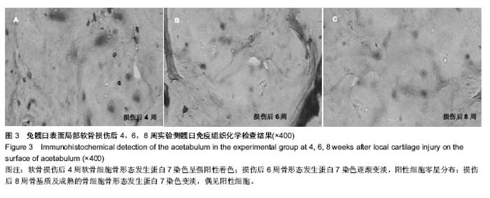
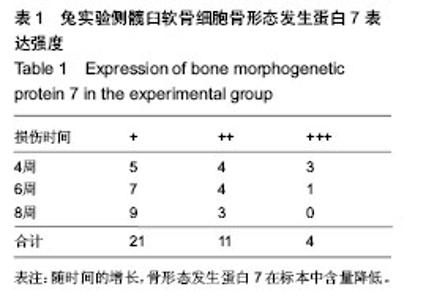
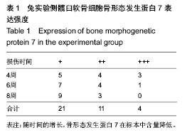
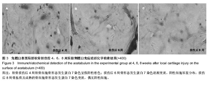
2.4 免疫组织化学检查结果 实验侧:术后4周软骨细胞骨形态发生蛋白7染色呈强阳性着色(图3A);术后6周骨形态发生蛋白7染色逐渐变淡,阳性染色的成骨细胞及软骨细胞零星分布(图3B);术后8周骨基质及成熟的骨细胞骨形态发生蛋白7染色变淡;偶见残余成骨细胞表达骨形态发生蛋白7(图3C)。应用SPSS17.0统计软件进行相关性分析表明骨形态发生蛋白7在标本中含量和时间之间存在负相关(r=-0.866,P < 0.05,表1),即随时间的增长,骨形态发生蛋白7在标本中含量降低。对照侧骨形态发生蛋白7阳性表达广泛分布于增殖的间充质细胞、新生软骨细胞、细胞外基质,表达部位位于胞浆和胞膜。随时间变化,其表达强度变化无显著差异。"
| [1]Klisic PJ. Congenital dislocation of the hip-amisleading term: Brief report. Bone Joint Surg.1991;71(1):136. [2]Gem M, Arslan H, Ozkul E, et al.One-stage bilateral open reduction using the anterior iliofemoral approach in developmental dysplasia of the hip. Acta Orthop elg. 2014; 80(2):211-215. [3]Desteli EE, Pi?kin A, Gülman AB, et al.Estrogen receptors in hip joint capsule and ligamentum capitis femoris of babies with developmental dysplasia of the hip. Acta Orthop Traumatol Turc.2013;47(3):158-161. [4]Zhang L, Yu LD, Yang GJ.Soft tissue balancing in the total hip arthroplasty for severe developmental dysplasia of the hip in adults. Zhonghua Wai Ke Za Zhi.2008;46 (17):1299-1302. [5]Ooishi T.Experimental study on the etiology of pathogenesis of acetabulardysplasia in congenital dislocation of the hip.Nippon Seikeigeka Gakkai Zasshi.1990;64(10):958-975. [6]毛宾尧.髋关节外科学[M].北京:人民卫生出版社,1998:331-353. [7]姜俊,麻宏伟,戴晓梅.先天性髋关节脱位的遗传流行病学研究[J].中国医科大学学报,2006,35(5):514-517. [8]Price KR, Dove R, Hunter JB.The use of X-ray at 5 months in a selective screening programme for developmental dysplasia of the hip.J Child Orthop.2011;5(3):195-200. [9]Ömero?lu H, Inan U.Inherited thrombophilia may be a causative factor for osteonecrosis of femoral head in male patients withdevelopmental dysplasia of the hip: a case series. Arch Orthop Trauma Surg.2012;132(9):1281-1285. [10]Wang E, Liu T, Li J,et al.Does swaddling influence developmental dysplasia of the hip?: An experimental study of the traditional straight-leg swaddling model in neonatal rats.J Bone Joint Surg Am. 2012;94(12):1071-1077. [11]Bachy M, Thevenin-Lemoine C, Rogier A, et al.Utility of magnetic resonance imaging (MRI) after closed reduction of developmental dysplasia of the hip. J Child Orthop. 2012;6 (1): 13-20. [12]Xu M, Gao S, Sun J, Yang Y, et al. Predictive values for the severity of avascular necrosis from the initial evaluation in closed reduction ofdevelopmental dysplasia of the hip. J Pediatr Orthop B. 2013;22(3):179-183. [13]Lee CB, Mata-Fink A, Millis MB, et al.Demographic differences in adolescent-diagnosed and adult-diagnosed acetabular dysplasia compared with infantile developmental dysplasia of the hip.J Pediatr Orthop.2013;33(2):107-111. [14]Rosendahl K, Tomà P.Reply to Riccabona et al. regarding screening for developmental dysplasia of the hip. Pediatr Radiol.2013;43(5):641-642. [15]Cook SD, Wolfe MW, Salkeld SL, et al.Effect of recombinant human osteogenic protein-1 on healing of segmental defects in non-human primate. J Bone Joint Surg Am.1995;77(5): 734. [16]Hidaka C, Goshi K, Rawlins B,et al.Enhancement of spine fusion using combined gene therapy and tissue engineering BMP-7 expressing bone marrow cells and allograft bone. Spine.2003;28(18): 2049. [17]Xu M, Gao S, Sun J, et al. Predictive values for the severity of avascular necrosis from the initial evaluation in closed reduction of developmental dysplasia of the hip. J Pediatr Orthop B.2013;22 (3):179-183. [18]Tréguier C, Baud C, Ferry M, et al. Irreducible developmental dysplasia of the hip due to acetabular roof cartilage hypertrophy. Diagnostic sonography in 15 hips.Orthop Traumatol Surg Res.2011;97 (6):629-633. [19]Azzopardi T, Van Essen P, Cundy PJ, et al. Late diagnosis of developmental dysplasia of the hip: an analysis of risk factors. J Pediatr Orthop B. 2011;20(1):1-7. [20]Sharpe P, Mulpuri K, Chan A, et al.Differences in risk factors between early and late diagnosed developmental dysplasia of the hip. Arch Dis Child Fetal Neonatal Ed. 2006;91(3): F158-162. [21]Shorter D, Hong T, Osborn DA. Screening programmes for developmental dysplasia of the hip in newborn infants. Cochrane Database Syst Rev. 2011;7(9): CD004595. [22]Ewald E, Kiesel E.Screening for developmental dysplasia of the hip in newborns.Am Fam Physician. 2013;87(1):10-11. [23]Roposch A, Ridout D, Protopapa E,et al.Osteonecrosis complicating developmental dysplasia of the hip compromises subsequent acetabular remodeling. Clin Orthop Relat Res. 2013;471(7):2318-2326. [24]Mahan ST, Katz JN, Kim YJ.To screen or not to screen? A decision analysis of the utility of screening for developmental dysplasia of the hip. J Bone Joint Surg Am. 2009;91(7): 1705-1719. [25]Rális Z, McKibbin B. Changes in shape of the human hip joint during its development and their relation to its stability.J Bone Joint Surg Br.1973;55(4):780-785. [26]Reddi AH, Anderson WA.Collagenous bone matrix induced endochondral ossification and hemopoiesis. J Cell Biol. 2006; 69:557-572. [27]Sionek A, Czubak J, Kornacka M, et al.Evaluation of risk factors in developmental dysplasia of the hip in children from multiple pregnancies: results of hip ultrasonography using Graf's method. Ortop Traumatol Rehabil. 2008;10(2): 115-130. [28]Yang G, Zhu Z, Wang Y, et al.Bone Morphogenetic Protein-7 inhibits ilica-induced pulmonary fibrosis in rats.Toxicol Lett. 2013;29:S0378-4274. [29]Odabas S, Feichtinger GA, Korkusuz P, et al. Auricular cartilage repair using cryogel scaffolds loaded with BMP-7-expressing primary chondrocytes. J Tissue Eng Regen Med. 2012, 26. doi: 10.1002/term.1634 [30]Kim HK, Kim JH, Park DS, et.al.Osteogenesis induced by a bone forming peptide from the prodomain region of BMP-7, Biomaterials. 2012; 33(29):7057-7063. [31]Soran N, Altindag O, Aksoy N,et al.The association of serum prolidase activity with developmental dysplasia of the hip. Rheumatol Int. 2013;33(8):1939-1942. |
| [1] | Zhang Chao, Lü Xin. Heterotopic ossification after acetabular fracture fixation: risk factors, prevention and treatment progress [J]. Chinese Journal of Tissue Engineering Research, 2021, 25(9): 1434-1439. |
| [2] | Chen Junming, Yue Chen, He Peilin, Zhang Juntao, Sun Moyuan, Liu Youwen. Hip arthroplasty versus proximal femoral nail antirotation for intertrochanteric fractures in older adults: a meta-analysis [J]. Chinese Journal of Tissue Engineering Research, 2021, 25(9): 1452-1457. |
| [3] | Wu Xun, Meng Juanhong, Zhang Jianyun, Wang Liang. Concentrated growth factors in the repair of a full-thickness condylar cartilage defect in a rabbit [J]. Chinese Journal of Tissue Engineering Research, 2021, 25(8): 1166-1171. |
| [4] | Li Jiacheng, Liang Xuezhen, Liu Jinbao, Xu Bo, Li Gang. Differential mRNA expression profile and competitive endogenous RNA regulatory network in osteoarthritis [J]. Chinese Journal of Tissue Engineering Research, 2021, 25(8): 1212-1217. |
| [5] | Geng Qiudong, Ge Haiya, Wang Heming, Li Nan. Role and mechanism of Guilu Erxianjiao in treatment of osteoarthritis based on network pharmacology [J]. Chinese Journal of Tissue Engineering Research, 2021, 25(8): 1229-1236. |
| [6] | He Xiangzhong, Chen Haiyun, Liu Jun, Lü Yang, Pan Jianke, Yang Wenbin, He Jingwen, Huang Junhan. Platelet-rich plasma combined with microfracture versus microfracture in the treatment of knee cartilage lesions: a meta-analysis [J]. Chinese Journal of Tissue Engineering Research, 2021, 25(6): 964-969. |
| [7] | Liu Xin, Yan Feihua, Hong Kunhao. Delaying cartilage degeneration by regulating the expression of aquaporins in rats with knee osteoarthritis [J]. Chinese Journal of Tissue Engineering Research, 2021, 25(5): 668-673. |
| [8] | Deng Zhenhan, Huang Yong, Xiao Lulu, Chen Yulin, Zhu Weimin, Lu Wei, Wang Daping. Role and application of bone morphogenetic proteins in articular cartilage regeneration [J]. Chinese Journal of Tissue Engineering Research, 2021, 25(5): 798-806. |
| [9] | Chen Lu, Zhang Jianguang, Deng Changgong, Yan Caiping, Zhang Wei, Zhang Yuan. Finite element analysis of locking screw assisted acetabular cup fixation [J]. Chinese Journal of Tissue Engineering Research, 2021, 25(3): 356-361. |
| [10] | Fan Yinuo, Guan Zhiying, Li Weifeng, Chen Lixin, Wei Qiushi, He Wei, Chen Zhenqiu. Research status and development trend of bibliometrics and visualization analysis in the assessment of femoroacetabular impingement [J]. Chinese Journal of Tissue Engineering Research, 2021, 25(3): 414-419. |
| [11] | Cheng Chongjie, Yan Yan, Zhang Qidong, Guo Wanshou. Diagnostic value and accuracy of D-dimer in periprosthetic joint infection: a systematic review and meta-analysis [J]. Chinese Journal of Tissue Engineering Research, 2021, 25(24): 3921-3928. |
| [12] | Chen Lei, Zheng Rui, Jie Yongsheng, Qi Hui, Sun Lei, Shu Xiong. In vitro evaluation of adipose-derived stromal vascular fraction combined with osteochondral integrated scaffold [J]. Chinese Journal of Tissue Engineering Research, 2021, 25(22): 3487-3492. |
| [13] | Tian Guangzhao, Yang Zhen, Zha Kangkang, Sun Zhiqiang, Li Xu, Sui Xiang, Huang Jingxiang, Guo Quanyi, Liu Shuyun. Regulatory effect of decellularized cartilage matrix on macrophage polarization [J]. Chinese Journal of Tissue Engineering Research, 2021, 25(22): 3545-3550. |
| [14] | Ren Wenbo, Liao Yuanpeng. Visualization analysis of traumatic osteoarthritis research hotspots and content based on CiteSpace [J]. Chinese Journal of Tissue Engineering Research, 2021, 25(21): 3374-3381. |
| [15] | Fu Panfeng, Shang Wei, Kang Zhe, Deng Yu, Zhu Shaobo. Efficacy of anterolateral minimally invasive approach versus traditional posterolateral approach in total hip arthroplasty: a meta-analysis [J]. Chinese Journal of Tissue Engineering Research, 2021, 25(21): 3409-3415. |
| Viewed | ||||||
|
Full text |
|
|||||
|
Abstract |
|
|||||
