Chinese Journal of Tissue Engineering Research ›› 2014, Vol. 18 ›› Issue (30): 4845-4850.doi: 10.3969/j.issn.2095-4344.2014.30.014
Previous Articles Next Articles
Effect of type I or type II collagen on biological characteristics of human chondrocytes
Jiang Ping, Wei Peng, Zhao Ming-cai, Chen Qiong, Wang Zi
- Department of Orthopedics, Affiliated Hospital of North Sichuan Medical College, Nanchong 637000, Sichuan Province, China
-
Revised:2014-05-21Online:2014-07-16Published:2014-08-08 -
Contact:Wei Peng, Chief physician, Department of Orthopedics, Affiliated Hospital of North Sichuan Medical College, Nanchong 637000, Sichuan Province, China -
About author:Jiang Ping, Master, Physician, Department of Orthopedics, Affiliated Hospital of North Sichuan Medical College, Nanchong 637000, Sichuan Province, China -
Supported by:a grant by Sichuan Provincial Health Ministry
CLC Number:
Cite this article
Jiang Ping, Wei Peng, Zhao Ming-cai, Chen Qiong, Wang Zi . Effect of type I or type II collagen on biological characteristics of human chondrocytes[J]. Chinese Journal of Tissue Engineering Research, 2014, 18(30): 4845-4850.
share this article

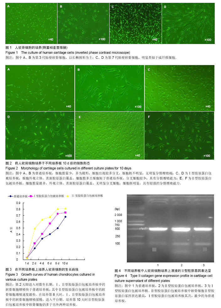
2.1 倒置相差显微镜观察软骨细胞形态 软骨细胞培养:贴壁的原代软骨细胞呈椭圆形、多角形;传代软骨细胞较原代细胞稍大,呈椭圆形、多角形或者不规则形;P3代软骨细胞和P1、P2代细胞相似,但细胞逐渐伸展,变成以长椭圆形为主(图1A,B);传至P7代软骨细胞明显类似于成纤维细胞(图1C,D)。 不同培养板培养的软骨细胞形态:培养10 d后,普通板中软骨细胞数量少,形态多为梭形,较细长(图2A);细胞出现较多分叉,细胞核不明显,无明显细胞分裂增殖相(图2B)。Ⅰ型板细胞表面被胶原蛋白纤维覆盖,细胞外观立体,细胞数多且细胞短于普通板(图2C),具有分叉的细胞较普通板少,分叉的细胞分支较少,60%细胞核明显,具有分裂增殖能力(图2D)。Ⅱ型板软骨细胞数明显较多,约长满培养板90%,且多为短小细胞,同Ⅰ型板细胞一样,细胞外观立体,表面胶原蛋白覆盖(图2E),短小细胞占80%,无明显分叉细胞,80%细胞核明显,具有较强的分裂增殖能力(图2F)。 2.2 不同培养板中软骨细胞的增殖速度 观察细胞生长曲线(图3),第2天即进入快速生长的对数生长期。Ⅰ、Ⅱ型板中的软骨细胞增殖快于普通板,其中Ⅱ型板中的软骨细胞增殖速度最快,为Ⅰ型板的2倍、普通板的5倍。在培养第8天时,Ⅰ、Ⅱ型板中的软骨细胞增殖缓慢,进入平台期,考虑细胞增殖快,接触抑制。由于Ⅱ型板中软骨细胞个体小于Ⅰ型和普通板,故培养第10天时Ⅱ型板中软骨细胞仍多于另外培养板。 2.3 不同培养板对软骨细胞生物学特性的影响 软骨细胞分泌Ⅰ、Ⅱ型胶原蛋白水平及定量检测:3种培养板中软骨细胞Ⅰ型胶原蛋白分泌量的差异均有显著性意义(P < 0.01,表1),再使用q检验进行两两比较显示,普通板中软骨细胞分泌Ⅰ型胶原蛋白量高于Ⅰ、Ⅱ型板,但不能认为Ⅰ、Ⅱ型板中软骨细胞分泌Ⅰ型胶原蛋白量不同。3种培养板中软骨细胞Ⅱ型胶原蛋白分泌量差异有显著性意义(P < 0.01),使用q检验进行两两比较后,显示Ⅰ、Ⅱ型板中软骨细胞分泌Ⅱ型胶原蛋白量均高于普通板,其中Ⅱ型板中软骨细胞分泌Ⅱ型胶原蛋白量最高。根据半定量PCR检测也得出Ⅱ型板中软骨细胞Ⅱ型胶原蛋白基因表达最高,Ⅰ型板其次,最少的为普通板(图4)。 软骨细胞分泌糖胺多糖的量:3种培养板中软骨细胞糖胺多糖分泌量差异有统计学意义(P < 0.01),使用q检验进行两两比较后显示,Ⅰ、Ⅱ型板中软骨细胞分泌糖胺多糖都高于普通板,其中Ⅱ型培养板中软骨细胞分泌量最高(表1)。"

| [1] Brittberg M,Lindahl A,Nilsson A,et al.Treatment of deep cartilage defects in the Knee with autologous chondrocytes transplantation.N Engl J Med. 1994;331(14):889-895. [2] Kosher RA, Church RL. Stimulation of in vitro somite chondrogenesis by procollagen and collagen. Nature. 1975;258(5533):327-330. [3] Shouldes MD,Raines RT.Collagen structure and stability.Annu Rev Biochem.2009;78:929-958. [4] 顾其胜.胶原蛋白的临床应用[J].中国修复重建外科杂志, 2006, 20(10):1052-1057. [5] Award H, Butler DL,Boivin GP,et al.Autologous mesenchymal stem cell-mediated repair of tendon.Tissue Eng. 1999;5(3): 267-277. [6] Wakitani S,Goto T,Young RG,et al.Repair of large fullthickness ar-ticular cartilage defects with all graft articular chondrocytes embedded in a collagen gel.Tissue Eng.1998;4(4):429-444. [7] Hunter W.Of the structure and diseases of articulating cartilages. Clin orthop Relat Res.1995;(317):3-6. [8] Schulz RM,Bader A.Cartilage tissue engineering and bioreactor systems for the cultivation and stimulaion of chondrocytes.Eur Biophys J.2007;36(4-5):539-568. [9] Buckwalter JA,Mankin HJ.Articular cartilage: tissue design and chondrocyte matrix interactions.AAOS Inst Course Lect. 1998;47(3):477-486. [10] Tew SR,Murdoch AD,Rauchenberg RP,et al.Cellular methods in cartilage research: primary human chondrocytes in culture and chondrogenesis in human bone marrow stem cells. Methods.2008;45(1):2-9. [11] Outerbridge HK,Outerbridge RE,Smith DE.Osteochondral defects in the knee, A treatment using lateral patella autografts.Clin Orthop Relat Res.2000;37(7):145-151. [12] Shapiro F,Koide S,Glimcher MJ.Cell origin and differentiation in the repair of full-thickness detects of articular cartilage.J Bone Joint Surg Am .1993;75(4):532-553. [13] Schulze-Tanzil G,de Souza P,Villegas Castrejon H,et al.Redifferentiation of dedifferentiated human chondrocytes in high-density cultures.Cell Tissue Res. 2002;308(3):371-379. [14] Watt F. Effect of seeding density on stability of the dedifferentiated phenotype of pig articular chondrocytes in culture.J Cell Sci.1988;89:373-378. [15] Temenoff JS,Mikos AG.Review: tissue engineering for regeneration of articular cartilage. Biomaterials. 2000;21(3):431-440. [16] Nerem RM,Sambanis A.Tissue engineering : from biology to biological substitutes.Tissue Eng.1995;1(1):3-13. [17] Benya PD,Shaffer JD.Dedifferentiated chondrocytes reexpress the differentiated collagen phenotype when cultured in agarose gels.Cell. 1982;30(1):215-224. [18] Kuriwaka M,Ochi M,Uchio Y,et al.Optimum combination of monolayer and three-dimensional cultures for cartilage-like tissue engineering.Tissue Eng.2003;9(1):41-49. [19] Pieper JS,Vander Kraan PM,Hafmans T,et al.Crosslinked type II collagen matrices:preparation,characterization,and potential for cartilage ngineering.Biomaterials.2002;23(15):3183-3192. [20] Van Beuningen HM,Stoop R,Buma P,et al.Phenotypic differences in murine chondrocyte cell lines derived from mature articular cartilage.Osteoarthritis Cartiage. 2002; 10(12): 977-986. [21] Ushida T,Furukawa K,Toita K,et al.Three-dimensional seeding of chondrocytes encapsulated in collagen gel into PLLA scaffolds.Cell Transplant.2002;11:489-494. [22] Ma,HL,Hung,SC,Lin SY,Chen YL,et al.Chonderogenesis of human mesenchymal stem cells encapsulated in alginate beads.J Biomed Mater Res.2003;64(2):273-281. [23] Kimura T,Yasui N,Ohsawa S,et al.Chondrocytes embedded in collagen gels maintain cartilage phenotype dur ing long term cultures.Clin Orthop Relat Res. 1984;(186):231-239. [24] Speer DP,Chvapil M,Volz R,et al.Enhancement of healing in osteochondral defects by collagen sponge implants.Clin Orthop Relat Res.1979;(144):326-335. [25] Ito Y,Ochi M,Adachi N,et al.Repair of osteochondral defect with tissue-engineered chondral plug in a rabbit model. Arthroscopy.2005;21(10):1155-1163. [26] Darting EM,Athanasiou KA.Retaining zonal chondrocyte phenotype by means of novel growth environments.Tissue Eng.2005;11(3-4):395-403. [27] Qi WN,Scully SP.Extracelluar collagen regulates expression of transforming growth factorth factor-betal gene.J Orthop Res.2000;18:928-932. [28] Qi WN,Scully SP.Effect of type II collagen in chondrocyte response to TGF-beta1 regulation.Exp Cell Res. 1998;241(1): 142-150. [29] Qui W,Scully S.Extracellular collagens demonstrate a type specific influence on cytokine regulation of articular chondrocytes.Proc of the ORS, Atlanta, GA; 1996:308. [30] Tew SR,Murdoch AD,Rauchenberg RP,et al.Cellular methods in cartilage research: primary human chondrocytes in culture and chondrogenesis in human bone marrow stem cells. Methods.2008;45(1):2-9. [31] 周强,李起鸿,戴刚.骺板软骨细胞复合三维支架体外构建组织工程软骨的研究[J].中国修复重建外科杂志,2004,18(2):92-95. [32] 林建华,陈晓东,邓凌霄,等.大鼠软骨细胞复制性老化的体外观察[J].中国修复重建外科杂志,2007,21(11):1228-1232. [33] 周强,李起鸿,戴刚.三步酶消化法高效分离兔原代关节软骨细胞及体外培养观察[J].中华外科杂志,2005,43(8):522-526. [34] Sandell LJ,Sugai JV,Trippel SB.Expression of collagens Ⅰ, Ⅱ, Ⅹ,and Ⅺ and aggrecan mRNAs by bovine growth plate chondrocytes insitu.J Orthop Res.1994;12(1):1-14. [35] Miot S,Woodfield T,Daniels AU,et al. Effects of scaffold composition and architecture on human nasal chondrocyte redifferentiation and cartilaginous matrix deposition. Biomaterials.2005;26(15):2479-2489. [36] 张艳,柴岗,刘伟,等.体外培养过程中去分化的人软骨细胞基因表达谱的变化[J].中华整形外科杂志,2007,23(4):331-334. [37] Karlsen TA,Shahdadfar A,Brinchmann JE.Human primary articular chondrocytes, chondroblasts-like cells, and dedifferentiated chondrocytes: differences in gene, microRNA,and protein expression and phenotype.Tissue Eng Part C Methods. 2011;17(2):219-227. [38] Binette F,McQuaid DP,Haudenschild,DR,et al.Expression of a stable articular cartilage phenotype without evidence of hypertrophy by adult human articular chondrocytes in vitro.J Orthop Res.1998;16:207-216. [39] Liu H, Lee YW, Dean,MF. Re-expression of differentiated proteoglycan phenotype by dedifferentiated human chondrocytes during culture in alginate beads.Biochim Biophys Acta.1998;1425:505-515. |
| [1] | Sun Kexin, Zeng Jinshi, Li Jia, Jiang Haiyue, Liu Xia. Mechanical stimulation enhances matrix formation of three-dimensional bioprinted cartilage constructs [J]. Chinese Journal of Tissue Engineering Research, 2023, 27(在线): 1-7. |
| [2] | Zhang Hui, Wang Jiayang, Wang Qian, Gan Hongquan, Wang Zhiqiang. Effects of hyaluronic acid combined with domestic porous tantalum on chondrocyte function under the dynamic environment [J]. Chinese Journal of Tissue Engineering Research, 2023, 27(3): 339-345. |
| [3] | Wang Kang, Zhi Xiaodong, Zhang Yuqiang, Gong Chao, Wang Chenliang, Wang Wei. Human amniotic epithelial cells regulate the proliferation, apoptosis and extracellular matrix synthesis of articular chondrocytes by activating the EGFR/ERK1 signaling axis [J]. Chinese Journal of Tissue Engineering Research, 2022, 26(31): 4967-4974. |
| [4] | Gao Zilong, Li Ting, Lyu Zheng, Shen Huarui. Psoralen combined with transforming growth factor beta 1 induces the differentiation of bone marrow mesenchymal stem cells into chondrocytes [J]. Chinese Journal of Tissue Engineering Research, 2022, 26(30): 4884-4888. |
| [5] | Liao Jun, Xu Pu. Effect of nanonized freshwater pearl powder on the expression of osteogenic related genes [J]. Chinese Journal of Tissue Engineering Research, 2022, 26(27): 4325-4329. |
| [6] | Xie Yan, Li Wuyin, Xing Weipeng, Pei Yuanyuan, Wang Na. Screening of drugs for inhibiting growth plate chondrocyte apoptosis and the effect of resveratrol on delaying epiphyseal closure in rats [J]. Chinese Journal of Tissue Engineering Research, 2022, 26(23): 3644-3649. |
| [7] | Han Zhi, Wang Zhimiao, Gaxi Sijia, Lu Qingling, Guo Tao. Tissue engineered cartilage constructed by polyurethane composite chondrocytes [J]. Chinese Journal of Tissue Engineering Research, 2022, 26(22): 3455-3459. |
| [8] | Guan Hong, Zhang Hongbo, Shao Yan, Guo Dong, Zhang Haiyan, Cai Daozhang. PDZ domain containing 1 deficiency promotes chondrocyte senescence in osteoarthritis [J]. Chinese Journal of Tissue Engineering Research, 2022, 26(2): 182-189. |
| [9] | Ma Dujun, Peng Liping, Zhou Ziqiong, Zhao Jing, Zhu Houjun, Jiang Shunwan, Zhong Jing, She Ruihao. Effect of RAB39B gene based on CRISPR/Cas9 technology on cartilage differentiation of bone marrow mesenchymal stem cells [J]. Chinese Journal of Tissue Engineering Research, 2022, 26(19): 2978-2984. |
| [10] | Shi Hang, Li Jia, Liu Xia, Jiang Haiyue. Heterogeneity of chondrocytes derived from human ribs based on single-cell transcriptome sequencing [J]. Chinese Journal of Tissue Engineering Research, 2022, 26(19): 3011-3017. |
| [11] | Yang Shuang, Yan Jinglong. Effects of SOX9 on chondrocyte differentiation [J]. Chinese Journal of Tissue Engineering Research, 2022, 26(14): 2279-2284. |
| [12] | Yang Tengyun, Li Yanlin, Liu Dejian, Wang Guoliang, Zheng Zhujun. Chondrogenic differentiation of peripheral blood-derived mesenchymal stem cells induced by transforming growth factor beta 3: a dose-effect relationship [J]. Chinese Journal of Tissue Engineering Research, 2022, 26(1): 45-51. |
| [13] | Feng Zhiguo, Sun Haibiao, Han Xiaoqiang. Regulation of proliferation, differentiation and apoptosis of bone-related cells by long-stranded non-coding RNA [J]. Chinese Journal of Tissue Engineering Research, 2022, 26(1): 112-118. |
| [14] | Liu Xiaogang, Li Tian, Zhang Duo. Effect and mechanism of the effective components of Chinese medicine on promoting the differentiation of bone marrow mesenchymal stem cells into chondrocytes [J]. Chinese Journal of Tissue Engineering Research, 2022, 26(1): 119-124. |
| [15] | Ma Zetao, Zeng Hui, Wang Deli, Weng Jian, Feng Song. MicroRNA-138-5p regulates chondrocyte proliferation and autophagy [J]. Chinese Journal of Tissue Engineering Research, 2021, 25(5): 674-678. |
| Viewed | ||||||
|
Full text |
|
|||||
|
Abstract |
|
|||||

