Chinese Journal of Tissue Engineering Research ›› 2014, Vol. 18 ›› Issue (10): 1477-1483.doi: 10.3969/j.issn.2095-4344.2014.10.001
Paracrine effects of bone marrow mesenchymal stem cells on biological function of osteoblasts
Li Cheng, Zhou Hai-bin
- Department of Orthopedics, the Second Affiliated Hospital, Soochow University, Suzhou 215004, Jiangsu Province, China
-
Online:2014-03-05Published:2014-03-05 -
Contact:Zhou Hai-bin, M.D., Chief physician, Department of Orthopedics, the Second Affiliated Hospital, Soochow University, Suzhou 215004, Jiangsu Province, China -
About author:Li Cheng, Master, Physician, Department of Orthopedics, the Second Affiliated Hospital, Soochow University, Suzhou 215004, Jiangsu Province, China
CLC Number:
Cite this article
Li Cheng, Zhou Hai-bin. Paracrine effects of bone marrow mesenchymal stem cells on biological function of osteoblasts [J]. Chinese Journal of Tissue Engineering Research, 2014, 18(10): 1477-1483.
share this article
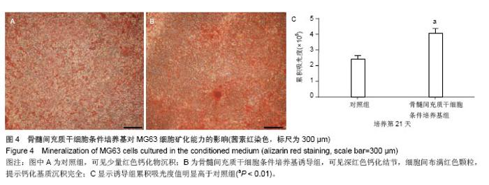
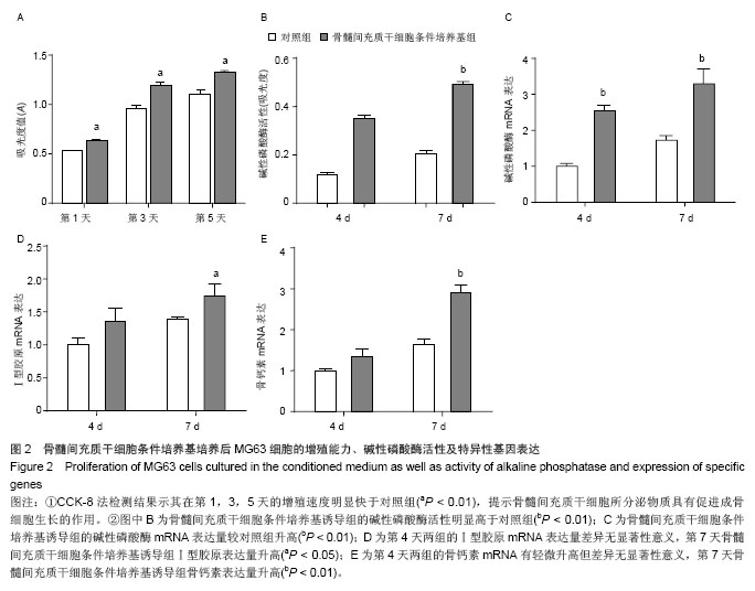
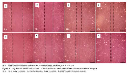
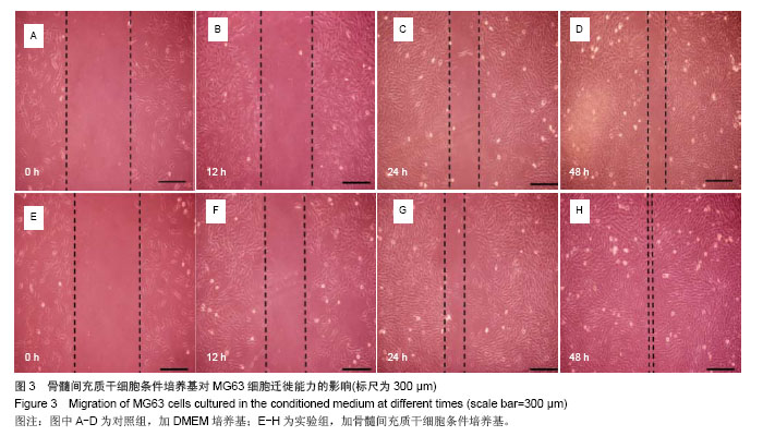
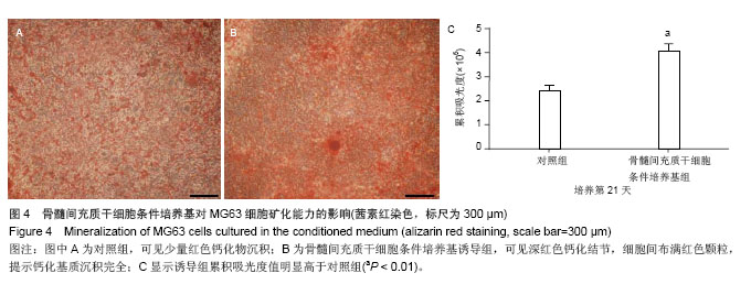
2.5 骨髓间充质干细胞条件培养基对MG63细胞分化及矿化能力的影响 分别于DEME培养基和骨髓间充质干细胞条件培养基条件下诱导成骨细胞。碱性磷酸酶活性检测结果显示,在第4,7天时,骨髓间充质干细胞条件培养基诱导组的碱性磷酸酶活性明显高于对照组(P < 0.01,图2B)。Real-time PCR结果显示,骨髓间充质干细胞条件培养基诱导组的碱性磷酸酶mRNA表达量也较对照组明显升高(P < 0.01),与碱性磷酸酶活性检测结果一致(图2C);在第4天时两组的Ⅰ型胶原mRNA表达量差异无显著性意义(P > 0.05),但于第7天时骨髓间充质干细胞条件培养基组Ⅰ型胶原表达量升高(P < 0.05);骨钙素mRNA表达量在第4天时有轻微升高但差异无显著性意义(P > 0.05,图2D),而至第7天时则明显升高(P < 0.01,图2E)。21 d后行茜素红染色DMEM对照组可见少量红色钙化物沉积(图4A),而骨髓间充质干细胞条件培养基诱导组可见深红色钙化结节,细胞间布满红色颗粒,提示钙化基质沉积完全(图4B)。IPP软件分析显示诱导组累积吸光度值明显高于对照组(P < 0.01,图4C)。"
| [1]Centeno CJ, Schultz JR, Cheever M,et al.Safety and complications reporting on the re-implantation of culture- expanded mesenchymal stem cells using autologous platelet lysate technique.Curr Stem Cell Res Ther. 2010;5(1):81-93.[2]Izadpanah R, Kaushal D, Kriedt C,et al. Long-term in vitro expansion alters the biology of adult mesenchymal stem cells. Cancer Res. 2008;68(11):4229-4238.[3]Ando W, Tateishi K, Hart DA,et al.Cartilage repair using an in vitro generated scaffold-free tissue-engineered construct derived from porcine synovial mesenchymal stem cells. Biomaterials. 2007;28(36):5462-5470.[4]Ando W, Tateishi K, Katakai D,et al.In vitro generation of a scaffold-free tissue-engineered construct (TEC) derived from human synovial mesenchymal stem cells: biological and mechanical properties and further chondrogenic potential. Tissue Eng Part A. 2008;14(12):2041-2049. [5]Anitua E, Tejero R, Zalduendo MM,et al.Plasma rich in growth factors promotes bone tissue regeneration by stimulating proliferation, migration, and autocrine secretion in primary human osteoblasts.J Periodontol. 2013;84(8): 1180-1190. [6]Anitua E, Troya M, Orive G.Plasma rich in growth factors promote gingival tissue regeneration by stimulating fibroblast proliferation and migration and by blocking transforming growth factor-β1-induced myodifferentiation.J Periodontol. 2012;83(8):1028-1037. [7]Smith PC, Cáceres M, Martinez J.Induction of the myofibroblastic phenotype in human gingival fibroblasts by transforming growth factor-beta1: role of RhoA-ROCK and c-Jun N-terminal kinase signaling pathways.J Periodontal Res. 2006;41(5):418-425.[8]Murphy JM, Fink DJ, Hunziker EB,et al.Stem cell therapy in a caprine model of osteoarthritis.Arthritis Rheum. 2003;48(12): 3464-3474.[9]Matsumoto T, Cooper GM, Gharaibeh B,et al.Cartilage repair in a rat model of osteoarthritis through intraarticular transplantation of muscle-derived stem cells expressing bone morphogenetic protein 4 and soluble Flt-1.Arthritis Rheum. 2009;60(5):1390-1405. [10]Kuroda R, Usas A, Kubo S,et al.Cartilage repair using bone morphogenetic protein 4 and muscle-derived stem cells. Arthritis Rheum. 2006;54(2):433-442.[11]Koga H, Muneta T, Ju YJ,et al.Synovial stem cells are regionally specified according to local microenvironments after implantation for cartilage regeneration.Stem Cells. 2007;25(3):689-696. [12]Shukunami C, Oshima Y, Hiraki Y.Chondromodulin-I and tenomodulin: a new class of tissue-specific angiogenesis inhibitors found in hypovascular connective tissues.Biochem Biophys Res Commun. 2005;333(2):299-307.[13]Tongers J, Losordo DW, Landmesser U.Stem and progenitor cell-based therapy in ischaemic heart disease: promise, uncertainties, and challenges.Eur Heart J. 2011;32(10): 1197-1206. [14]Besler C, Doerries C, Giannotti G,et al.Pharmacological approaches to improve endothelial repair mechanisms.Expert Rev Cardiovasc Ther. 2008;6(8):1071-1082.[15]Ahmadi H, Baharvand H, Ashtiani SK,et al.Safety analysis and improved cardiac function following local autologous transplantation of CD133(+) enriched bone marrow cells after myocardial infarction.Curr Neurovasc Res. 2007;4(3): 153-160.[16]Smith RR, Barile L, Cho HC,et al.Regenerative potential of cardiosphere-derived cells expanded from percutaneous endomyocardial biopsy specimens.Circulation. 2007;115(7): 896-908. [17]Mirotsou M, Jayawardena TM, Schmeckpeper J,et al. Paracrine mechanisms of stem cell reparative and regenerative actions in the heart.J Mol Cell Cardiol. 2011; 50(2):280-289.[18]Bruno S, Grange C, Deregibus MC,et al.Mesenchymal stem cell-derived microvesicles protect against acute tubular injury. J Am Soc Nephrol. 2009;20(5):1053-1067.[19]Smith RR, Barile L, Cho HC,et al.Regenerative potential of cardiosphere-derived cells expanded from percutaneous endomyocardial biopsy specimens.Circulation. 2007;115(7): 896-908.[20]Block GJ, DiMattia GD, Prockop DJ.Stanniocalcin-1 regulates extracellular ATP-induced calcium waves in human epithelial cancer cells by stimulating ATP release from bystander cells. PLoS One. 2010;5(4):e10237.[21]Markel TA, Wang Y, Herrmann JL,et al.VEGF is critical for stem cell-mediated cardioprotection and a crucial paracrine factor for defining the age threshold in adult and neonatal stem cell function.Am J Physiol Heart Circ Physiol. 2008; 295(6):H2308-H2314. [22]Allan R, Kass M, Glover C,et al.Cellular transplantation: future therapeutic options.Curr Opin Cardiol. 2007;22(2):104-110.[23]Burchfield JS, Iwasaki M, Koyanagi M,et al.Interleukin-10 from transplanted bone marrow mononuclear cells contributes to cardiac protection after myocardial infarction. Circ Res. 2008;103(2):203-211.[24]Garbade J, Dhein S, Lipinski C,et al.Bone marrow-derived stem cells attenuate impaired contractility and enhance capillary density in a rabbit model of Doxorubicin-induced failing hearts.J Card Surg. 2009;24(5):591-599. [25]Parekkadan B, van Poll D, Suganuma K, et al. Mesenchymal stem cell-derived molecules reverse fulminant hepatic failure. PLoS One. 2007 Sep 26;2(9):e941.[26]Mayr-Wohlfart U, Waltenberger J, Hausser H, et al.Vascular endothelial growth factor stimulates chemotactic migration of primary human osteoblasts.Bone. 2002;30(3):472-477.[27]Nakajima A, Nakajima F, Shimizu S,et al.Spatial and temporal gene expression for fibroblast growth factor type I receptor (FGFR1) during fracture healing in the rat.Bone. 2001;29(5): 458-466.[28]Loots GG, Keller H, Leupin O, et al.TGF-β regulates sclerostin expression via the ECR5 enhancer.Bone. 2012;50(3): 663- 669. [29]Kato M, Patel MS, Levasseur R,et al.Cbfa1-independent decrease in osteoblast proliferation, osteopenia, and persistent embryonic eye vascularization in mice deficient in Lrp5, a Wnt coreceptor.J Cell Biol. 2002;157(2):303-314. [30]Leupin O, Piters E, Halleux C,et al.Bone overgrowth-associated mutations in the LRP4 gene impair sclerostin facilitator function.J Biol Chem. 2011;286(22): 19489-19500. [31]Loots GG, Kneissel M, Keller H,et al.Genomic deletion of a long-range bone enhancer misregulates sclerostin in Van Buchem disease.Genome Res. 2005;15(7):928-935. [32]Santibañez JF, Quintanilla M, Bernabeu C.TGF-β/TGF-β receptor system and its role in physiological and pathological conditions.Clin Sci (Lond). 2011;121(6):233-251. [33]Kamiya N, Ye L, Kobayashi T,et al.BMP signaling negatively regulates bone mass through sclerostin by inhibiting the canonical Wnt pathway.Development. 2008;135(22):3801- 3811.[34]Bissell DM, Roulot D, George J.Transforming growth factor beta and the liver.Hepatology. 2001;34(5):859-867.[35]Ominsky MS, Vlasseros F, Jolette J,et al.Two doses of sclerostin antibody in cynomolgus monkeys increases bone formation, bone mineral density, and bone strength.J Bone Miner Res. 2010;25(5):948-959. [36]Mohammad KS, Chen CG, Balooch G,et al. Pharmacologic inhibition of the TGF-beta type I receptor kinase has anabolic and anti-catabolic effects on bone.PLoS One. 2009;4(4): e5275. [37]Atkins GJ, Rowe PS, Lim HP,et al.Sclerostin is a locally acting regulator of late-osteoblast/preosteocyte differentiation and regulates mineralization through a MEPE-ASARM-dependent mechanism.J Bone Miner Res. 2011;26(7):1425-1436. [38]Frank O, Heim M, Jakob M,et al.Real-time quantitative RT-PCR analysis of human bone marrow stromal cells during osteogenic differentiation in vitro.J Cell Biochem. 2002;85(4): 737-746.[39]Lindström M, Thornell LE.New multiple labelling method for improved satellite cell identification in human muscle: application to a cohort of power-lifters and sedentary men. Histochem Cell Biol. 2009;132(2):141-157. [40]Otsuru S, Tamai K, Yamazaki T,et al.Circulating bone marrow-derived osteoblast progenitor cells are recruited to the bone-forming site by the CXCR4/stromal cell-derived factor-1 pathway.Stem Cells. 2008;26(1):223-234.[41]Schulz TJ, Huang TL, Tran TT,et al.Identification of inducible brown adipocyte progenitors residing in skeletal muscle and white fat.Proc Natl Acad Sci U S A. 2011;108(1):143-148.[42]Yeh LC, Lee JC.Co-transfection with the osteogenic protein (OP)-1 gene and the insulin-like growth factor (IGF)-I gene enhanced osteoblastic cell differentiation.Biochim Biophys Acta. 2006;1763(1):57-63. [43]Zhao M, Zhao Z, Koh JT,et al.Combinatorial gene therapy for bone regeneration: cooperative interactions between adenovirus vectors expressing bone morphogenetic proteins 2, 4, and 7.J Cell Biochem. 2005;95(1):1-16.[44]Shoba LN, Lee JC.Inhibition of phosphatidylinositol 3-kinase and p70S6 kinase blocks osteogenic protein-1 induction of alkaline phosphatase activity in fetal rat calvaria cells.J Cell Biochem. 2003;88(6):1247-1255. |
| [1] | Pu Rui, Chen Ziyang, Yuan Lingyan. Characteristics and effects of exosomes from different cell sources in cardioprotection [J]. Chinese Journal of Tissue Engineering Research, 2021, 25(在线): 1-. |
| [2] | Lin Qingfan, Xie Yixin, Chen Wanqing, Ye Zhenzhong, Chen Youfang. Human placenta-derived mesenchymal stem cell conditioned medium can upregulate BeWo cell viability and zonula occludens expression under hypoxia [J]. Chinese Journal of Tissue Engineering Research, 2021, 25(在线): 4970-4975. |
| [3] | Hou Jingying, Yu Menglei, Guo Tianzhu, Long Huibao, Wu Hao. Hypoxia preconditioning promotes bone marrow mesenchymal stem cells survival and vascularization through the activation of HIF-1α/MALAT1/VEGFA pathway [J]. Chinese Journal of Tissue Engineering Research, 2021, 25(7): 985-990. |
| [4] | Shi Yangyang, Qin Yingfei, Wu Fuling, He Xiao, Zhang Xuejing. Pretreatment of placental mesenchymal stem cells to prevent bronchiolitis in mice [J]. Chinese Journal of Tissue Engineering Research, 2021, 25(7): 991-995. |
| [5] | Liang Xueqi, Guo Lijiao, Chen Hejie, Wu Jie, Sun Yaqi, Xing Zhikun, Zou Hailiang, Chen Xueling, Wu Xiangwei. Alveolar echinococcosis protoscolices inhibits the differentiation of bone marrow mesenchymal stem cells into fibroblasts [J]. Chinese Journal of Tissue Engineering Research, 2021, 25(7): 996-1001. |
| [6] | Fan Quanbao, Luo Huina, Wang Bingyun, Chen Shengfeng, Cui Lianxu, Jiang Wenkang, Zhao Mingming, Wang Jingjing, Luo Dongzhang, Chen Zhisheng, Bai Yinshan, Liu Canying, Zhang Hui. Biological characteristics of canine adipose-derived mesenchymal stem cells cultured in hypoxia [J]. Chinese Journal of Tissue Engineering Research, 2021, 25(7): 1002-1007. |
| [7] | Geng Yao, Yin Zhiliang, Li Xingping, Xiao Dongqin, Hou Weiguang. Role of hsa-miRNA-223-3p in regulating osteogenic differentiation of human bone marrow mesenchymal stem cells [J]. Chinese Journal of Tissue Engineering Research, 2021, 25(7): 1008-1013. |
| [8] | Lun Zhigang, Jin Jing, Wang Tianyan, Li Aimin. Effect of peroxiredoxin 6 on proliferation and differentiation of bone marrow mesenchymal stem cells into neural lineage in vitro [J]. Chinese Journal of Tissue Engineering Research, 2021, 25(7): 1014-1018. |
| [9] | Zhu Xuefen, Huang Cheng, Ding Jian, Dai Yongping, Liu Yuanbing, Le Lixiang, Wang Liangliang, Yang Jiandong. Mechanism of bone marrow mesenchymal stem cells differentiation into functional neurons induced by glial cell line derived neurotrophic factor [J]. Chinese Journal of Tissue Engineering Research, 2021, 25(7): 1019-1025. |
| [10] | Duan Liyun, Cao Xiaocang. Human placenta mesenchymal stem cells-derived extracellular vesicles regulate collagen deposition in intestinal mucosa of mice with colitis [J]. Chinese Journal of Tissue Engineering Research, 2021, 25(7): 1026-1031. |
| [11] | Pei Lili, Sun Guicai, Wang Di. Salvianolic acid B inhibits oxidative damage of bone marrow mesenchymal stem cells and promotes differentiation into cardiomyocytes [J]. Chinese Journal of Tissue Engineering Research, 2021, 25(7): 1032-1036. |
| [12] | Li Cai, Zhao Ting, Tan Ge, Zheng Yulin, Zhang Ruonan, Wu Yan, Tang Junming. Platelet-derived growth factor-BB promotes proliferation, differentiation and migration of skeletal muscle myoblast [J]. Chinese Journal of Tissue Engineering Research, 2021, 25(7): 1050-1055. |
| [13] | Liu Cong, Liu Su. Molecular mechanism of miR-17-5p regulation of hypoxia inducible factor-1α mediated adipocyte differentiation and angiogenesis [J]. Chinese Journal of Tissue Engineering Research, 2021, 25(7): 1069-1074. |
| [14] | Wang Xianyao, Guan Yalin, Liu Zhongshan. Strategies for improving the therapeutic efficacy of mesenchymal stem cells in the treatment of nonhealing wounds [J]. Chinese Journal of Tissue Engineering Research, 2021, 25(7): 1081-1087. |
| [15] | Wang Shiqi, Zhang Jinsheng. Effects of Chinese medicine on proliferation, differentiation and aging of bone marrow mesenchymal stem cells regulating ischemia-hypoxia microenvironment [J]. Chinese Journal of Tissue Engineering Research, 2021, 25(7): 1129-1134. |
| Viewed | ||||||
|
Full text |
|
|||||
|
Abstract |
|
|||||
