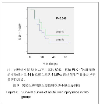
Chinese Journal of Tissue Engineering Research ›› 2013, Vol. 17 ›› Issue (53): 9139-9144.doi: 10.3969/j.issn.2095-4344.2013.53.007

Previous Articles     Next Articles

Transplantation of fetal liver FLK-1+ cells in treatment of acute liver injury

Li Na1, Ding Wei-rong2, Wu Bo-qing3, Liu Ting-ting2, Cheng Hong-bo2, Jin Cheng-hao2   

  1. 1Medical College of Nanchang University, Nanchang  330006, Jiangxi Province, China; 2Department of Hematology, 3Department of Clinical Laboratories, Jiangxi Provincial People’s Hospital, Nanchang  330006, Jiangxi Province, China
  • Revised:2013-09-19 Online:2013-12-31 Published:2013-12-31
  • Contact: Jin Cheng-hao, Ph.D., Chief physician, Master’s supervisor, Department of Hematology, Jiangxi Provincial People’s Hospital, Nangchang 330006, Jiangxi Province, China inch227@yahoo.com.cn
  • About author:Li Na★, Master, Physician, Medical College of Nanchang University, Nanchang 330006, Jiangxi Province, China 634624912@qq.com Ding Wei-rong☆, M.D., Associate chief physician, Department of Hematology, Jiangxi Provincial People’s Hospital, Nanchang 330006, Jiangxi Province, China dingwr97@163.com Li Na and Ding Wei-rong contributed equally to this work.
  • Supported by:

    the National Natural Science Foundation of China, No. 30860115*; the Natural Science Foundation of Jiangxi Province in China, No. 2008GZY0084*

Abstract:

BACKGROUND: Previous studies of our research group have shown that FLK-1+ cells existed in the mouse fetal liver at embryonic 17-19 days, and they expressed embryonic stem cells and presented multi-differentiation potentials.

OBJECTIVE: To evaluate the therapeutic effect of the transplantation of FLK-1+ cells derived from mouse fetal liver on acute liver injury in mice.
METHODS: The FLK-1+ fraction were enriched from the fetal liver with immunomagnetic beads method and detected by flow cytometry. The Oct-3/4 and Rex-1 genes in FLK-1+ cells were detected by reverse transcription-polymerase chain reaction. The FLK-1+ cells were induced to differentiate into liver cells by hepatocyte growth factor and epidermal growth factor. The model of acute liver injury in mice was established by intraperitoneally injecting carbon tetrachloride 4, and was randomly divided into two groups. In the control group, mouse models with acute liver injury were infused normal saline via tail vein; in the experimental group, mouse models with acute liver injury were infused induced FLK-1+ cells (1×10⁶) via tail vein. The blood was collected at 16 hours and liver functions were detected. The mortality of mice was observed at 64 hours.
RESULTS AND CONCLUSION: The fetal liver FLK-1+ cells highly expressed Oct-3/4 and Rex-1 mRNA, and albumin expressing rate in FLK-1+ cells was 0.6%. After induced by hepatocyte growth factor for 3 days, FLK-1 did not express. After induced by hepatocyte growth factor and epidermal growth factor for 3 days, Oct-3/4 and Rex-1 mRNA expression was significantly reduced or disappeared in FLK-1+ cells, and 96.38% FLK-1+ cells expressed albumin. After FLK-1+ cells induced for 3 days were transplanted to mouse models with acute liver injury for 16 hours, the serum glutamic-oxalacetic transaminease and glutamic-pyruvic transaminase were significantly lower (P < 0.05), but serum albumin was significantly higher than the control group (P < 0.05). However, serum total bilirubin and fibrinogen showed no significant differences between two groups (P > 0.05). The mortality rate in the control group was 80% and the mortality rate in the experimental group was 61.5% at 64 hours, with no significant difference between two groups (P > 0.05). Experimental findings indicate that, transplanting fetal liver FLK-1+ cells induced by hepatocyte growth factor and epidermal growth factor for 3 days can improve liver cells function in mice with acute liver damage.


中国组织工程研究
杂志出版内容重点:肾移植肝移植移植;心脏移植;组织移植;皮肤移植;皮瓣移植;血管移植;器官移植组织工程


全文链接:

Key words: hepatocyte growth factor, epidermal growth factor, aspartate aminotransferase, alanine aminotransferase

CLC Number: