Chinese Journal of Tissue Engineering Research ›› 2013, Vol. 17 ›› Issue (53): 9132-9138.doi: 10.3969/j.issn.2095-4344.2013.53.006
Previous Articles Next Articles
Intrasplenic delivery of xenogeneic hepatic progenitor cells ameliorates acute liver injury in rats
Wan Zhen1, 2, Zhang Xiao-gang1, 2, Zheng Xing-long1, 2, Ma Feng1, 2, Ma Jia1, 2, Xiang Jun-xi1, 2, Wang Hao-hua1, 2, Lü Yi1, 2
- 1Department of Hepatobiliary Surgery, First Affiliated Hospital of Xi’an Jiaotong University, Xi’an 710061, Shaanxi Province, China; 2Institute of Advanced Surgery Technology and Engineering, Xi’an Jiaotong University, Xi’an 710061, Shaanxi Province, China
-
Revised:2013-09-24Online:2013-12-31Published:2013-12-31 -
Contact:Lü Yi, Ph.D., Professor, Doctoral supervisor, Department of Hepatobiliary Surgery, First Affiliated Hospital of Xi’an Jiaotong University, Xi’an 710061, Shaanxi Province, China; Institute of Advanced Surgery Technology and Engineering, Xi’an Jiaotong University, Xi’an 710061, Shaanxi Province, China luyi169@126.com -
About author:Wan Zhen☆, Studying for doctorate, Department of Hepatobiliary Surgery, First Affiliated Hospital of Xi’an Jiaotong University, Xi’an 710061, Shaanxi Province, China; Institute of Advanced Surgery Technology and Engineering, Xi’an Jiaotong University, Xi’an 710061, Shaanxi Province, China -
Supported by:The National Natural Science Foundation of China, No. 30600575*, 30830099*
CLC Number:
Cite this article
Wan Zhen1, 2, Zhang Xiao-gang1, 2, Zheng Xing-long1, 2, Ma Feng1, 2, Ma Jia1, 2, Xiang Jun-xi1, 2, Wang Hao-hua1, 2, Lü Yi1, 2. Intrasplenic delivery of xenogeneic hepatic progenitor cells ameliorates acute liver injury in rats[J]. Chinese Journal of Tissue Engineering Research, 2013, 17(53): 9132-9138.
share this article
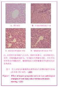
2.1 实验动物数量分析 60只大鼠全部进入结果分析,无脱失。 2.2 肝上皮样前体细胞形态 肝上皮样前体细胞呈上皮样细胞形态,细胞核大而明显。肝上皮样前体细胞生长旺盛,倍增时间平均为24 h,多在贴壁后两三天达到约80%的汇合度,胰酶消化传代。可在体外培养>6个月时间,传代50次以上。连续传代对肝上皮样前体细胞细胞形态和增殖能力无明显影响。取对数生长期肝上皮样前体细胞消化重悬得到单细胞悬液,锥虫蓝染色示大于90%细胞拒染呈无色透明状,用于下一步移植实验。 2.3 肝脏病理学表现 在肝切除后1,5,14和21 d取肝脏组织行苏木精-伊红染色检查。假手术组无明显肝组织学异常。CCl4腹腔注射联合2/3肝切除处理诱导的急性肝损伤表现为肝细胞广泛水肿,体积明显增大,肝血窦腔受压变窄,肝细胞胞浆疏松化,可见散在的嗜酸性变性,并有点状坏死伴炎症细胞浸润。细胞移植组大鼠肝细胞水肿较肝损伤组显著改善。在细胞移植后 21 d,肝细胞肿胀消失,大部分肝细胞结构正常,无炎症细胞浸润,见图1。"

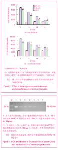
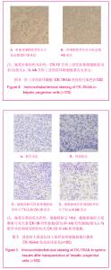
2.4 血清转氨酶变化 肝损伤组大鼠肝切除后1 d丙氨酸转氨酶升高至(6 808.03±123.36) nkat/L,远远高于细胞移植组(6 024.54±215.04) nkat/L的水平(P < 0.05)。此后,细胞移植组大鼠丙氨酸转氨酶较肝损伤组下降更明显,差异有显著性意义(P < 0.05)。天门冬氨酸转氨酶变化趋势与丙氨酸转氨酶类似,即细胞移植组大鼠天门冬氨酸转氨酶峰值较肝损伤组低,下降更迅速。各个时间点丙氨酸转氨酶和天门冬氨酸转氨酶值及动态变化见图2。 2.5 肝上皮样前体细胞在大鼠脾脏中的定植 提取肝切除后1,5,14和21 d细胞移植组脾脏DNA行PCR反应,PCR产物琼脂糖凝胶电泳可见402 bp大小的条带,提示小鼠Y染色体特异性序列Sry存在于脾脏中。假手术组和肝损伤组未见阳性结果,见图3。 CK-19是肝前体细胞特异性分子标志物,在肝上皮样前体细胞胞浆内表达(图4),在脾脏内无表达,可用来监测移植的肝上皮样前体细胞在大鼠脾脏实质的定植。细胞移植组大鼠脾脏可见大量CK-19阳性细胞,而假手术组和肝损伤组无阳性细胞,见图5。 2.6 肝上皮样前体细胞在大鼠脾脏实质中分化为肝细胞样细胞 Alb是肝细胞特异性分子标志物,免疫组化显示在肝上皮样前体细胞胞浆内无表达(图4),在大鼠脾脏内同样无表达,可用来评价移植肝上皮样前体细胞在大鼠脾脏实质中向肝细胞方向分化的能力。Alb阳性细胞在细胞移植后5 d首次出现,而且移植后14 d和21 d阳性细胞数目逐渐增多。假手术组和肝损伤组未见到Alb阳性细胞,见图5。"

"

| [1] Jindal A, Kumar M, Sarin SK. Management of acute hepatitis B and reactivation of hepatitis B. Liver Int. 2013;33 Suppl 1: 164-175.[2] Karsan HA, Parekh S. Management of alcoholic hepatitis: Current concepts. World J Hepatol. 2013;4(12):335-341.[3] Suk KT, Kim DJ. Drug-induced liver injury: present and future. Clin Mol Hepatol. 2012;18(3):249-257.[4] Bernal W, Hyyrylainen A, Gera A, et al. Lessons from look-back in acute liver failure? A single centre experience of 3300 patients. J Hepatol. 2013;59(1):74-80.[5] Jelnes P, Santoni-Rugiu E, Rasmussen M, et al. Remarkable heterogeneity displayed by oval cells in rat and mouse models of stem cell-mediated liver regeneration. Hepatology. 2007;45(6):1462-1470.[6] Wang X, Foster M, Al-Dhalimy M, et al. The origin and liver repopulating capacity of murine oval cells. Proc Natl Acad Sci U S A. 2003;100 suppl 1:11881-11888.[7] Duncan AW, Dorrell C, Grompe M. Stem cells and liver regeneration. Gastroenterology. 2009;137(2):466-481.[8] Zhang W, Tucker-Kellogg L, Narmada BC, et al. Cell-delivery therapeutics for liver regeneration. Adv Drug Deliv Rev. 2010; 62(7-8):814-826.[9] Gupta S, Rajvanshi P, Sokhi R, et al. Entry and integration of transplanted hepatocytes in rat liver plates occur by disruption of hepatic sinusoidal endothelium. Hepatology. 1999;29(2): 509-519.[10] Rajvanshi P, Kerr A, Bhargava KK, et al. Studies of liver repopulation using the dipeptidyl peptidase IV-deficient rat and other rodent recipients: cell size and structure relationships regulate capacity for increased transplanted hepatocyte mass in the liver lobule. Hepatology. 1996;23(3): 482-496.[11] Tirnitz-Parker JE, Tonkin JN, Knight B, et al. Isolation, culture and immortalisation of hepatic oval cells from adult mice fed a choline-deficient, ethionine-supplemented diet. Int J Biochem Cell Biol. 2007;39(12):2226-2239.[12] Tanaka M, Miyajima A. Identification and isolation of adult liver stem/progenitor cells. Methods Mol Biol. 2011;826:25-32.[13] Dolle L, Best J, Empsen C, et al. Successful isolation of liver progenitor cells by aldehyde dehydrogenase activity in naive mice. Hepatology. 2011;55(2):540-552.[14] Cheng K, Benten D, Bhargava K, et al. Hepatic targeting and biodistribution of human fetal liver stem/progenitor cells and adult hepatocytes in mice. Hepatology. 2009;50(4): 1194-1203.[15] Maganto P, Cienfuegos JA, Santamaria L, et al. Effect of ciclosporin on allogeneic hepatocyte transplantation: a morphological study. Eur Surg Res. 1988;20(4):248-253.[16] Chen L, Davis GJ, Crabb DW, et al. Intrasplenic transplantation of isolated periportal and perivenous hepatocytes as a long-term system for study of liver-specific gene expression. Hepatology. 1994;19(4):989-998.[17] Gupta S, Vemuru RP, Lee CD, et al. Hepatocytes exhibit superior transgene expression after transplantation into liver and spleen compared with peritoneal cavity or dorsal fat pad: implications for hepatic gene therapy. Hum Gene Ther. 1994; 5(8):959-967.[18] Li WL, Su J, Yao YC, et al. Isolation and characterization of bipotent liver progenitor cells from adult mouse. Stem Cells. 2006;24(2):322-332.[19] Martins PNA, Theruvath TP, Neuhaus P. Rodent models of partial hepatectomies. Liver Int. 2008;28(1):3-11.[20] Lambert JF, Benoit BO, Colvin GA, et al. Quick sex determination of mouse fetuses. J Neurosci Methods. 2000; 95(2):127-132.[21] Senior JR. Alanine aminotransferase: a clinical and regulatory tool for detecting liver injury-past, present, and future. Clin Pharmacol Ther. 92(3):332-339.[22] Lee WM, Squires RH Jr, Nyberg SL, et al. Acute liver failure: Summary of a workshop. Hepatology. 2008;47(4):1401-1415.[23] Zarrinpar A, Busuttil RW. Liver transplantation: past, present and future. Nat Rev Gastroenterol Hepatol. 2013;10(7): 434-440.[24] Michalopoulos GK. Liver regeneration after partial hepatectomy: critical analysis of mechanistic dilemmas. Am J Pathol. 2010;176(1):2-13.[25] Zhang H, Liu Z, Li R, et al. Transplantation of embryonic small hepatocytes induces regeneration of injured liver in adult rat. Transplant Proc. 2009;41(9):3887-3892.[26] Mochizuki S, Kawashita Y, Eguchi S, et al. Liver repopulation by transplanted hepatocytes in a rat model of acute liver failure induced by carbon tetrachloride and a partial hepatectomy. Ann Transplant. 2010;15(4):49-55.[27] Farber E. Similarities in the sequence of early histological changes induced in the liver of the rat by ethionine, 2-acetylamino-fluorene, and 3'-methyl-4-dimethylaminoazobenzene. Cancer Res. 1956; 16(2):142-148.[28] Yasui O, Miura N, Terada K, et al. Isolation of oval cells from Long-Evans Cinnamon rats and their transformation into hepatocytes in vivo in the rat liver. Hepatology. 1997;25(2): 329-334.[29] Song S, Witek RP, Lu Y, et al. Ex vivo transduced liver progenitor cells as a platform for gene therapy in mice. Hepatology. 2004;40(4):918-924.[30] Oertel M, Shafritz DA. Stem cells, cell transplantation and liver repopulation. Biochim Biophys Acta. 2008;1782(2): 61-74.[31] Roskams TA, Theise ND, Balabaud C, et al. Nomenclature of the finer branches of the biliary tree: canals, ductules, and ductular reactions in human livers. Hepatology. 2004;39(6): 1739-1745.[32] Libbrecht L, Roskams T. Hepatic progenitor cells in human liver diseases. Semin Cell Dev Biol. 2002;13(6):389-396.[33] Ahmad TA. Study of the survival of hepatocytes transplanted to the spleen in rats. Hepatogastroenterology. 2011;58 (107-108): 892-895.[34] Fukuda K, Sugihara A, Nakasho K, et al. The origin of biliary ductular cells that appear in the spleen after transplantation of hepatocytes. Cell Transplant. 2004;13(1):27-33.[35] Nagata H, Ito M, Cai J, et al. Treatment of cirrhosis and liver failure in rats by hepatocyte xenotransplantation. Gastroenterology. 2003;124(2):422-431.[36] Ito H, Kamiya A, Ito K, et al. In vitro expansion and functional recovery of mature hepatocytes from mouse adult liver. Liver Int. 2012;32(4):592-601.[37] Takayama K, Kawabata K, Nagamoto Y, et al. 3D spheroid culture of hESC/hiPSC-derived hepatocyte-like cells for drug toxicity testing. Biomaterials. 2012;34(7):1781-1789.[38] Wu K, Ding J, Chen C, et al. Hepatic transforming growth factor beta gives rise to tumor-initiating cells and promotes liver cancer development. Hepatology. 2012;56(6): 2255-2267.[39] Wang P, Cong M, Liu TH, et al. Primary isolated hepatic oval cells maintain progenitor cell phenotypes after two-year prolonged cultivation. J Hepatol. 2010;53(5):863-871.[40] Piscaglia AC, Shupe TD, Pani G, et al. Establishment of cancer cell lines from rat hepatocholangiocarcinoma and assessment of the role of granulocyte-colony stimulating factor and hepatocyte growth factor in their growth, motility and survival. J Hepatol. 2009;51(1):77-92. |
| [1] | Wang Jian-ji, Yang Long, Li Jing, Sun Qi, Zuo Wei-min, Ren Qi-feng, Sun Yu, Wu Zhan-yu, Zou Qiang, Ma Min-xian, Ye Chuan. Development and application of special-purpose grafter by femoral head decompression combined with bone marrow mesenchymal stem cells transplantation based on three-dimensional printing technology [J]. Chinese Journal of Tissue Engineering Research, 2016, 20(44): 6636-6642. |
| [2] | Li Su-juan, Yuan Wen-chang, Mai Yun-pei, Hou Ning. Adenoviral vectors carrying Brahma-related gene 1 attenuates oxidative stress-induced apoptosis of cardiomyocytes [J]. Chinese Journal of Tissue Engineering Research, 2016, 20(40): 6021-6027. |
| [3] | Jing Cai-xia, Liu Chang-kui, Tan Xin-ying, Luo Jin-chao, Hu Min. Bone mesenchymal stem cells with allogeneic bone to repair canine mandibular defects: detection of osteogenic ability [J]. Chinese Journal of Tissue Engineering Research, 2015, 19(14): 2138-2143. |
| [4] | Rao Li-jia, Li Qi-meng, Li Jin-ling, Xu Qiong. Expression pattern of ten-eleven translocation family during differentiation of human dental pulp cells [J]. Chinese Journal of Tissue Engineering Research, 2015, 19(14): 2261-2266. |
| [5] | Lv Pin-lei, Su Yue-han, Cao Yun, Wang Zheng. Treatment of osteoarthritis using colony-forming cells in stromal vascular fraction of adipose tissue [J]. Chinese Journal of Tissue Engineering Research, 2015, 19(14): 2149-2154. |
| [6] | Ruan Guang-ping, Yao Xiang, Liu Ju-fen, Wang Jin-xiang, Hu Yuan-yuan, Li Zi-an, Yang Jian-yong, Pang Rong-qing, Pan Xing-hua. Transplantation of human umbilical cord mesenchymal stem cells in the treatment of systemic lupus erythematosus [J]. Chinese Journal of Tissue Engineering Research, 2015, 19(14): 2172-2178. |
| [7] | Jiang Yan-jie, Mao Cheng-gang, Ning Xian-feng, Li Rong, Li Zi-pu. Human umbilical cord mesenchymal stem cells via intramuscular injection influence the expression of cytokines related to dilated cardiomyopathy in rats [J]. Chinese Journal of Tissue Engineering Research, 2015, 19(14): 2179-2185. |
| [8] | Lai Qin, Yu Lian, Qiu Yong-rong, Chen Long-tian, Huang Jian-qing, Li Yu-min, Zhang Li, Wu Wei-hao, Wu Ai-yu, Luo Bi-hua, Tian Pan. Umbilical cord blood mesenchymal stem cells transplantion for polymyositis/dermatomyositis: variation of Th cytokines [J]. Chinese Journal of Tissue Engineering Research, 2015, 19(14): 2186-2191. |
| [9] |
Liu Yu-liang, Li Jun, He Yu-qin, Zhuo Feng, Wei Kai-bin.
Transplantation of human umbilical cord derived-mesenchymal stem cells by different ways for the treatment of spinal cord injury
|
| [10] | Zhao Xiao-jian, Lu Cai-ping, Chu Wei-wei, Zhang Ya-xiao, Zhang Bing, Zhen Qiang, Tan Guo-liang, Wang Ren-feng, Liu Jia-bao, Wu Lin. Bone marrow mesenchymal stem cell transplantation for treatment of emphysema: intravenous versus intratracheal approach [J]. Chinese Journal of Tissue Engineering Research, 2015, 19(14): 2211-2215. |
| [11] | Liang Jian-ji, He Zhi-yong, Liu Kang, Li Xiao-ling, Cheng Wei-min, Yu Xin-ping, Chen Er-dong. Intraarticular injection of autologous bone marrow mesenchymal stem cells for mild-to-moderate osteoarthritis [J]. Chinese Journal of Tissue Engineering Research, 2015, 19(14): 2216-2223. |
| [12] | Du Qing-hua, Cao Jun-kai, Dong Xi-xi, E Ling-ling, Wei Li-jun. Osteogenic differentiation of pluripotent stem cells induced by akermanite extracts [J]. Chinese Journal of Tissue Engineering Research, 2015, 19(14): 2236-2242. |
| [13] | Liang Liang, Xu Tao, Song Yang, Sheng Wei-bin. siRNA lentiviral vectors carrying telomerase reverse transcriptase gene hasten astrocytes apoptosis [J]. Chinese Journal of Tissue Engineering Research, 2015, 19(11): 1707-1711. |
| [14] | Zhao Xiao-jian, Lu Cai-ping, Chu Wei-wei, Zhen Qiang, Tan Guo-liang, Zhang Ya-xiao, Wang Ren-feng, Zhang Bing, Liu Jia-bao. Bone marrow mesenchymal stem cell transplantation suppresses emphysema-induced inflammation and apoptosis [J]. Chinese Journal of Tissue Engineering Research, 2014, 18(6): 906-911. |
| [15] | Qu Xin, Wang Xin-chao, Han Lu, Zhang Hai-chao. Different sources of mesenchymal stem cells in the treatment of liver fibrosis in rats [J]. Chinese Journal of Tissue Engineering Research, 2014, 18(6): 926-931. |
| Viewed | ||||||
|
Full text |
|
|||||
|
Abstract |
|
|||||