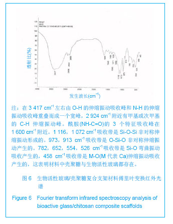
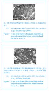
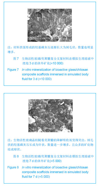
| [1]Nakahara H,Coldberg VM,Caplan AI.Culture expanded periosteal—derived cells exigbit ostechondeogenic potential in porous calcium phosphate ceramics in vivo.Clin Orthop Relat Res.1992;(276):291-298.[2]Borden M,Attawia M,Khan Y,et al.Tissue engineered bone formation in vivo using a novel sintered polymeric microsphere mat rix.J Bone Joint Surg Br. 2004;86(8):1200.[3]Langer R,Vacanti JP.Tissue engineerin.Science.1993;260: 920-926. [4]Griffith LG.Polymeric biomaterials.Acta Mater.2000;48: 263-277.[5]Agrawal CM, Ray RB.Biodegradable polymer scaffolds for musculoskeletal tissue engineering.J Biomed Mater Res. 2001;55(2):141-150. [6]Hayashi T.Biodegradable polymers for biomedical uses.Prog Polym Sci.1994;19: 663-702. [7]Reis RL.Natural-based polymers for biomedical applications.Woodhead Publishing,Cambridge,2008.[8]Lee KY,Mooney DJ.Hydrogels for tissue engineering.Chem Rev.2001;101(7):1869-1878.[9]Varghese S,Elisseeff JH.Hydrogels for musculoskeletal tissue engineering.Adv Polym Sci.2006;203:95-144.[10]Kokubo T, Takadama H.How useful is SBF in predicting in vivo bone bioactivity?Biomaterials.2006;27(15): 2907-2915.[11]Bohner M, Lemaitre J.Can bioactivity be tested in vitro with SBF solution?Biomaterials.2009;30(12):2175-2179. [12]Rohanová D, Boccaccini AR, Yunos DM,et al.TRIS buffer in simulated body fluid (SBF) distorts the assessment of glass-ceramic scaffold bioactivity.Acta Biomater.2011;7(6): 2623-2630.[13]Huang W,Day DE,Kittiratanapiboon K,et al.Kinetics and mechanisms of the conversion of silicate (45S5), borate, and borosilicate glasses to hydroxyapatite in dilute phosphate solution.J Mater Sci Mater Med.2006;17:583-596. [14]Huang W,Rahaman MN,Day DE,et al.Mechanisms for converting bioactive silicate, borate, and borosilicate glasses to hydroxyapatite in dilute phosphate solution.Phys Chem Glasses:Eur J Glass Sci Technol B.2006;47(6):647-658. [15]Hench LL,Splinter RJ,Allen WC,et al.Bonding mechanisms at the interface of ceramic prosthetic materials.J Biomed Mater Res.1971;5:117-141.[16]Hench LL,Polak JM.Third-generation biomaterials.Science. 2002; 295:1014-1017.[17]Lai W, Garino J, Ducheyne P. Silicon excretion from bioactive glass implanted in rabbit bone.Biomaterials.2002;23:213-217.[18]Chen ZQ,Thompson ID,Boccaccini AR.45S5 Bioglass®-derived glass-ceramic scaffold for bone tissue engineering.Biomaterials2006;27:2414-2425.[19]Austin K.Scaffold Design: Use of Chitosan in cartilage tissue engineering.MMG 445 Basic Biotech. eJ. 2007;3(1):62.[20]赖琛,唐绍裘.金属用耐磨耐高温陶瓷涂料的研究[J].陶瓷工程, 2000,34(6):41-43. |