Chinese Journal of Tissue Engineering Research
Previous Articles Next Articles
Secondary fracture risk assessment: Bone mineral density and bone metabolism of elderly women within 6-12 months after hip fractures
Zhang Zi-jun1, Zhao Wen1, Zhao Xi1, 2, Peng Hai-zhou1
- 1Beijing Aerospace General Hospital, Beijing 100076, China; 2Zunyi Medical University, Zunyi 563003, Guizhou Province, China
-
Revised:2013-11-22Online:2013-12-10Published:2013-12-10 -
Contact:Zhao Wen, Professor, Master’s supervisor, Beijing Aerospace General Hospital, Beijing 100076, China zw20897@hotmail.com -
About author:Zhang Zi-jun★, Master, Physician, Beijing Aerospace General Hospital, Beijing 100076, China 249758585@qq.com -
Supported by:the Health Fund of Fengtai District, Beijing, No. 2010-28*
CLC Number:
Cite this article
Zhang Zi-jun, Zhao Wen, Zhao Xi, Peng Hai-zhou . Secondary fracture risk assessment: Bone mineral density and bone metabolism of elderly women within 6-12 months after hip fractures[J]. Chinese Journal of Tissue Engineering Research, doi: 10.3969/j.issn.2095-4344.2013.50.003.
share this article
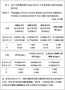
2.1 参与者数量分析 在伤后随访1年的过程中, 86例老年女性髋部骨折患者中有38例因各种原因脱落,48例观察对象按要求完成了随访,并进入结果分析。 2.2 老年女性髋部骨折患者骨密度及骨代谢指标在骨折愈合后的变化 见表1,2。"
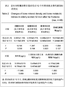
如表1所示,患者在伤后6个月,即骨折完全愈合时,患侧全髋部骨密度显著低于基线值(t=-8.221,P=0.000);腰椎骨密度明显低于基线值(t=-2.531, P=0.015);健侧全髋骨密度与基线值差异无显著性意义(t=-1.880,P=0.066);患者骨代谢指标骨碱性磷酸酶、骨钙素、Ⅰ型胶原交联C末端肽、血清抗酒石酸酸性磷酸酶5b水平均显著高于基线值(P < 0.05)。 如表2所示,患者在伤后12个月,即骨折完全愈合6个月,患侧全髋部骨密度显著低于基线值(t=-6.154, P=0.000);腰椎骨密度低于基线值(t=-2.30,P=0.020);健侧全髋骨密度与基线值差异无显著性意义(t=-1.347,P=0.184)。骨钙素水平明显高于基线值(t=-11.845,P=0.000),其余3项骨代谢指标与基线值差异无显著性意义(P > 0.05)。"

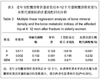
2.3 老年女性髋部骨折患者骨折愈合后患侧全髋部骨密度与各骨代谢指标血清水平的多元线性回归分析 见表3。"
| [1] 孟迅吾. 原发性骨质疏松的风险因素和风险评估[J].诊断学理论与实践,2012,11(1):1-4.[2] 蔡树鹏,周先来,黄坚霖,等.老年人髋部骨折与骨质疏松发病率的相关性研究[J].中国当代医药,2013,22(20):30-31.[3] Smulders E, Weerdesteyn V, Groen BE, et al. Efficacy of a short multidisciplinary falls prevention program for elderly persons with osteoporosis and a fall history a randomized controlled trial. Arch Phys Med Rehabil. 2010;91(11): 1705-1711. [4] Yesil Y,Ulger Z ,Halil M, et al. Coexistence of osteoporosis OP and coronary artery disease CAD in the elderly it is not just a by chance event. Arch Gerontol Geriatr. 2012;54(3):473-476. [5] Kouda K, Iki M, Fujita Y, et al. Alcohol intake and bone status in elderly Japanese men baseline data from the Fujiwarakyo osteoporosis sk in men FORMEN study. Bone. 2011;49(2): 275-280. [6] 陆铁,仲维佳,周君琳,等.全身振动对预防老年女性骨质疏松性骨折的作用[J].中华临床医师杂志:电子版,2012,6(5):1113-1116. [7] 杨涛涛,吕晓红,任凤华,等.老年骨质疏松性骨折患者的危险因素与干预措施[J].现代预防医学,2012,39(11):2756-2760. [8] Nguyen ND, Pongchaikayul C, Center JR, et al. Identification of high-risk individuals for hip fracture: a 14-year prospective study. J Bone Miner Res. 2005;20:1921-1928.[9] Xia WB, He SL, Xu L, et al. Rapidly increasing rates of hip fracture in Beijing, China. J Bone Miner Res. 2012;27: 125-129. [10] Starr AJ, Nakatani T, Reinert CM, et al. Superior pubic ramus fractures fixed with percutaneous screws : what predicts fixation failure? J Orthop Trauma. 2008;22(2):81-87. [11] Cooper C, Campion G, Melton LR. Hip fractures in the elderly: a world- wide projection. Osteoporos Int. 1992; 6: 285-289.[12] 李涛,刘智,孙天胜,等.老年髋部骨折后对侧髋部的再骨折[J]. 中国组织工程研究与临床康复,2012,16(4):752-756. [13] 李涛,于涛,张虎翼, 等.长春地区部分老年人群髋部骨质疏松性骨折发病特点初步调查与分析[J].中国骨质疏松杂志,2011, 17(5): 428-430. [14] 段国庆,张元民,赵晓伟, 等.手术治疗70岁以上老年骨质疏松性髋部骨折疗效分析[J].中国医师进修杂志: 综合版,2011, 34(29): 24-26.[15] 庞家容.心理干预对老年髋部骨折术后负性情绪及生活质量的影响[J].临床护理杂志,2013,12(1):20-21. [16] 王艳,吴新,汤根芬.家庭访视护理干预对老年髋部骨折术后病人生活质量的影响[J] .齐齐哈尔医学院学报,2012,33(13): 1803-1805. [17] 王晓伟, 孙天胜,刘智,等.老年髋部骨折手术疗效的危险因素分析[J].中华创伤骨科杂志,2011,13(9): 811-816.[18] Pillai A,Eranki V,Shenoy R,et al.Age related incidence and early-outcomes of hip fractures: a prospective cohort study of 1177 patients. J Orthop Surg Res. 2011;6: 5.[19] 宋洪强,玄燕华,董军,等.绝经后妇女骨代谢指标与骨折风险骨密度相关性研究[J].中国矫形外科杂志,2013,21(10):1005-1009.[20] 曹燕明,刘训志.骨代谢指标在老年骨质疏松性骨折后变化的临床应用[J].中国骨质疏松杂志,2011,11(1):18-23. [21] 石磊,薛庆云.骨代谢指标和骨密度在骨折后变化意义的评价[J].中国骨质疏松杂志,2009,15(5),377-380.[22] Yoshimura N,Muraki S,Oka H,et al. Biochemical markers of bone turnover as predictors of osteoporosis and osteoporotic fractures in men and women: 10- year follow- up of the Taiji cohort. Mod Rheumatol. 2011;6: 608-620.[23] Kaji H,Yamauchi M,Yamaguchi T,et al. Urinary deoxypyridinoline is a BMD- independent marker for prevalent vertebral fractures in postmenopausal women treated with glucocorticoid. Osteoporos Int. 2010;9: 1585-1590.[24] Wendeberg B. Mineral metabolism of fractures of the tibia in man studied with external counting of Sr85. Acta Orthop Scand Suppl.1961;(52):1-79.[25] Ingle BM, Hay SM, Bottjer HM, et al. Changes in Bone Mass and Bone Turnover Following Ankle Fracture. Osteoporos Int. 1999;10:408-415.[26] Veitch SW, Findlay SC, Hamer AJ, et al. Changes in bone mass and bone turnover following tibial shaft fracture. Osteoporos Int. 2006;17: 364-372.[27] Ivaska KK, Gerdhem P, Åkesson K,et al. Effect of Fracture on Bone Turnover Markers: A Longitudinal Study Comparing Marker Levels Before and After Injury in 113 Elderly Women. J Bone Miner Res. 2007;22(8):1155-1164.[28] Segal E, Raichlin V. Sophia Rimbrot. Hip fractures in the elderly in Israel-Possible impact of preventable conditions. Arch Gerontol Geriatr. 2009;48(2):182-185.[29] Puntus T, Schneider B, Meran J, et al . Influence of age and gender on associations of body mass index with bone mineral density, bone turnover markers and circulating calcium-regulating and bone-active sex hormones. Bone. 2011;49(4):824-829.[30] Chopin F, Biver E, Funck-Brentano T. Prognostic interest of bone turnover markers in the management of postmenopausal osteoporosis. Joint Bone Spine. 2012;79(1): 26-31.[31] 张萌萌,李亚刚,马倩倩,等.860例女性PTH、25(OH)D3、CT、骨代谢标志物与BMD相关性[J]. 中国妇幼保健,2012,27(32): 5054-5056. [32] Stofell K, Engler H, Kuster M, et al. Changes in Biochemical Markers after Lower Limb Fractures. Clin Chem. 2007;(53): 131-134. [33] Herade S, Roden GA. Control of osteoblast function and regulation of bone mass. Nature. 2003;(423):349-355.[34] 袁绍辉,刘伟,吴滨奇,等.骨质疏松性骨折愈合过程中Ⅰ,Ⅱ型胶原蛋白表达与其力学性能[J]. 中国组织工程研究与临床康复, 2011,15(2):208-212.[35] Ramonda R, Lorenzin M, Modesti V. Serological markers of erosive hand osteoarthritis. Eur J Intl Med. 2013;24(1):11-15.[36] Watts NB. Clinical utility of biochemical markers of bone remodeling. Clin Chem. 1999;45(8):1359-1368.[37] 何威,杨力,蔡德鸿.骨形成与吸收过程中的代谢生化标记物[J]. 中国临床康复,2005,9(42):118-120. [38] 夏维波. 骨转换生化标志物的检测及临床评价[J]. 医师进修杂志: 内科版,2005,28(5):3-6.[39] Nakasato YR, Janckila AJ, Halleen JM, et al. Clinical significance of immunoassays for type-5 tartrate-resistant acid phosphatase. Clin Chem. 1999;45(12):2150-2157.[40] Obrant KJ, Merle B, Bejui J, et al. Serum Bone-Gla Protein After Fracture. Clin Orthop Relat Res. 1990;(258):300-303.[41] Frost HM. The biology of fracture healing. An overviewfor clinicians. Part I. Clin Orthop. 1989;(248): 283-293.[42] Obrant KJ, Nilsson BE. Histomorphologic changes in the Tibial epiphysis after diaphyseal fracture. Clin Orthop. 1984; (185): 270-275.[43] Stofell K, Engler H, Kuster M, et al. Changes in Biochemical Markers after Lower Limb Fractures. Clin Chem. 2007;(53): 131-134. [44] Sato Y, Kaji M, Higuchi F, et al. Changes in Bone and Calcium Metabolism Following Hip Fracture in Elderly Patients. Osteop Int. 2001;12(6):445-449. |
| [1] | Li Xiaoqun, Xu Kaihang, Ji Fang. Corylin inhibits osteoclastogenesis and attenuates postmenopausal osteoporosis in mice [J]. Chinese Journal of Tissue Engineering Research, 2020, 24(在线): 4-. |
| [2] | Zhong Yuanming, Luo Man, Tang Fubo, Tang Cheng. Relationship between a linear black signal area of STIR image in MRI of osteoporotic thoracolumbar fracture and the size of external force [J]. Chinese Journal of Tissue Engineering Research, 2020, 24(9): 1400-1404. |
| [3] | Chen Qiang, Zhuo Hongwu, Xia Tian, Ye Zhewei . Toxic effects of different-concentration isoniazid on newborn rat osteoblasts in vitro [J]. Chinese Journal of Tissue Engineering Research, 2020, 24(8): 1162-1167. |
| [4] | Xu Shaoce, Wang Shiyao, Zhou Jianwei, Pan Yixin, Wang Yuliang. Fibroblast growth factor receptor 3 regulation and mechanism in callus formation [J]. Chinese Journal of Tissue Engineering Research, 2020, 24(7): 1083-1088. |
| [5] | Liu Zhendong, Qin Sihe. Four-dimensional space events of fracture healing [J]. Chinese Journal of Tissue Engineering Research, 2020, 24(6): 903-910. |
| [6] | Liu Qun, Sun Dongdong, Gao Lilan, He Zhijiang, Sun Minglin. Risk factors for fractures secondary to percutaneous kyphoplasty: a meta-analysis [J]. Chinese Journal of Tissue Engineering Research, 2020, 24(6): 976-984. |
| [7] | Cao Guolong, Tian Faming, Liu Jiayin. Lovastatin combined with insulin effects on fracture healing in rat models of bilateral ovariectomized type 2 diabetic mellitus [J]. Chinese Journal of Tissue Engineering Research, 2020, 24(5): 673-681. |
| [8] | Yu Peiyuan, Zhang Zhida, Liang Lichang, Yang Zhidong, Huang Jinjing, Peng Jiancheng, Liang Ziyang, He Jiahui, Zhao Wenhua, Yu Fuyong, Chen Guifeng, Liang De, Jiang Xiaobing . Difference in the effect of metformin on bone strength of lumbar vertebrae and hind limbs in diabetic rats [J]. Chinese Journal of Tissue Engineering Research, 2020, 24(5): 657-661. |
| [9] | Wu Qi, Liao Ying, Sun Guanghua, Zhou Guijuan, Liao Yuan, Liu Jing, Zhong Peirui, Cheng Guo, Deng Chengyuan, Wang Tiantian. Changes of subchondral bone in rat models of knee osteoarthritis treated by elcatonin [J]. Chinese Journal of Tissue Engineering Research, 2020, 24(5): 709-715. |
| [10] | Luo Peijie, Yuan Kai, Li Daxing, Zhang Shuncong, Guo Huizhi, Tang Yongchao, Zhou Tengpeng, Guo Danqing, Li Yongxian, Mo Guoye. Comparison of the short-segment and long-segment cement-augmented pedicle screw fixation for osteoporotic thoracolumbar fracture: a finite element study [J]. Chinese Journal of Tissue Engineering Research, 2020, 24(3): 342-347. |
| [11] | Yang Shun, Chen Keyi, Cheng Yabo, Xiang Wang, Zhang Jing, Gu Hongji, Chi Haotian. Wrist arthroscopy-assisted titanium internal fixator for the treatment of complex distal radius fractures [J]. Chinese Journal of Tissue Engineering Research, 2020, 24(3): 366-371. |
| [12] | Liu Ruizhen, Wang Wangren, Hao Chen, Liang Dongmu, Guan Haishan. Effect of bone cement distribution on adjacent vertebral body fracture after unilateral percutaneous vertebroplasty for single segment osteoporotic vertebral compression fracture [J]. Chinese Journal of Tissue Engineering Research, 2020, 24(28): 4498-4504. |
| [13] | Xie Hui, Chen Haopeng, Wang Benjie, Fu Weimin, Zhao Dewei. Effect of different distribution types of bone cement after percutaneous kyphoplasty on osteoporotic vertebral compression fractures at different sites [J]. Chinese Journal of Tissue Engineering Research, 2020, 24(28): 4505-4510. |
| [14] | Yao Shunhan, Wei Huacheng, Qin Jiagang, Liao Liang. Mogroside V stimulates osteoblast proliferation and differentiation by promoting LncRNA TUG1 expression [J]. Chinese Journal of Tissue Engineering Research, 2020, 24(26): 4129-4134. |
| [15] |
Li Shaoshuo, Gu Yidan, Shao Yang, Yin Heng, Wang Jianwei.
Visualization analysis of CiteSpace knowledge maps regarding prevention and treatment of postmenopausal osteoporosis with traditional Chinese medicine [J]. Chinese Journal of Tissue Engineering Research, 2020, 24(26): 4224-4230. |
| Viewed | ||||||
|
Full text |
|
|||||
|
Abstract |
|
|||||