Chinese Journal of Tissue Engineering Research ›› 2013, Vol. 17 ›› Issue (47): 8162-8168.doi: 10.3969/j.issn.2095-4344.2013.47.004
Previous Articles Next Articles
Preparation of polyethylenimine-chitosan/DNA nanoparticles for transfecting articular chondrocytes in vitro
Lu Hua-ding, Dai Yu-hu, Lian Li-yi, Lü Lu-lu, Zhao Hui-qing
- Department of Orthopaedics, the Third Affiliated Hospital of Sun Yat-san University, Guangzhou 510630, Guangdong Province, China
-
Revised:2013-10-22Online:2013-11-19Published:2013-11-19 -
About author:Lu Hua-ding☆, M.D., Associate professor, Department of Orthopaedics, the Third Affiliated Hospital of Sun Yat-san University, Guangzhou 510630, Guangdong Province, China johnnielu@126.com -
Supported by:the National Natural Science Foundation of China, No. 82172040*, 30600632*; the Science and Technology Project of Guangdong Province, No. 2012B031800451*; the Natural Science Foundation of Guangdong Province, No. S2011010004808*
CLC Number:
Cite this article
Lu Hua-ding, Dai Yu-hu, Lian Li-yi, Lü Lu-lu, Zhao Hui-qing. Preparation of polyethylenimine-chitosan/DNA nanoparticles for transfecting articular chondrocytes in vitro[J]. Chinese Journal of Tissue Engineering Research, 2013, 17(47): 8162-8168.
share this article

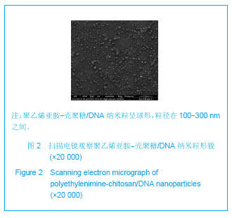
2.1 聚乙烯亚胺-壳聚糖/DNA纳米粒的形态特征 见图2所示,扫描电镜下观察聚乙烯亚胺-壳聚糖/DNA纳米粒呈球形,粒径在100-300 nm之间。经激光粒度分析仪检测,在pH=5.5时,粒径为(154.6±18.6) nm,Zeta电位为(24.68±6.82) mV,分散度为0.284±0.078。"

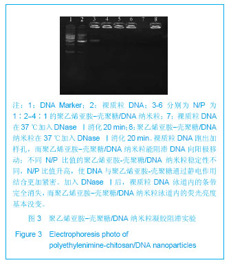
2.2 凝胶电泳阻滞实验结果 见图3所示,裸质粒DNA跑出加样孔,而聚乙烯亚胺-壳聚糖/DNA纳米粒能阻滞DNA向阳极移动;不同N/P比值的聚乙烯亚胺-壳聚糖/ DNA 纳米粒稳定性不同,N/P比值升高,使DNA与聚乙烯亚胺-壳聚糖通过静电作用结合更加紧密。加入DNaseⅠ后,裸质粒DNA 泳道内的条带完全消失,而聚乙烯亚胺-壳聚糖/DNA纳米粒泳道内的荧光亮度基本没变。"

2.3 聚乙烯亚胺-壳聚糖/DNA纳米粒入胞、入核考察 见图4所示,图中绿色部分为FITC标记的DNA,在1 h时,激光共聚焦显微镜检测可见绿色部分已经入胞,大部分在胞浆中,围绕着蓝色DAPI标记的细胞核周围; 2 h后绿色部分与蓝色已开始重叠,说明DNA已经入核。"

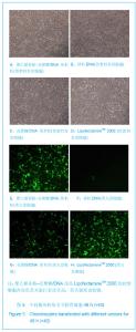
2.4 体外基因转染实验结果 软骨细胞转染后24 h,聚乙烯亚胺-壳聚糖/DNA纳米粒组在荧光显微镜下有较少细胞表达绿色荧光蛋白,壳聚糖/DNA纳米粒组只有少数细胞表达弱的绿色荧光,至48 h细胞的转染率增高,荧光强度也增强;裸质粒DNA组只有极个别的细胞表达绿色荧光蛋白蛋白阳性,聚乙烯亚胺-壳聚糖/DNA纳米粒组在48 h阳性细胞的比例明显增加,荧光强度也增强,见图5,LipofectamineTM 2000转染软骨细胞24 h 后,荧光显微镜下已有表达绿色荧光蛋白的阳性细胞,转染48 h后细胞的转染率更高。"


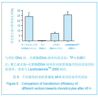
见图6所示,4组之间对软骨细胞的转染效率(绿色荧光蛋白表达阳性率)存在明显差别(F=258.234,P=0);组间两两比较采用LSD-t 法检验,聚乙烯亚胺-壳聚糖/ DNA纳米粒与LipofectamineTM 2000组均与裸质粒DNA组及壳聚糖/DNA纳米粒组存在明显差别(P < 0.001),但聚乙烯亚胺-壳聚糖/DNA纳米粒与LipofectamineTM 2000之间差别不显著(P=0.522)。"
| [1] Rekha MR,Sharma CP.Polymers for gene delivery: current status and future perspectives.Recent Pat DNA Gene Seq. 2012;6(2):98-107.
[2] Tiera MJ,Shi Q,Winnik FM,et al.Polycation-based gene therapy: current knowledge and new perspectives.Curr Gene Ther.2011;11(4):288-306.
[3] Tripathi SK,Goyal R,Kumar P,et al.Linear polyethylenimine-graft-chitosan copolymers as efficient DNA/siRNA delivery vectors in vitro and in vivo. Nanomedicine.2012;8(3):337-345.
[4] Mao S,Sun W,Kissel T.Chitosan-based formulations for delivery of DNA and siRNA.Adv Drug Deliv Rev.2010; 62(1):12-27.
[5] Gao JQ,Zhao QQ,Lv TF,et al.Gene-carried chitosan-linked-PEI induced high gene transfection efficiency with low toxicity and significant tumor-suppressive activity.Int J Pharm.2010;387(1-2):286-294.
[6] 卢华定,赵慧清,吕璐璐,等.壳聚糖-负载增强型绿色荧光蛋白基因的质粒DNA纳米微球体外转染软骨细胞的能力及影响因素[J].中华创伤骨科杂志,2011,13(11):64-69.
[7] Steinert AF,Nöth U,Tuan RS.Concepts in gene therapy for cartilage repair. Injury.2008;39 Suppl 1:S97-113.
[8] Lehrman S.Virus treatment questioned after gene therapy death. Nature.1999;401(6753):517-518.
[9] Liu Q,Muruve DA.Molecular basis of the inflammatory response to adenovirus vectors.Gene Ther.2003;10(11): 935-940.
[10] 张世浩,朱立新,靳安民,等.软骨细胞复合胶原/壳聚糖/β-磷酸三钙层状梯度修复体的体外培养[J].中国组织工程研究与临床康复,2008,12(41):8033-8036.
[11] Jayakumar R,Chennazhi KP,Muzzarelli RAA,et al.Chitosan conjugated DNA nanoparticles in gene therapy.Carbohydrate Polymers.2010;79(1):1-8.
[12] Aiba S.Studies on chitosan: 2. Solution stability and reactivity of partially N-acetylated chitosan derivatives in aqueous media. Int J Biol Macromol.1989;11(4): 249-252.
[13] Wong K,Sun G,Zhang X,et al.PEI-g-chitosan, a novel gene delivery system with transfection efficiency comparable to polyethylenimine in vitro and after liver administration in vivo. Bioconjug Chem.2006;17(1): 152-158.
[14] Lavertu M,Méthot S,Tran-Khanh N,et al. High efficiency gene transfer using chitosan/DNA nanoparticles with specificcombinationsof molecular weight and degree of deacetylation. Biomaterials.2006;27(27):4815-4824.
[15] Chang KL,Higuchi Y,Kawakami S,et al. Efficient gene transfection by histidine-modified chitosan through enhancement ofendosomal escape.Bioconjug Chem. 2010; 21(6):1087-1095.
[16] Song B,Zhang W,Peng R,et al.Synthesis and cell activity of novel galactosylated chitosan as a gene carrier.Colloids Surf B Biointerfaces. 2009;70(2):181-186.
[17] Xu Z,Wan X,Zhang W,et al.Synthesis of biodegradable polycationic methoxy poly (ethylene glycol)– polyethylenimine–chitosan and its potential as gene carrier. Carbohydr Polym.2009;78:46-53.
[18] Jiang HL,Kim YK,Arote R,et al.Chitosan-graft- polyethylenimine as a gene carrier.J Control Release.2007; 117(2):273-280.
[19] Gao Y,Xu Z,Chen S,et al. Arginine-chitosan/DNA self-assemble nanoparticles for gene delivery: In vitro characteristics and transfection efficiency.Int J Pharm.2008; 359(1-2):241-246.
[20] Germershaus O,Mao S,Sitterberg J,et al.Gene delivery using chitosan, trimethyl chitosan or polyethylenglycol-graft- trimethyl chitosan block copolymers: establishment of structure-activity relationships in vitro.J Control Release.2008; 125(2):145-154.
[21] Morris VB,Neethu S,Abraham TE,et al.Studies on the condensation of depolymerized chitosans with DNA for preparing chitosan-DNA nanoparticles for gene delivery applications.J Biomed Mater Res B Appl Biomater.2009;89(2): 282-292.
[22] 陈斌,马世坤,高娴.巯基化壳聚糖-质粒 DNA 纳米粒的制备及相关性质的研究[J]. 天津医科大学学报,2008,14(4):466-469.
[23] Knudson W,Chow G,Knudson CB.CD44-mediated uptake and degradation of hyaluronan.Matrix Biol.2002;21(1):15-23.
[24] Campo GM,Avenoso A,Campo S,et al.Small hyaluronan oligosaccharides induce inflammation by engaging both toll-like-4 and CD44 receptors in human chondrocytes.Biochem Pharmacol.2010;80(4):480-490.
[25] Lu HD,Zhao HQ,Wang K,et al.Novel hyaluronic acid-chitosan nanoparticles as non-viral gene delivery vectors targeting osteoarthritis.Int J Pharm.2011; 420(2):358-365.
[26] Sezer AD,Akbu?a J.Comparison on in vitro characterization of fucospheres and chitosan microspheres encapsulated plasmid DNA (pGM-CSF): formulation design and release characteristics.AAPS Pharm Sci Tech.2009;10(4):1193-1199.
[27] 金才益,王斌,曾忠友,等.壳聚糖基因转移载体在关节软骨细胞中表达的定量分析[J].中国组织工程研究与临床康复,2007,11(45): 9035-9038.
[28] Hashimoto M,Morimoto M,Saimoto H,et al.Lactosylated chitosan for DNA delivery into hepatocytes: the effect of lactosylation on the physicochemical properties and intracellular trafficking of pDNA/chitosan complexes.Bioconjug Chem.2006;17(2):309-316.
[29] Feng M,Lee D,Li P.Intracellular uptake and release of poly(ethyleneimine)-co-poly(methyl methacrylate) nanoparticle/pDNA complexes for gene delivery.Int J Pharm. 2006;311(1-2):209-214.
[30] 卢华定,吕璐璐,赵慧清,等.转化生长因子β1基因缓释的壳聚糖纳米粒制备及体外检测[J].中国组织工程研究,2012,16(12): 2120-2124. |
| [1] | He Yan-ping, Ma De-chun, Li Lei, Zhang Li, Zheng Shuang, Dong Ke-xin. Hemocompatibility and surface modification of artificial blood vessel materials [J]. Chinese Journal of Tissue Engineering Research, 2015, 19(8): 1272-1276. |
| [2] | Li Bing-ting, Jia Ying-zhen, Liu Zhi-fang, Song Yuan, Hou Xiao-wei. Effect of freeze-dried bone xenograft and platelet-rich fibrin compound on osteogenesis and osseointegration of alveolar bone defects [J]. Chinese Journal of Tissue Engineering Research, 2014, 18(52): 8376-8381. |
| [3] | Wu Tao, Wu Jin-hui, Zheng Guo-dong, Lu Zhi-qin, Zeng Hai-yan, Lv Jun, Xing Jian-zhou. Release behavior of icariin-chitosan/hydroxyapatite scaffolds in vitro [J]. Chinese Journal of Tissue Engineering Research, 2014, 18(52): 8399-8404. |
| [4] | Wang Xiao-dong, Zhang Yong-hong. Preparation and performance of recombinant human bone morphogenetic protein-2-poly(hydroxybutyrate-co-hydroxyoctanoate) nanospheres [J]. Chinese Journal of Tissue Engineering Research, 2014, 18(52): 8405-8408. |
| [5] | Feng Chao, Li Zhe, Lv Xiang-guo, Xu Yue-min, Fu Qiang. In vitro preparation and biochemical evaluation of oxygen generative keratin/silk fibroin compound biomaterial [J]. Chinese Journal of Tissue Engineering Research, 2014, 18(52): 8480-8486. |
| [6] | Li Tian-shi, Zeng Wen-ni, He Jun-jun. Chitin and its derivatives inhibit scar formation [J]. Chinese Journal of Tissue Engineering Research, 2014, 18(52): 8504-8508. |
| [7] | Su Da-ming, Zhao Jun-hua, Huang Zhi-yuan, Li Da-lian, Liu Jian-jun, Wang Min, Li Hua. Effect of Jianxi Qianggu Pill on the pathological changes of articular cartilage in knee osteoarthritis rabbits [J]. Chinese Journal of Tissue Engineering Research, 2014, 18(5): 657-662. |
| [8] | Ma Song-feng, Cao Hui, Zheng Feng, Qiao Jun, Zhang Guo-ming. Concomitant cardiac valve replacement and coronary artery bypass grafting [J]. Chinese Journal of Tissue Engineering Research, 2014, 18(5): 699-704. |
| [9] | Liu Xue-guang, Qiu Yong, Sun Zhen-zhong, Qian Bang-ping, Wang Shou-feng. Culture and identification of chondrocytes isolated from the vertebral endplate of patients with type I neurofibromatosis associated with atrophic changes in vitro [J]. Chinese Journal of Tissue Engineering Research, 2014, 18(46): 7396-7400. |
| [10] | Xu Jing1, Wang Li-ming, Zhou Li-dong, Wu Mei, Cui Hui, Zhao Jing, Zeng Du-juan, Zhang Zhong-wen, Liu Ai-bing. Platelet-rich plasma plus human umbilical cord mesenchymal stem cells for cartilage repair [J]. Chinese Journal of Tissue Engineering Research, 2014, 18(41): 6633-6638. |
| [11] | Ning Xiao-ting, Shao Bo, Gong Zhong-cheng, Liu Hui, Ling Bin, Keremu Abass, Lin Zhao-quan, . Chondrogenesis of synovial mesenchymal stem cells co-cultured with chondrocytes on the three-dimensional scaffold [J]. Chinese Journal of Tissue Engineering Research, 2014, 18(34): 5434-5440. |
| [12] | Lu Hua-ding, Lian Li-yi, Chen Ming-wei, Dai Yu-hu. Cloning and expression of Asperguillus endo-chitosanase gene in Escherichia coli [J]. Chinese Journal of Tissue Engineering Research, 2014, 18(34): 5490-5496. |
| [13] | Fan Zhong-bao, Rong Da-qing, Liu Qing-feng. Prolene hernia system versus polypropylene mesh plug in tension-free hernia repair [J]. Chinese Journal of Tissue Engineering Research, 2014, 18(34): 5535-5539. |
| [14] | Li A-li. Absorbable ligating clip in laparoscopic hysterectomy [J]. Chinese Journal of Tissue Engineering Research, 2014, 18(34): 5561-5565. |
| [15] | Dai Bing, Xu Hai-ting, Jin Hai-dong, Chen Hui, Cai Jian-wu, Fan Shi-yang, Pan Jun. Hypoxia effects on the chondrogenic differentiation of three-dimensional co-cultured adipose-derived stem cells and articular chondrocytes [J]. Chinese Journal of Tissue Engineering Research, 2014, 18(29): 4630-4635. |
| Viewed | ||||||
|
Full text |
|
|||||
|
Abstract |
|
|||||