Chinese Journal of Tissue Engineering Research
Previous Articles Next Articles
Establishment of human-rhesus chimeric liver using adult bone marrow mesenchymal stem cells
He Bao-li1, 2, Ma Li-hua3, Chen Li-ling1, 2, Liu Ru-wen2, Yang Ren-hua2
- 1School of Veterinary Medicine, Yangzhou University, Yangzhou 225009, Jiangsu Province, China; 2Department of Experimental Animals, Kunming Medical University, Kunming 650500, Yunnan Province, China; 3Department of Clinical Laboratory, Kunming General Hospital of Chengdu Military Region, Kunming 650032, Yunnan Province, China
-
Revised:2013-08-01Online:2013-11-05Published:2013-11-05 -
Contact:Yang Ren-hua, Master, Department of Experimental Animals, Kunming Medical University, Kunming 650500, Yunnan Province, China Yangrhua@hotmail.com -
About author:He Bao-li☆, Studying for doctorate, School of Veterinary Medicine, Yangzhou University, Yangzhou 225009, Jiangsu Province, China; Department of Experimental Animals, Kunming Medical University, Kunming 650500, Yunnan Province, China hbl117@sina.com Ma Li-hua, Department of Clinical Laboratory, Kunming General Hospital of Chengdu Military Region, Kunming 650032, Yunnan Province, China He Bao-li and Ma Li-hua contributed equally to this work. -
Supported by:the Scientific and Technological Plan of Yunnan Province, No. 2013FZ071*
CLC Number:
Cite this article
He Bao-li, Ma Li-hua, Chen Li-ling, Liu Ru-wen, Yang Ren-hua. Establishment of human-rhesus chimeric liver using adult bone marrow mesenchymal stem cells[J]. Chinese Journal of Tissue Engineering Research, doi: 10.3969/j.issn.2095-4344.2013.45.002.
share this article
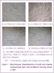
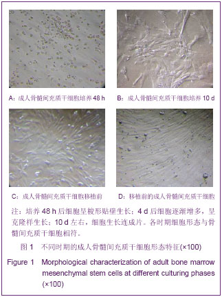
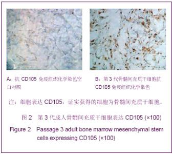
2.1 成人骨髓间充质干细胞分离、培养及鉴定结果 见图1,2。"
"

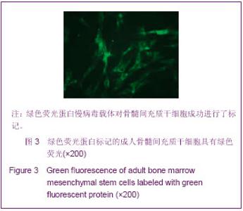
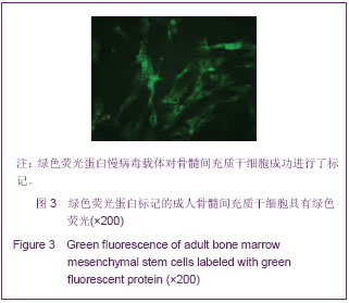
骨髓细胞经1.073 g/mL的人淋巴细胞分离液分离后,细胞大多呈圆球形,大小差异很大。培养48 h后,可以看到有细胞呈梭形贴壁生长,见图1A。4 d后,细胞逐渐增多,呈克隆样生长,见图1B;10 d左右,细胞生长连成片,见图1C,覆盖瓶底超过80%,用胰蛋白酶消化后传代。细胞传至6代,细胞呈典型的梭形状态,生长极性较为一致。 小鼠抗人CD34及抗人CD105对第3代骨髓间充质干细胞经免疫细胞化学染色,细胞表达CD105而不表达CD34,见图2。 2.2 绿色荧光蛋白标记成人骨髓间充质干细胞 成人骨髓间充质干细胞用包含绿色荧光蛋白慢病毒载体的培养液培养48 h后,在荧光显微镜下绿色荧光清晰可见,主要集中于细胞核周围的细胞质内,远离细胞核,荧光逐渐减弱,见图3。"
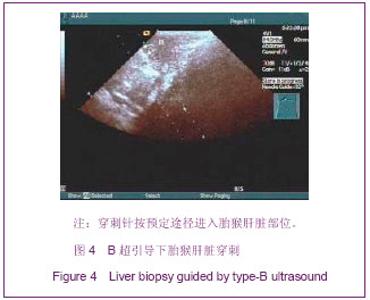
2.3 B超引导下胎猴肝脏穿刺结果 在B超引导下,穿刺针准确进入胎猴的肝脏内,见图4,图中箭头示穿刺针进入胎猴肝脏的位置。"
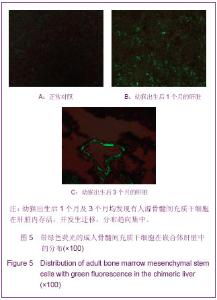
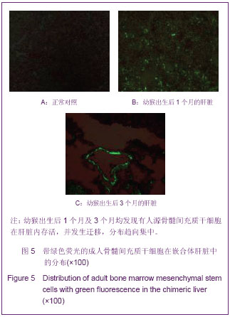
2.4 成人骨髓间充质干细胞在嵌合体肝脏中的分布 对照组未移植成人骨髓间充质干细胞,在荧光显微镜下未发现具有绿色荧光的细胞,见图5A。 绿色荧光蛋白标记的细胞进入胎猴肝脏形成人-猴嵌合体肝脏,荧光显微镜下可以看到幼猴出生1个月后标记绿色荧光的成人骨髓间充质干细胞细胞在猴肝脏内散在分布较为均匀,见图5B;出生后3个月,标有绿色荧光的骨髓间充质干细胞分布较为集中,亮度明显增强,见图5C。"
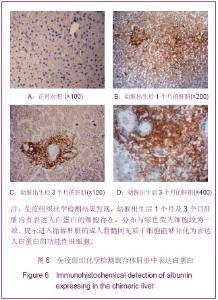
2.5 人-猴嵌合体肝脏中白蛋白的表达 成人骨髓间充质干细胞在人-猴嵌合体肝脏中的分布较为一致,对照组未移植人骨髓间充质干细胞,在猴肝脏内没有发现人源白蛋白表达,见图6A。 而在移植了成人骨髓间充质干细胞的实验组人-猴嵌合体肝脏内,幼猴出生1个月就发现有人的白蛋白表达。从免疫组织化学染色结果来看,表达人白蛋白阳性细胞在出生后1个月的幼猴嵌合体肝脏内散在分布,大部分呈弱阳性表达,小部分表达阳性较强,见图6B。 3个月后,表达人白蛋白的阳性细胞分布集中,而且表达量有所增加,见图6C;从放大的显微照片上可以看出白蛋白表达集中于细胞质内,特异性很好,见图6D。"

| [1] Salamon A, Toldy E, Nagy L, et al. The role of adult bone marrow derived mesenchymal stem cells in the repair of tissue injuries. Orv Hetil. 2012;153(46):1807-1815. [2] Hanson S, Thibeault SL, Hematti P. Clinical applications of mesenchymal stem cells in laryngotracheal reconstruction. Curr Stem Cell Res Ther. 2010;5(3):268-272. [3] Bruckner S, Tautenhahn HM, Winkler S, et al. Isolation and hepatocyte differentiation of mesenchymal stem cells from porcine bone marrow-"surgical waste" as a novel msc source. Transplant Proc. 2013;45(5):2056-2058. [4] Zompi S, Harris E. Animal models of dengue virus infection. Viruses. 2012;4(1):62-82. [5] Kelschenbach JL, Saini M, Hadas E, et al. Mice chronically infected with chimeric hiv resist peripheral and brain superinfection: A model of protective immunity to hiv. J Neuroimmune Pharmacol. 2012;7(2):380-387. [6] Kakuni M, Morita M, Matsuo K, et al. Chimeric mice with a humanized liver as an animal model of troglitazone-induced liver injury. Toxicol Lett. 2012;214(1):9-18. [7] Tee R, Morrison WA, Dusting GJ, et al. Transplantation of engineered cardiac muscle flaps in syngeneic rats. Tissue Eng Part A. 2012;18(19-20):1992-1999. [8] 邹一丰,张珑娟,吴小剑,等.猪-人-鼠嵌合体动物模型的建立及介导人t细胞对猪异种抗原的耐受[J].中国病理生理杂志,2009, 25(2):318-322. [9] 周文君,张连峰.人干细胞实验动物嵌合体模型的研究进展[J]. 中国比较医学杂志,2007,17(11):694-696,699. [10] Almeida-Porada G, El Shabrawy D, Porada C, et al. Differentiative potential of human metanephric mesenchymal cells. Exp Hematol. 2002;30(12):1454-1462. [11] Liechty KW, MacKenzie TC, Shaaban AF, et al. Human mesenchymal stem cells engraft and demonstrate site-specific differentiation after in utero transplantation in sheep. Nat Med. 2000;6(11):1282-1286. [12] 薛丽香,霍名赫,闫章才,等.建设动物模型平台,推动医学科学发展——科学基金“疾病动物模型”项目介绍与资助分析[J]. 中国科学基金,2013,(2):79-82. [13] Zhang L, Peng L P, Wu N, et al. Development of bone marrow mesenchymal stem cell culture in vitro. Chin Med J (Engl). 2012;125(9):1650-1655. [14] De Schauwer C, Meyer E, Van de Walle GR, et al. Markers of stemness in equine mesenchymal stem cells: A plea for uniformity. Theriogenology. 2011;75(8):1431-1443. [15] Liu H, Zhang J, Liu CY, et al. Bone marrow mesenchymal stem cells can differentiate and assume corneal keratocyte phenotype. J Cell Mol Med. 2012;16(5):1114-1124. [16] Heo YT, Lee SH, Kim T, et al. Production of somatic chimera chicks by injection of bone marrow cells into recipient blastoderms. J Reprod Dev. 2012;58(3):316-322. [17] 马丽花,赵熠,蔡学敏,等.绿色荧光蛋白标记成人骨髓间充质干细胞的超微结构特征[J].中国组织工程研究与临床康复,2011, 15(6): 959-962. [18] 刘春喜,李大庆,王荣,等.血管组织自发荧光的去除方法及应用[J]. 中华病理学杂志,2008,37(4):276-277. [19] 陈加玲,宋志军,李瑗.肝细胞移植嵌合体动物模型研究进展[J]. 医学综述,2009,15(2):177-179. [20] Xu H, Ramsey DM, Wu S, et al. Simultaneous bone marrow and composite tissue transplantation in rats treated with nonmyeloablative conditioning promotes tolerance. Transplantation. 2013;95(2):301-308. [21] Xu Q, Chen LL, Ruan X, et al. The draft genome of sweet orange (citrus sinensis). Nat Genet. 2013;45(1):59-66. [22] Tachibana M, Sparman M, Ramsey C, et al. Generation of chimeric rhesus monkeys. Cell. 2012;148(1-2):285-295. [23] Coulombel L. ES cells and postnatal chimeric rhesus monkeys. Med Sci (Paris). 2012;28(4):370-372. [24] Sachs DH, Sykes M, Kawai T, et al. Immuno-intervention for the induction of transplantation tolerance through mixed chimerism. Semin Immunol. 2011;23(3):165-173. [25] 胡彬,刘晓华,赵林.骨髓间充质干细胞的分离培养与鉴定[J]. 中国组织工程研究与临床康复,2011,15(27):5108-5111. [26] 王忠,高毅,汪艳,等.人骨髓间充质干细胞的分离培养与鉴定[J]. 中华神经医学杂志,2006,5(10):973-977. [27] 范萍,刘永琦,吴晓晶,等.不同条件下骨髓间充质干细胞分离培养及其生物学特性研究[J].细胞与分子免疫学杂志,2010,26(4): 322-324. [28] Li C, Zheng Y, Wang X, et al. Bone marrow-derived stem cells contribute skin regeneration in skin and soft tissue expansion. J Cell Physiol. 2011;226(11):2834-2840. [29] 王耘川. Bmscs联合骨髓移植建立嵌合体并诱导免疫耐受形成的机制研究[D]. 第四军医大学,2012. [30] Leu YW, Huang TH, Hsiao SH. Epigenetic reprogramming of mesenchymal stem cells. Adv Exp Med Biol. 2013;754: 195-211. [31] Zhang Y, Khan D, Delling J, et al. Mechanisms underlying the osteo- and adipo-differentiation of human mesenchymal stem cells. Scientific World J. 2012;2012:793823. [32] Zhang L, Ye JS, Decot V, et al. Research on stem cells as candidates to be differentiated into hepatocytes. Biomed Mater Eng. 2012;22(1-3):105-111. [33] Zhao MT, Yang X, Lee K, et al. The in vivo developmental potential of porcine skin-derived progenitors and neural stem cells. Stem Cells Dev. 2012;21(14):2682-2688. [34] Zou J, Wei X. Transplantation of gfp-expressing blastomeres for live imaging of retinal and brain development in chimeric zebrafish embryos. J Vis Exp. 2010;(41). pii: 1924. [35] 石永乾. 慢病毒载体介导的绿色荧光蛋白转基因标记与猪嵌合体制作研究[D]. 东北农业大学, 2012. [36] 谭云鹤,铁种,平杨,等.人骨髓间充质干细胞生物学特性及腺病毒介导的gfp标记[J].西安交通大学学报: 医学版, 2012,5(5): 587-592. [37] Lutgehetmann M, Bornscheuer T, Volz T, et al. Hepatitis b virus limits response of human hepatocytes to interferon-alpha in chimeric mice. Gastroenterology. 2011; 140(7):2074-2083. [38] Chayama K, Hayes CN, Hiraga N, et al. Animal model for study of human hepatitis viruses. J Gastroenterol Hepatol. 2011;26(1):13-18. [39] Wu Y, Zhao RC. The role of chemokines in mesenchymal stem cell homing to myocardium. Stem Cell Rev. 2012;8(1): 243-250. [40] Li H, Fu X. Mechanisms of action of mesenchymal stem cells in cutaneous wound repair and regeneration. Cell Tissue Res. 2012;348(3):371-377. [41] Wise AF, Ricardo SD. Mesenchymal stem cells in kidney inflammation and repair. Nephrology (Carlton). 2012;17(1): 1-10. |
| [1] | Kong Desheng, He Jingjing, Feng Baofeng, Guo Ruiyun, Asiamah Ernest Amponsah, Lü Fei, Zhang Shuhan, Zhang Xiaolin, Ma Jun, Cui Huixian. Efficacy of mesenchymal stem cells in the spinal cord injury of large animal models: a meta-analysis [J]. Chinese Journal of Tissue Engineering Research, 2020, 24(在线): 3-. |
| [2] | Chen Jinsong, Wang Zhonghan, Chang Fei, Liu He. Tissue engineering methods for repair of articular cartilage defect under special conditions [J]. Chinese Journal of Tissue Engineering Research, 2020, 24(8): 1272-1279. |
| [3] | Liu Chundong, Shen Xiaoqing, Zhang Yanli, Zhang Xiaogen, Wu Buling. Effects of strontium-modified titanium surfaces on adhesion, migration and proliferation of bone marrow mesenchymal stem cells and expression of bone formation-related genes [J]. Chinese Journal of Tissue Engineering Research, 2020, 24(7): 1009-1015. |
| [4] | Lin Ming, Pan Jinyong, Zhang Huirong. Knockout of NIPBL gene down-regulates the abilities of proliferation and osteogenic differentiation in mouse bone marrow mesenchymal stem cells [J]. Chinese Journal of Tissue Engineering Research, 2020, 24(7): 1002-1008. |
| [5] | Zhang Wen, Lei Kun, Gao Lei, Li Kuanxin. Neuronal differentiation of rat bone marrow mesenchymal stem cells via lentivirus-mediated bone morphogenetic protein 7 transfection [J]. Chinese Journal of Tissue Engineering Research, 2020, 24(7): 985-990. |
| [6] | Wu Zhifeng, Luo Min. Biomechanical analysis of chemical acellular nerve allograft combined with bone marrow mesenchymal stem cell transplantation for repairing sciatic nerve injury [J]. Chinese Journal of Tissue Engineering Research, 2020, 24(7): 991-995. |
| [7] | Huang Yongming, Huang Qiming, Liu Yanjie, Wang Jun, Cao Zhenwu, Tian Zhenjiang, Chen Bojian, Mai Xiujun, Feng Enhui. Proliferation and apoptosis of chondrocytes co-cultured with TDP43 lentivirus transfected-human umbilical cord mesenchymal stem cells [J]. Chinese Journal of Tissue Engineering Research, 2020, 24(7): 1016-1022. |
| [8] | Qin Xinyu, Zhang Yan, Zhang Ningkun, Gao Lianru, Cheng Tao, Wang Ze, Tong Shanshan, Chen Yu. Elabela promotes differentiation of Wharton’s jelly-derived mesenchymal stem cells into cardiomyocyte-like cells [J]. Chinese Journal of Tissue Engineering Research, 2020, 24(7): 1046-1051. |
| [9] | Liu Mengting, Rao Wei, Han Bing, Xiao Cuihong, Wu Dongcheng. Immunomodulatory characteristics of human umbilical cord mesenchymal stem cells in vitro [J]. Chinese Journal of Tissue Engineering Research, 2020, 24(7): 1063-1068. |
| [10] |
Cen Yanhui, Xia Meng, Jia Wei, Luo Weisheng, Lin Jiang, Chen Songlin, Chen Wei, Liu Peng, Li Mingxing, Li Jingyun, Li Manli, Ai Dingding, Jiang Yunxia.
Baicalein inhibits the biological behavior of hepatocellular
carcinoma stem cells by downregulation of Decoy receptor 3 expression |
| [11] | Zhang Peigen, Heng Xiaolai, Xie Di, Wang Jin, Ma Jinglin, Kang Xuewen. Electrical stimulation combined with neurotrophin 3 promotes proliferation and differentiation of endogenous neural stem cells after spinal cord injury in rats [J]. Chinese Journal of Tissue Engineering Research, 2020, 24(7): 1076-1082. |
| [12] | Huang Cheng, Liu Yuanbing, Dai Yongping, Wang Liangliang, Cui Yihua, Yang Jiandong. Transplantation of bone marrow mesenchymal stem cells overexpressing glial cell line derived neurotrophic factor gene for spinal cord injury [J]. Chinese Journal of Tissue Engineering Research, 2020, 24(7): 1037-1045. |
| [13] | Han Bo, Yang Zhe, Li Jing, Zhang Mingchang . Regulation of limbal stem cells via Wnt signaling in the treatment of limbal stem cell deficiency [J]. Chinese Journal of Tissue Engineering Research, 2020, 24(7): 1057-1062. |
| [14] | Li Jia, Tang Ying, Zhu Qi, Zhang Yanping, Zhou Peigang, Gu Yongchun. Transplantation of human stem cells from the apical papilla for treating dextran sulfate sodium-induced experimental colitis [J]. Chinese Journal of Tissue Engineering Research, 2020, 24(7): 1069-1075. |
| [15] | Zhang Shengmin, Liu Chao. Research progress in osteogenic differentiation of adipose-derived stem cells induced by bioscaffold materials [J]. Chinese Journal of Tissue Engineering Research, 2020, 24(7): 1107-1116. |
| Viewed | ||||||
|
Full text |
|
|||||
|
Abstract |
|
|||||