Chinese Journal of Tissue Engineering Research ›› 2013, Vol. 17 ›› Issue (44): 7771-7776.doi: 10.3969/j.issn.2095-4344.2013.44.019
Previous Articles Next Articles
Conventional ultrasonography and contrast-enhanced ultrasonography of normal rabbit biliary ducts
-
Online:2013-10-29Published:2013-10-31 -
About author:Zheng Bo-wen★, Master, Physician, Department of Ultrasound, the Third Affiliated Hospital of Sun Yat-sen University, Guangzhou 510630, Guangdong Province, China bovenzu@126.com -
Supported by:National Natural Science Foundation of China, No. 30901465*; Special Funds for Fundamental Scientific Research of Central Universities*; University Students Innovative Experimental Projects of Guangdong Province, No. 201002267*
CLC Number:
Cite this article
share this article
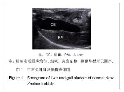
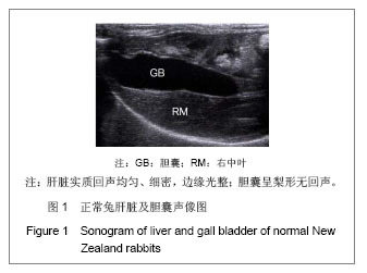
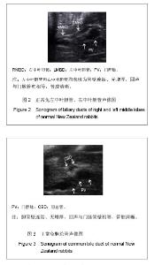
2.1 实验动物数量分析 参加实验动物数量为10只,均进入结果分析,中途无死亡、脱落。 2.2 正常兔肝胆系统常规超声表现 肝脏实质回声均匀、细密,边缘光整;左外叶、左中叶、右外叶及右中叶均清楚可辨,尾状叶及方形叶位于右中叶后方,因体积小且被胃遮挡无法显示。胆囊呈梨形无回声,囊壁光滑呈线状高回声,见图1。"
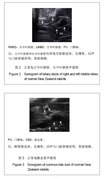
所有胆总管(100%)均可显示。各叶胆管显示率不同,中叶胆管显示率(左、右中叶均为100%),显著高于外叶胆管(左外叶50%,右外叶30%)(P < 0.001)。叶以下胆管分支均不能显示。门脉管径/胆管管径比值为3.59±0.54,各叶胆管及胆总管间该比值,差异无显著性意义(P=0.295),见表1。"
所有胆管均表现为管壁连续、无增厚,回声与门脉管壁相等,管腔清晰,见图2,3。"
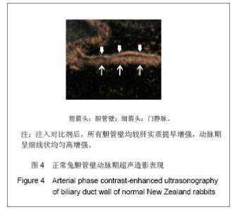
2.3 正常兔胆管壁超声造影表现 见图4,5。"
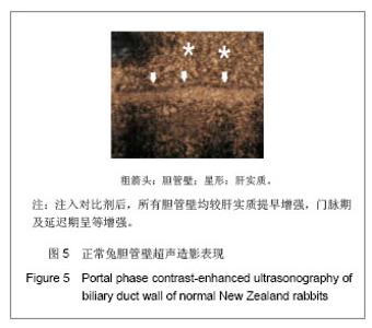
所有胆总管管壁均可显示,显示率为100%。各叶胆管壁显示率不同,见表1,中叶胆管壁显示率(左中叶90%,右中叶50%)显著高于外叶胆管壁(左外叶20%,右外叶33%)(P=0.007)。中叶以下胆管分支管壁均不能显示。 肝动脉增强始增时间为(8.94±2.84) s,PT为(14.13±4.26) s;门脉增强始增时间为(13.38±3.54) s,PT为(22.44±6.63)s。超声造影时相划分为:动脉期10-20 s,门脉期21-30 s,延迟期31-180 s。注入对比剂后,所有胆管壁均较肝实质提早增强,动脉期呈细线状均匀高增强,门脉期及延迟期呈等增强。"

| [1]Sherlock S. Overview of chronic cholestatic conditions in adults: terminology and definitions. Clin Liver Dis. 1998;2(2): 217-233. [2]Deltenre P, Valla DC. Ischemic cholangiopathy. Semin Liver Dis. 2008;28(3):235-246. [3]Skaro AI, Jay CL. The impact of ischemic cholangiopathy in liver transplantation using donors after cardiac death: The untold story. Surgery. 2009;146(4):543-553. [4]Sanchez-Urdazpal L, Gores GJ, Ward EM, et al. Diagnostic features and clinical outcome of ischemic-type biliary complications after liver transplantation. Hepatology. 1993; 17(4):605-609. [5]Hintze RE, Abou-Rebyeh H, Adler A, et al. Endoscopic therapy of ischemia-type biliary lesions in patients following orthotopic liver transplantation. Z Gastroenterol. 1999;37(1):13-20. [6]Thethy S, Thomson B, Pleass H, et al. Management of biliary tract complications after orthotopic liver transplantation. Clin Transplant. 2004;18(6):647-653. [7]Nakamura N, Nishida S, Neff GR, et al. Intrahepatic biliary strictures without hepatic artery thrombosis after liver transplantation: an analysis of 1,113 liver transplantations at a single center. Transplantation. 2005;79(4):427-432. [8]董家鸿,张雷达,王曙光,等.肝移植术后缺血型胆道病变的预防和治疗[J].中华医学杂志,2006,86(18): 1236-1239. [9]陆敏强.肝移植术后胆道狭窄的原因和治疗[J].中国实用外科杂志,2006,26(3):169-171. [10]Williams ED, Draganov PV. Endoscopic management of biliary strictures after liver transplantation. World J Gastroenterol. 2009;15(30): 3725-3733. [11]Catalano G, Urbani L, Biancofiore G, et al. Hepatresection after liver transplantation as a graft-saving procedure: indication criteria, timing and outcome. Transplant Proc. 2004; 36(3):545-546. [12]Schlitt HJ, Meier PN, Nashan B, et al. Reconstructive surgery for ischemic-type lesions at the bile duct bifurcation after liver transplantation. Ann Surg. 1999;229 (1):137-145. [13]Takasaki S, Hano H. Three-dimensional observations of the human hepatic artery (arterial system in the liver). J Hepatol. 2001;34(3):455-466. [14]任杰,郑荣琴,吕明德,等.肝移植缺血性胆道病变的超声造影研究[J].中华超声影像学杂志,2008,17(7):587-589. [15]Ren J, Lu MD, Zheng RQ, et al. Evaluation of the microcirculatory disturbance of biliary ischemia after liver transplantation with contrast-enhanced ultrasound: preliminary experience. Liver Transpl. 2009;15(12):1703- 1708. [16]Sheng QS, Chen DZ. Establishment of an animal model of ischemic type intrahepatic biliary lesion in rabbits. World J Gastroenterol. 2009;15(6):732-736. [17]Yamamoto K, Sherman I. Three-dimensional observations of the hepatic arterial terminations in rat, hamster and human liver by scanning electron microscopy of microvascular casts. Hepatology. 1985;5(3):452-456. [18]何炜,王维,周平,等.兔肝VX2肿瘤超声造影时相划分:与多排螺旋CT增强扫描对照实验研究[J].中华超声影像学杂志,2010, 19(1):65-69. [19]章雅琴,何炜,李丛蕊,等.兔肝VX-2肿瘤磁共振灌注扫描与超声造影增强特征及其时相划分对比研究[J].中国医学影像学杂志, 2011,19(12): 942-945. [20]端学军,林礼务,薛恩生,等.不同肝实质背景超声造影时相变化的实验研究[J].中国超声医学杂志,2009,25(1):8-11. [21]刘燕萍.肝硬化超声造影时相变化的实验性研究[J].全科医学临床与教育,2011,9(4):407-408,417. [22]陈曼,詹维伟,周建桥,等.肝纤维化动物实验超声造影评估[J].中国超声医学杂志,2008,24(6): 495-498. [23]李杰,董宝玮,于晓玲,等.低机械指数灰阶造影对肝VX2瘤动态期相性变化的研究[J].中华超声影像学杂志,2005,14(9):702-705. [24]李杰,董宝玮,于晓玲,等.灰阶超声造影对兔荷VX2瘤前后肝实质血流灌注的对照研究[J].中华超声影像学杂志,2005,14(2): 144-146. [25]李银燕,王学梅,欧国成.家兔正常肝脏超声造影两种大小感兴趣区定量参数的比较[J].中国医学影像技术,2011,27(7): 1348-1350. [26]中华人民共和国科学技术部.关于善待实验动物的指导性意见. 2006-09-30. [27]Ichikawa T, Erturk SM, Araki T. Multiphasic contrast-enhanced multidetector-row CT of liver: contrast-enhancement theory and practical scan protocol with a combination of fixed injection duration and patients' body-weight-tailored dose of contrast material. Eur J Radiol. 2006;58(2):165-176. [28]董家鸿,段恒春,韩本立,等.兔自身对照性胆原性脓毒症模型的建立[J].第三军医大学学报,1996,18(1):52-55. [29]南开大学实验动物解剖学编写组.实验动物解剖学[M].北京:高等教育出版社,1983:14-15. [30]王太一,韩子玉编.实验动物解剖图谱[M].辽宁科学技术出版社, 2000:283-284. [31]任杰,廖梅,曾婕,等.超声造影检测正常人及正常移植肝肝门部胆管微循环的对比研究[J].中国超声医学杂志,2012,28(7): 633-635. [32]董晓秋,刘新颖,毕伟, 等. 定量分析肝脏局限性低回声小病灶的超声造影结果并与增强CT诊断的对比研究[J]. 中国超声医学杂志,2009,25(3):290-293. [33]雷志辉,陈文卫,刘艳,等. 超声造影成像参数在肝脏良恶性局灶性病变中的诊断价值[J]. 武汉大学学报:医学版,2012,33(2): 224-227. [34]Imanieh MH, Dehghani SM, Bagheri MH, et al. Triangular cord sign in detection of biliary atresia: is it a valuable sign? Dig Dis Sci. 2010;55(1):172-175. [35]Sugimoto K, Moriyasu F, Negishi Y, et al. Quantification in molecular ultrasound imaging: a comparative study in mice between healthy liver and a human hepatocellular carcinoma xenograft. J Ultrasound Med. 2012;31(12):1909-1916. [36]华兴,李锐,郭燕丽,等.经静脉声学造影后肝脏实质时间-强度曲线分析诊断肝硬化[J].第三军医大学学报,2004,26(16): 1463-1465. [37]周路遥,谢晓燕,徐辉雄,等.经皮胆管超声造影与X线造影对肝门部胆管癌进行分型的比较研究[J].中华超声影像学杂志,2010, 18(12):1047-1050. |
| [1] | Wang Shuo, Liu Wenying, Lü Chaofan, Li Jiacong, Geng Yi, Zhao Yungang. Cardioprotective effect of 3-nitro-N-methyl salicylamide on the isolated rat heart under cold ischemia preservation [J]. Chinese Journal of Tissue Engineering Research, 2022, 26(8): 1194-1201. |
| [2] | Li Zhiyi, He Pengcheng, Bian Tianyue, Xiao Yuxia, Gao Lu, Liu Huasheng. Bibliometric and visualized analysis of ferroptosis mechanism research [J]. Chinese Journal of Tissue Engineering Research, 2022, 26(8): 1202-1209. |
| [3] | Yang Shenglin, Pu Xingwei, Luo Chunshan, Yang Jianwen. Neuroprotective effects of tetrandrine preconditioning in rabbits with spinal cord ischemia-reperfusion injury [J]. Chinese Journal of Tissue Engineering Research, 2022, 26(8): 1223-1227. |
| [4] | Xuan Juanjuan, Bai Hongtai, Zhang Jixiang, Wang Yaoquan, Chen Guoyong, Wei Sidong. Role of regulatory T cell subsets in liver transplantation and progress in clinical application [J]. Chinese Journal of Tissue Engineering Research, 2022, 26(7): 1143-1148. |
| [5] | Dong Miaomiao, Lai Han, Li Manling, Xu Xiuhong, Luo Meng, Wang Wenhao, Zhou Guoping. Effect of electroacupuncture on expression of nucleotide binding oligomerization domain-like receptor protein 3/cysteinyl aspartate specific proteinase 1 in rats with cerebral ischemia/reperfusion injury [J]. Chinese Journal of Tissue Engineering Research, 2022, 26(5): 749-755. |
| [6] | Li Wanhai, Dong Yuhang, Shi Chao, Jiang Yiyao, Liu Ge, Diao Wenjie, Wu Zhen. Ligustrazine furoxan complex protects against renal ischemia-reperfusion injury in mice [J]. Chinese Journal of Tissue Engineering Research, 2022, 26(2): 205-210. |
| [7] | Lai Han, Wang Jiao, Dong Miaomiao, Luo Meng, Wang Wenhao, Zhou Guoping. Electroacupuncture intervenes with changes of mitogen-activated protein kinase pathway in a rat model of cerebral ischemia/reperfusion due to middle cerebral artery occlusion [J]. Chinese Journal of Tissue Engineering Research, 2022, 26(2): 225-231. |
| [8] | Zhao Xu, Mao Xin, Li Chuntian, Wang Feng. Effect of mesenchymal stem cells on myocardial ischemia-reperfusion injury [J]. Chinese Journal of Tissue Engineering Research, 2022, 26(1): 130-134. |
| [9] | Wang Shiqi, Zhang Jinsheng. Effects of Chinese medicine on proliferation, differentiation and aging of bone marrow mesenchymal stem cells regulating ischemia-hypoxia microenvironment [J]. Chinese Journal of Tissue Engineering Research, 2021, 25(7): 1129-1134. |
| [10] | Yang Xin, Jin Zhe, Feng Xu, Lu Bing. The current situation of knowledge and attitudes towards organ, eye tissue, body donation of residents in Shenyang [J]. Chinese Journal of Tissue Engineering Research, 2021, 25(5): 779-784. |
| [11] | Li Jie, Ma Yuewen, Kang Nan, Zhang Jing, Zhang Yu. Radial extracorporeal shock wave therapy promotes the proliferation of neural stem cells in hippocampus of cerebral infarction rats and inhibits miR-124 expression [J]. Chinese Journal of Tissue Engineering Research, 2021, 25(31): 4981-4987. |
| [12] | Xue Qing, Tong Liangcheng, Yang Zhiwei, Wang Jianling, Zhao Lei, Zhou Sheng, Peng Sai, Li Ying. Epigallocatechin gallate alleviates skeletal muscle ischemia-reperfusion injury in rats [J]. Chinese Journal of Tissue Engineering Research, 2021, 25(26): 4145-4149. |
| [13] | Lu Tan, Chang Yaohui, Wei Na. Lipoxanthin A4 plays a neuroprotective role against spinal cord ischemia-reperfusion injury in rats [J]. Chinese Journal of Tissue Engineering Research, 2021, 25(26): 4175-4179. |
| [14] | Chen Shi, Du Chao, He Xuemei, Zhou Xiangyu. Effect of proanthocyanidins on proliferation and differentiation of skeletal muscle satellite cells under hypoxic-ischemic condition [J]. Chinese Journal of Tissue Engineering Research, 2021, 25(26): 4130-4136. |
| [15] | Fan Junchao, Chen Yong, Song Junjie. Sevoflurance combined with xenon pretreatment protects against spinal cord ischemia-reperfusion injury in a rat model [J]. Chinese Journal of Tissue Engineering Research, 2021, 25(23): 3660-3665. |
| Viewed | ||||||
|
Full text |
|
|||||
|
Abstract |
|
|||||