Chinese Journal of Tissue Engineering Research ›› 2013, Vol. 17 ›› Issue (35): 6281-6286.doi: 10.3969/j.issn.2095-4344.2013.35.010
Previous Articles Next Articles
Magnetic resonance imaging of T2 mapping in rabbit lumbar intervertebral disc
Wei Wen-jiang1, Zhou Zhi-yang2, Guo Wen-bo1, Zhu Pan2, Wu Zhi-qiang1, Zhuang Wen-quan1
- 1Department of Interventional Radiology, the First Affiliated Hospital of Sun Yat-sen University, Guangzhou 510080, Guangdong Province, China; 2Department of Radiology, the Sixth Affiliated Hospital of Sun Yat-sen University, Guangzhou 510655, Guangdong Province, China
-
Received:2013-05-15Revised:2013-06-10Online:2013-08-27Published:2013-08-27 -
Contact:Zhuang Wen-quan, Professor, Master’s supervisor, Department of Interventional Radiology, the First Affiliated Hospital of Sun Yat-sen University, Guangzhou 510080, Guangdong Province, China zwq6233@126.com -
About author:Wei Wen-jiang★, Studying for master’s degree, Department of Interventional Radiology, the First Affiliated Hospital of Sun Yat-sen University, Guangzhou 510080, Guangdong Province, China 147weiwenjiang@sina.com -
Supported by:National Natural Science Foundation of China, No. 81272041*; Guangdong National Science Foundation, No. 10151008901000202*
CLC Number:
Cite this article
Wei Wen-jiang, Zhou Zhi-yang, Guo Wen-bo, Zhu Pan, Wu Zhi-qiang, Zhuang Wen-quan. Magnetic resonance imaging of T2 mapping in rabbit lumbar intervertebral disc[J]. Chinese Journal of Tissue Engineering Research, 2013, 17(35): 6281-6286.
share this article
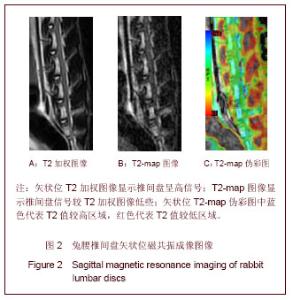
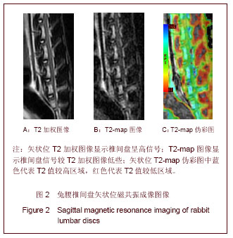
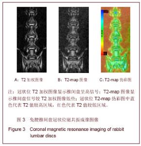
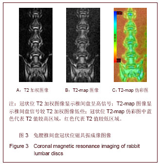
2.1 实验动物数量分析 实验共纳入15只新西兰大白兔,1只新西兰大白兔主要用于初步探索扫描条件,因花时间较长需再次麻醉。另1只兔用于探索脊柱最佳扫描体位,经过多次调整体位与重复扫描,找到满意的扫描体位为足先入右侧卧位。余13只兔进入结果分析。 2.2 兔腰椎间盘的常规T2WI和T2-map表现 根据前2只兔磁共振成像扫描经验,接着13只兔均在一次麻醉和摆位的情况下完成所需序列扫描并获得满意的图像,见图2,3。"
"
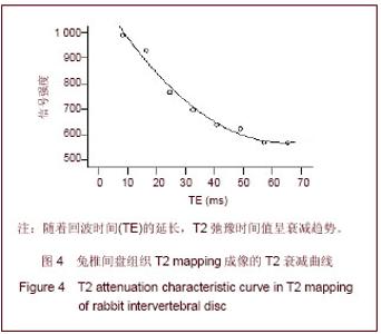
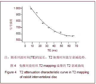
2.3 兔腰椎间盘T2 mapping成像的T2衰减情况 实验分别设定TE:8.1 ms、16.3 ms、24.4 ms、32.5 ms、40.7 ms、48.8 ms、57.0 ms和65.1 ms进行扫描,结果发现,随着TE的延长,T2值逐渐减小。T2 mapping成像中组织区域内某点的T2衰减曲线见图4。"
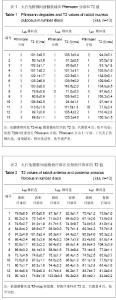
2.4 兔腰椎间盘髓核组织、腹侧纤维环和背侧纤维环感兴趣区的T2值和Pfirrmann分级结果 13只实验兔的L4/5、L5/6和L6/7椎间盘前、中、后3等份的感兴趣区T2值和Pfirrmann分级见表1,2。可见绝大部分椎间盘为PfirrmannⅠ级,属于正常椎间盘,仅少数表现为Ⅱ或Ⅲ级。PfirrmannⅠ级的L4/5、L5/6和L6/7椎间盘髓核组织(中央部)的T2值分别为(104.6±14.0) ms,(109.1±13.8) ms和(109.5±15.1) ms;L4/5、L5/6和L6/7椎间盘腹侧纤维环(前部)的T2值分别为(82.1±9.5) ms,(80.4±11.2) ms和(79.9±10.6) ms;L4/5、L5/6和L6/7椎间盘背侧纤维环(后部)的T2值分别为(85.8±11.9) ms,(85.1±12.1) ms和(85.3±9.3) ms。L4/5、L5/6和L6/7椎间盘组间的前、中、后部的T2值均符合正态分布且方差齐,经随机区组设计资料的方差分析,组间腹侧纤维环、髓核组织及背侧纤维环的T2值差异均无显著性意义(P > 0.05)。 L4/5、L5/6和L6/7椎间盘组内前、中、后部的T2值符合正态分布且方差齐,经随机区组设计资料的方差分析,发现椎间盘内的腹侧纤维环、髓核组织和背侧纤维环的T2值差异有显著性意义(P < 0.01);进一步比较发现,同一椎间盘内髓核组织的T2值明显高于腹侧纤维环或背侧纤维环的T2值(P < 0.01),而腹侧纤维环与背侧纤维环的T2值比较差异无显著性意义(P > 0.05)。"

| [1] Sowa G, Vadalà G, Studer R, et al. Characterization of intervertebral disc aging: longitudinal analysis of a rabbit model by magnetic resonance imaging, histology, and gene expression. Spine (Phila Pa 1976). 2008;33(17):1821-1828.[2] Kim KS, Yoon ST, Li J, et al. Disc degeneration in the rabbit: a biochemical and radiological comparison between four disc injury models. Spine (Phila Pa 1976). 2005;30(1):33-37.[3] Blumenkrantz G, Zuo J, Li X, et al. In vivo 3.0-tesla magnetic resonance T1rho and T2 elaxation mapping in subjects with interbertebral disc degeneration and clinical symptoms. Magn Reson Med. 2010;63(5):1193-1200.[4] Blumenkrantz G, Li X, Han ET, et al. A feasibility study of in vivo T1rho imaging of the intervertebral disc. Magn Reson Imaging. 2006;24(8):1001-1007.[5] Johannessen W, Auerbach JD, Wheaton AJ, et al. Assessment of human disc degeneration and proteoglycan content using T1rho-weighted magnetic resonance imaging. Spine (Phila Pa 1976). 2006;31(11):1253-1257.[6] Auerbach JD, Johannessen W, Borthakur A, et al. In vivo quantification of human lumbar disc degeneration using T(1rho)-weighted magnetic resonance imaging. Eur Spine J. 2006;15 Suppl 3:S338-344.[7] Nguyen AM, Johannessen W, Yoder JH, et al. Noninvasive quantification of human mucleus pulposus pressure with use of T1rho-weighted magnetic resonance imaging. J Bone Joint Surg Am. 2008;90(4):796-802.[8] Zobel BB, Vadala G, Del Vescovo R, et al. T1ρmagnetic resonance imaging quantification of early lumbar intervertebral disc degeneration in healthy young adults. Spine (Phila Pa 1976). 2012;37(14):1224-1230.[9] Apprich S, Welsch GH, Mamisch TC, et al. Detection of degenerative cartilage disease: comparison of high-resolution morphological MR and quantitative T2 mapping at 3.0 Tesla. Osteoarthritis Cartilage. 2010;18(9):1211-1217.[10] Wei ZM, Du XK, Huo TL, et al. Quantitative T2 mapping evaluation for articular cartilage lesions in a rabbit model of anterior cruciate ligament ransaction osteoarthritis. Chin Med J (Engl). 2012;125(5):843-850.[11] Apprich S, Mamisch TC, Welsch GH, et al. Quantitative T2 mapping of the patella at 3.0T is sensitive to early cartilage degeneration, but also to loading of the knee. Eur J Radiol. 2012;81(4):e438-443.[12] Marik W, Apprich S, Welsch GH, et al. Biochemical evaluation of articular cartilage in patients with osteochondrosis dissecans by means of quantitative T2-and T2-mapping at 3 T MRI: a feasibility study. Eur J Radiol. 2012;81(5):923-927.[13] Moon CH, Jacobs L, Kim JH, et al. Quantitative proton T2 and sodium magnetic resonance imaging to assess intervertebral disc degeneration in a rabbit model. Spine (Phila Pa 1976). 2012;37(18):E1113-1119. [14] Trattniq S, Stelzeneder D, Goed S, et al. Lumbar intervertebral disc abnormalities: comparison of quantitative T2 mapping with conventional MR at 3.0T. Eur Radiol. 2010; 20(11): 2715-2722.[15] Takashima H, Takebayashi T, Yoshimoto M, et al. Correlation between T2 relaxation time and intervertebral disk degeneration. Skeletal Radiol. 2012;41(2):163-167.[16] Jazini E, Sharan AD, Morse LJ, et al. Alterations in T2 relaxation magnetic resonance imaging of the ovine intervertebral disc due to nonenzymatic glycation. Spine (Phila Pa 1976). 2012;37(4):E209-215.[17] Hoppe S, Quirbach S, Mamisch TC, et al. Axial T2 mapping in intervertebral disc: a new technique for assessment of intervertebral disc degeneration. Eur Radiol. 2012;22(9): 2013-2019.[18] Watanabe A, Benneker LM, Boesch C, et al. Classification of interbertebral disk degeneration with axial T2 mapping. AJR Am J Roentqenol. 2007;189(4):936-942.[19] Stelzeneder D, Kovacs BK, Goed S, et al. Effect of short-term unloading on T2 relaxation time in the lumbar intervertebral disc-in vivo magnetic resonance imaging study at 3.0 tesla. Spine J. 2012;12(3):257-264.[20] Stelzeneder D, Welsch GH, Kovacs BK, et al. Quantitative T2 evaluation at 3.0T compared to morphological grading of the lumbar intervertebral disc: a standardized evaluation approach in patients with low back pain. Eur J Radiol. 2012; 81(2):324-330.[21] Marinelli NL, Hauqhton VM, Munoz A, et al. T2 relaxation times of intervertebral disc tissue correlated with water content and proteoglycan content. Spine (Phila Pa 1976). 2009;34(5):520-524.[22] 中华人民共和国科学技术部.关于善待实验动物的指导性意见. 2006-09-30.[23] Pfirrmann CW, Metzdorf A, Zanetti M, et al. Magnetic resonance classification of lumber intervertebral disk degeneration. Spine (Phila Pa 1976). 2001;26(17):1873-1878.[24] 李鹤平,庄文权,杨建勇,等.家兔脊椎与脊髓磁共振成像方法和表现[J].中国临床康复,2002,6(24):3711-3712.[25] 康宁,俎栋林,张宏杰.自旋密度ρ、驰豫时间T1和T2定量磁共振成像[J].中国医学影像技术,2004,20(12):1944-1947.[26] 李雅芬,牛爱青,康立丽.磁共振T2-map的单、双指数拟合方法[J].中国医学影像技术,2010,26(2):347-350. |
| [1] | Chen Ziyang, Pu Rui, Deng Shuang, Yuan Lingyan. Regulatory effect of exosomes on exercise-mediated insulin resistance diseases [J]. Chinese Journal of Tissue Engineering Research, 2021, 25(25): 4089-4094. |
| [2] | Jiang Xiaoyan, Zhu Haifei, Lin Haiqi, Lin Wentao. Cold therapy promotes self-limited recovery of delayed-onset muscle soreness [J]. Chinese Journal of Tissue Engineering Research, 2021, 25(23): 3609-3613. |
| [3] | Xie Jingshu, Zhang Xianglin, Liu Jinlei, Wen Jing. Application of High Resolution reconstruction algorithm in precision CT scans of the middle and inner ears [J]. Chinese Journal of Tissue Engineering Research, 2021, 25(23): 3614-3618. |
| [4] | Liu Jinwei, Chen Yunzhen, Wan Chunyou. Changes of osteogenic growth factors in the broken end of bone nonunion under stress [J]. Chinese Journal of Tissue Engineering Research, 2021, 25(23): 3619-3624. |
| [5] | Luo Anyu, Liu Hanlin, Xie Xiaofei, Huang Chen. Effect of antioxidant mixture on structural degeneration of an osteoarthritis rat model [J]. Chinese Journal of Tissue Engineering Research, 2021, 25(23): 3625-3629. |
| [6] | Zhou Wu, Wang Binping, Wang Yawen, Cheng Yanan, Huang Xieshan. Transforming growth factor beta combined with bone morphogenetic protein-2 induces the proliferation and differentiation of mouse MC3T3-E1 cells [J]. Chinese Journal of Tissue Engineering Research, 2021, 25(23): 3630-3635. |
| [7] | Gao Kun, Chen Dayu, Zhang Yong, Liu Weidong, Sun Shufen, Lai Wenqiang, Ma Dujun, Wu Yihong, Lin Zhanpeng, Jiang Yinglu, Yu Weiji. Achyranthes bidentata alcohol extract inhibits extracellular matrix degradation of the cartilage by regulating synovial fibroblast exosomes [J]. Chinese Journal of Tissue Engineering Research, 2021, 25(23): 3636-3640. |
| [8] | Liang Meifu, Qu Shuhua. Optimal power load forecasting of the skeletal muscle based on back propagation neural network [J]. Chinese Journal of Tissue Engineering Research, 2021, 25(23): 3641-3647. |
| [9] | Bai Shengchao, Gao Yang, Wang Bo, Li Junping, Wang Ruiyuan. Dynamic changes of mitochondrial function of the skeletal muscle after acupuncture intervention in rats with heavy load exercise-induced injury [J]. Chinese Journal of Tissue Engineering Research, 2021, 25(23): 3648-3653. |
| [10] | Yang Caihui, Liu Qicheng, Dong Ming, Wang Lina, Zuo Meina, Lu Ying, Niu Weidong. Serine/threonine protein kinases can promote bone destruction in mouse models of chronic periapical periodontitis [J]. Chinese Journal of Tissue Engineering Research, 2021, 25(23): 3654-3659. |
| [11] | Fan Junchao, Chen Yong, Song Junjie. Sevoflurance combined with xenon pretreatment protects against spinal cord ischemia-reperfusion injury in a rat model [J]. Chinese Journal of Tissue Engineering Research, 2021, 25(23): 3660-3665. |
| [12] | Zuo Zhenkui, Han Jiarui, Ji Shuling, He Lulu. Pretreatment with ginkgo biloba extract 50 alleviates radiation-induced acute intestinal injury in mice [J]. Chinese Journal of Tissue Engineering Research, 2021, 25(23): 3666-3671. |
| [13] | Zhang Liang, Ma Xiaoyan, Wang Jiahong. Regulatory mechanism of Shenshuai Yin on cell apoptosis in the kidney of chronic renal failure rats [J]. Chinese Journal of Tissue Engineering Research, 2021, 25(23): 3672-3677. |
| [14] | Cheng Yanan, Wu Yucong, Mao Qiuhua, Chen Ling, Lu Liying, Xu Pu. Restoration effect and stability of resin infiltration combined with bioactive glass on demineralized tooth enamel [J]. Chinese Journal of Tissue Engineering Research, 2021, 25(22): 3522-3526. |
| [15] | Bi Qingwei, Liu Chengpu, Li Yan, Zhao Wenwen, Han Mei. Structure analysis of platelet-rich fibrin derived from two centrifugation procedures [J]. Chinese Journal of Tissue Engineering Research, 2021, 25(22): 3534-3539. |
| Viewed | ||||||
|
Full text |
|
|||||
|
Abstract |
|
|||||