Chinese Journal of Tissue Engineering Research ›› 2013, Vol. 17 ›› Issue (23): 4256-4263.doi: 10.3969/j.issn.2095-4344.2013.23.011
Previous Articles Next Articles
Transplantation of neuron-like cells from bone marrow mesenchymal stem cells for treatment of spinal cord injury
Gao Ping1,2, Sun Zhan-sheng1, Wang Bo-min1, Li Lian-xin1, Wang Fu1, Mu Le-ming1
- 1 Department of Traumatic Orthopedics, Provincial Hospital Affiliated to Shandong University, Jinan 250021, Shandong Province, China
2 Medical College of Shandong University, Jinan 250012, Shandong Province, China
-
Online:2013-06-04Published:2013-06-04 -
About author:Gao Ping★, Studying for master’s degree, Department of Traumatic Orthopedics, Provincial Hospital Affiliated to Shandong University, Jinan 250021, Shandong Province, China; Medical College of Shandong University, Jinan 250012, Shandong Province, China Gaopinggp125@163.com -
Supported by:Key Scientific and Technological Project of Shandong Province, No. 2006GG3202046*
CLC Number:
Cite this article
Gao Ping, Sun Zhan-sheng, Wang Bo-min, Li Lian-xin, Wang Fu, Mu Le-ming. Transplantation of neuron-like cells from bone marrow mesenchymal stem cells for treatment of spinal cord injury[J]. Chinese Journal of Tissue Engineering Research, 2013, 17(23): 4256-4263.
share this article
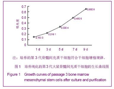
2.1 实验动物数量分析 实验选用Wistar大鼠48只,分为4组,实验过程无脱失,全部进入结果分析。 2.2 骨髓间充质干细胞的鉴定 见图1。"
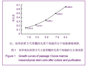
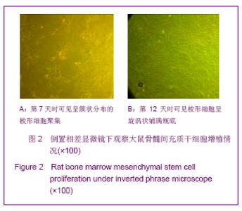
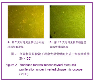
培养纯化的第3代骨髓间充质干细胞以1×10⁸ L-1浓度接种,在第1,3,5,7,9天分别测得MTT实验的平均值为0.153 3,0.218 1,0.330 4,0.493 5,0.650 8。绘制成细胞生长曲线,符合干细胞增殖规律。 在倒置像差显微镜下,逐日观察骨髓间充质干细胞,开始细胞呈散在集落,第7天时可见呈簇状分布的梭形细胞聚集,第12天时可见梭形细胞呈旋涡状铺满瓶底。见图2。"
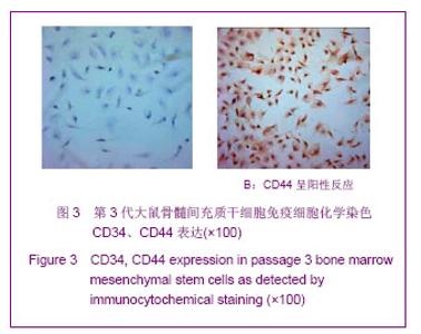
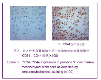
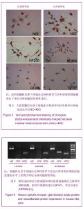
细胞免疫化学染色在显微镜下可以见到CD44呈阳性反应,整个细胞呈棕黄色;而CD34呈阴性反应,细胞没有着色。符合骨髓间充质干细胞表面特异性标志的特点[10] 。见图3。"
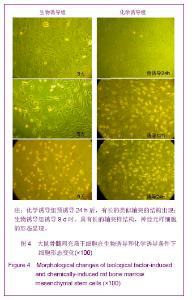
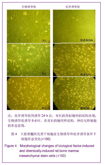
2.3 骨髓间充质干细胞来源的神经元样细胞的鉴定 细胞形态学观察:在诱导培养基条件下,持续镜下观察细胞形态的变化。化学诱导组:预诱导24 h后,细胞形态改变不明显;正式诱导12 h后,细胞形态发生明显改变,胞体变圆、变小,折光性增强,多个突起产生,24 h后,有长的类似轴突的结构出现。生物诱导组:开始诱导后3 d,细胞由原来的长梭形逐渐变为三角形、多边形;5 d后,大部分细胞胞体收缩,折光性增强,有多个长的突起形成;9 d时,细胞胞体变圆,有数个突起,具有长的轴突样结构,交织成网状,神经元样细胞的形态显现。见图4。"
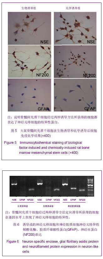
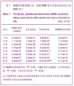
免疫细胞化学法鉴定:化学诱导组和生物诱导组细胞,免疫细胞化学染色结果相似,显示神经元特异性烯醇化酶和神经丝蛋白表达阳性,而胶质纤维酸性蛋白表达为阴性。说明骨髓间充质干细胞经过两种诱导方法所获得的细胞都表达了神经元样细胞的特异性蛋白。见图5。 PCR检测:化学诱导组和生物诱导组细胞,PCR结果相似,神经元特异性烯醇化酶、神经丝蛋白在电泳图像中表现为阳性,胶质纤维酸性蛋白为阴性。以神经胶质细胞作为对照,其神经元特异性烯醇化酶、胶质纤维酸性蛋白、神经丝蛋白在电泳图像中均为阳性表达。说明骨髓间充质干细胞经过两种诱导方法定向诱导所获得的细胞在基因水平上表现了神经元样细胞的特异性。见图6。"
2.4 动物模型行为学评分 造模前,各大鼠活动正常,BBB评分21分[9,11] 。造模后表现右后肢及尾无活动,针刺皮肤无反应,排尿障碍,排便未观察到明显异常。伤后1-12周BBB评分见表1。"
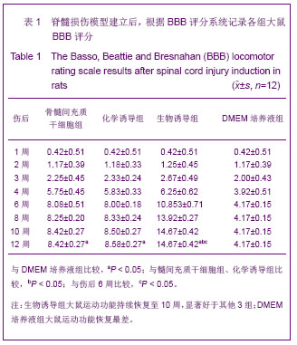
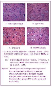
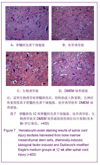
伤后1-4周:伤后1周,针刺右后肢皮肤有回缩反应,伤后2周偶见髋关节活动,伤后3周髋膝关节皆有活动。伤后4周,骨髓间充质干细胞组和化学诱导组可见到后肢三关节活动,而DMEM培养液组踝关节未见活动。 伤后6周:骨髓间充质干细胞组和化学诱导组见掌面非承重着地,生物诱导组掌面承重着地,但后肢活动不协调,DMEM培养液组较前恢复不明显。 伤后8周:骨髓间充质干细胞组和化学诱导组见掌面承重着地,但活动不协调,生物诱导组有持续性掌面承重移动和前后肢协调动作,常见掌面移动,持续型前后肢协调动作,偶有爪背侧移动,DMEM培养液组无继续恢复。 伤后10周:骨髓间充质干细胞组、化学诱导组、生物诱导组3组较前略有恢复但不明显,DMEM培养液组无继续恢复。伤后12周,4组均无继续恢复,骨髓间充质干细胞组、化学诱导组与DMEM培养液组比较差异有显著性意义(t=22.81/23.70,P < 0.05);生物诱导组与骨髓间充质干细胞组、化学诱导组比较差异有显著性意义(t=26.08/25.38,P < 0.05);化学诱导组与骨髓间充质干细胞组比较差异无显著意义(t=0.792 8,P=0.436 3)。 在伤后的前4-6周,随着损伤部位水肿和炎症的消退及周围轻度受损神经元的恢复,部分神经传导重新建立,模型动物的运动功能也部分恢复。6周后,DMEM培养液组功能再无继续恢复;骨髓间充质干细胞组和化学诱导组继续恢复但不明显,伤后12周与伤后6周比较差异无显著性意义(t=1.586/3.023,P=0.127 1/0.063 0);生物诱导组继续恢复至10周,且恢复明显,伤后12周与伤后6周比较差异有显著意义(t=13.70,P < 0.05)。见表1。 脊髓切片苏木精-伊红染色:见图7。 损伤12周后损伤部位脊髓切片苏木精-伊红染色显示DMEM培养液组脊髓灰质与白质分界不清,结构混乱,且有较多大的坏死空洞存在,神经元样细胞崩解广泛,胶质细胞增生不明显。骨髓间充质干细胞组与化学诱导组相似,灰质与白质分界清楚,坏死空洞较DMEM培养液组小且少,神经元样细胞崩解较DMEM培养液组轻,胶质细胞增生。说明骨髓间充质干细胞组和化学诱导组功能恢复较DMEM培养液组好。生物诱导组脊髓结构清晰,坏死空洞较其他3组都少,胶质细胞增生明显,神经元样细胞崩解少。说明生物诱导组脊髓损伤后,较快的进入恢复期,且神经恢复程度高于骨髓间充质干细胞组、化学诱导组和DMEM培养液组。"
| [1]Wright KT, Masri W, Osman A,et al.Concise Review: Bone Marrow for the Treatment of Spinal Cord Injury: Mechanisms and Clinical Applications.J Stem Cells.2011; 29(2):169-178.[2]Huang Y,Jia X,Bai K,et al.Effect of fluid shear stress on cardiomyogenic differentiation of rat bone marrow mesenchymal stem cells. J Arch Med Res.2010;41(7):497- 505.[3]Woodbury D, Schwarz EJ, Prockop DJ,et al.Adult rat and human bone marrow stromal cells differentiate into neurons.J Neurosci Res.2000;61:364-370.[4]Radtke C, Schmitz B, Spies M, et al.Peripheral glial cell differentiation from neurospheres derived from adipose mesenchymal stem cells.Int J Devl Neuroscience.2009; 27(8):817-823.[5]Wislet-Gendebien S, Laudet E, Neirinckx V,et al.Adult Bone Marrow: Which Stem Cells for Cellular Therapy Protocols in Neurodegenerative Disorders? J Biomed Biotechnol. 2012; 2012:601560.[6]Mrówczyński W, Celichowski J, Krutki P,et al.Changes of the force-frequency relationship in the rat medial gastrocnemius muscle after total transection and hemisection of the spinal cord. J Neurophysiol.2011;105(6):2943-2950.[7]Hofstetter CP, Sehwarz EJ, Hess D,et al. Marrow stromal cells from guiding strands in the injured spinal cord and promote recovery.Proc Nad Aead Sei USA.2002;99(4):2199- 2204.[8]Ankeny DP, McTique DM, Jakeman LB,et al.Bone marrow transplants provide tissue protection and directional guidance for axon after contusive spinal cord injury in rats.J Exp Neurol. 2004;190(1):17-31.[9]Scheff SW, Saucier DA, Cain ME. A statistical method for analyzing rating scale data: the BBB locomotor score.J Neurotrauma.2002;19(10):1251-1260.[10]Mafi P, Hindocha S, Mafi R,et al.Adult Mesenchymal Stem Cells and Cell Surface Characterization - A Systematic Review of the Literature.J Open ORTHOP J. 2011; 5(Suppl 2): 253-260.[11]Basso DM, Beattie MS and Bresnahan JC.A sensitive and reliable locomotor rating scale for open field testing in rats.J Neurotrauma.1995;12(1):1-21.[12]Pittenger MF, Mackay AM, Beck SC,et al.Multilineage potential of adult human mesenchymal stem cells.J Science. 1999;284(5411):143-147.[13]Arinzeh TL. Mesenchymal stem cells for bone repair: preclinical studies and potential orthopedic applications.J Foot Ankle Clin.2005;10(4):651-665.[14]Caplan AI. Adult mesenchymal stem cells for tissue engineering versus regenerative medicine. J Cell Physiol. 2007;213(2):341-347.[15]Barry FP, Murphy JM.Mesenchymal stem cells: clinical applications and biological characterization.J Int J Biochem Cell Biol.2004;36(4):568-584.[16]Helder MN, Knippenberg M, Klein-Nulend J,et al.Stem cells from adipose tissue allow challenging new concepts for regenerative medicine. Tissue Engineering.2007;13(8): 1799-1808.[17]Trubiani O, Orsini G, Caputi S,et al.Adult mesenchymal stem cells in dental research: a new approach for tissue engineering.Int J Immunopathol Pharmacol.2006;19(3):451- 460.[18]Kopen GC, Prockop DJ, Phinney DG.Marrow stromal cells migrate throughout forebrain and cerebellum, and they differentiate into astrocytes after injection into neonatal mouse brains.J Proc Natl Acad Sci USA.1999;96(19):10711-10716.[19]Abouelfetouh A, Kondoh T, Ehara K,et al.Morphological differentiation of bone marrow stromal cells into neuron-like cells after co-culture with hippocampal slice.J Brain Res. 2004; 1029(1):114-119.[20]Jin K, Mao XO, Batteur S,et al.Induction of neuronal markers in bone marrow cells: differential effects of growth factors and patterns of intracellular expression. Exp Neurol.2003;1841: 78-89.[21]Zhou J, Tian GP, Wang JE, et al. In vitro differentiation of adipose-derived stem cells and bone marrow-derived stromal stem cells into neuronal-like cells. Neural Regen Res. 2011; 6(19):1467-1472.[22]Li Q, Geng YJ, Lu L, et al. Platelet-rich fibrin-induced bone marrow mesenchymal stem cell differentiation into osteoblast-like cells and neural cells. Neural Regen Res. 2011;6(31):2419-2423.[23]Neuhuber B, Himes BT, Shumsky JS,et al.Axon growth and recovery of function supported by human bone marrow stromal cells in the injured spinal cord exhibit donor variations. J Brain Res.2005;1035:73-85.[24]Crigler L, Robey RC, Asawachaicharn A,et al. Human mesenchymal stem cell subpopulations express a variety of neuro-regulatory molecules and promote neuronal cell survival and neuritogenesis. Exp Neurol. 2006;198:54-64.[25]Poll D, Parekkadan B, Borel Rinkes IHM,et al.Mesenchymal Stem Cell Therapy for Protection and Repair of Injured Vital Organs. Cellular and Molecular Bioengineering.2008;1:42-50.[26]Uccelli A, Benvenuto F, Laroni A,et al.Neuroprotective features of mesenchymal stem cells.J Best Pract Res Clin Haematol.2011;24:59-64.[27]da Silva Meirelles L, Fontes AM, Covas DT,et al.Mechanisms involved in the therapeutic properties of mesenchymal stem cells.J Cytokine Growth Factor Rev.2009;20:419-427.[28]Caplan AI, Dennis JE.Mesenchymal stem cells as trophic mediators. Journal of Cellular Biochemistry. 2006;98:1076- 1084.[29]Wu S, Suzuki Y, Ejiri Y,et al.Bone marrow stromal cells enhance differentiation of co-cultured neurosphere cells and promote regeneration of the injured spinal cord.J Neurosci Res. 2003;72:343-351.[30]Barnabé GF, Schwindt TT, Calcagnotto ME,et al. Chemically- Induced RAT Mesenchymal Stem Cells Adopt Molecular Properties of Neuronal-Like Cells but Do Not Have Basic Neuronal Functional Properties.PLoS ONE.2009;4(4): e5222.[31]Tarasenko YI, Gao J, Nie L, Johnson KM,et al.Human fetal neural stem cells grafted into contusion-injured rat spinal cords improve behavior.J Neurosci Res.2007;85:47-57.[32]Cai PQ, Sun GY, Cai PS, et al. Survival of transplanted neurotrophin-3 expressing human neural stem cells and motor function in a rat model of spinal cord injury. Neural Regen Res. 2009;4(7):485-491.[33]Osaka M, Honmou O, Murakami T,et al.Intravenous administration of mesenchymal stem cells derived from bone marrow after contusive spinal cord injury improves functional outcome.J Brain Res.2010;1343:226-235.[34]Li L, Lü G, Wang YF, et al. Glial cell-derived neurotrophic factor mRNA expression in a rat model of spinal cord injury following bone marrow stromal cell transplantation. Neural Regen Res 2008;3(10):1056-1059.[35]Li BC, Li Y, Chen LF,et al.Olfactory ensheathing cells can reduce the tissue loss but not the cavity formation in contused spinal cord of rats.J Neurol Sci.2011;303:67-74. |
| [1] | Pu Rui, Chen Ziyang, Yuan Lingyan. Characteristics and effects of exosomes from different cell sources in cardioprotection [J]. Chinese Journal of Tissue Engineering Research, 2021, 25(在线): 1-. |
| [2] | Jiang Hongying, Zhu Liang, Yu Xi, Huang Jing, Xiang Xiaona, Lan Zhengyan, He Hongchen. Effect of platelet-rich plasma on pressure ulcers after spinal cord injury [J]. Chinese Journal of Tissue Engineering Research, 2021, 25(8): 1149-1153. |
| [3] | Zhang Xiumei, Zhai Yunkai, Zhao Jie, Zhao Meng. Research hotspots of organoid models in recent 10 years: a search in domestic and foreign databases [J]. Chinese Journal of Tissue Engineering Research, 2021, 25(8): 1249-1255. |
| [4] | Wang Zhengdong, Huang Na, Chen Jingxian, Zheng Zuobing, Hu Xinyu, Li Mei, Su Xiao, Su Xuesen, Yan Nan. Inhibitory effects of sodium butyrate on microglial activation and expression of inflammatory factors induced by fluorosis [J]. Chinese Journal of Tissue Engineering Research, 2021, 25(7): 1075-1080. |
| [5] | Wang Xianyao, Guan Yalin, Liu Zhongshan. Strategies for improving the therapeutic efficacy of mesenchymal stem cells in the treatment of nonhealing wounds [J]. Chinese Journal of Tissue Engineering Research, 2021, 25(7): 1081-1087. |
| [6] | Wan Ran, Shi Xu, Liu Jingsong, Wang Yansong. Research progress in the treatment of spinal cord injury with mesenchymal stem cell secretome [J]. Chinese Journal of Tissue Engineering Research, 2021, 25(7): 1088-1095. |
| [7] | Liao Chengcheng, An Jiaxing, Tan Zhangxue, Wang Qian, Liu Jianguo. Therapeutic target and application prospects of oral squamous cell carcinoma stem cells [J]. Chinese Journal of Tissue Engineering Research, 2021, 25(7): 1096-1103. |
| [8] | Xie Wenjia, Xia Tianjiao, Zhou Qingyun, Liu Yujia, Gu Xiaoping. Role of microglia-mediated neuronal injury in neurodegenerative diseases [J]. Chinese Journal of Tissue Engineering Research, 2021, 25(7): 1109-1115. |
| [9] | Li Shanshan, Guo Xiaoxiao, You Ran, Yang Xiufen, Zhao Lu, Chen Xi, Wang Yanling. Photoreceptor cell replacement therapy for retinal degeneration diseases [J]. Chinese Journal of Tissue Engineering Research, 2021, 25(7): 1116-1121. |
| [10] | Jiao Hui, Zhang Yining, Song Yuqing, Lin Yu, Wang Xiuli. Advances in research and application of breast cancer organoids [J]. Chinese Journal of Tissue Engineering Research, 2021, 25(7): 1122-1128. |
| [11] | Wang Shiqi, Zhang Jinsheng. Effects of Chinese medicine on proliferation, differentiation and aging of bone marrow mesenchymal stem cells regulating ischemia-hypoxia microenvironment [J]. Chinese Journal of Tissue Engineering Research, 2021, 25(7): 1129-1134. |
| [12] | Zeng Yanhua, Hao Yanlei. In vitro culture and purification of Schwann cells: a systematic review [J]. Chinese Journal of Tissue Engineering Research, 2021, 25(7): 1135-1141. |
| [13] | Kong Desheng, He Jingjing, Feng Baofeng, Guo Ruiyun, Asiamah Ernest Amponsah, Lü Fei, Zhang Shuhan, Zhang Xiaolin, Ma Jun, Cui Huixian. Efficacy of mesenchymal stem cells in the spinal cord injury of large animal models: a meta-analysis [J]. Chinese Journal of Tissue Engineering Research, 2021, 25(7): 1142-1148. |
| [14] | Hou Jingying, Yu Menglei, Guo Tianzhu, Long Huibao, Wu Hao. Hypoxia preconditioning promotes bone marrow mesenchymal stem cells survival and vascularization through the activation of HIF-1α/MALAT1/VEGFA pathway [J]. Chinese Journal of Tissue Engineering Research, 2021, 25(7): 985-990. |
| [15] | Shi Yangyang, Qin Yingfei, Wu Fuling, He Xiao, Zhang Xuejing. Pretreatment of placental mesenchymal stem cells to prevent bronchiolitis in mice [J]. Chinese Journal of Tissue Engineering Research, 2021, 25(7): 991-995. |
| Viewed | ||||||
|
Full text |
|
|||||
|
Abstract |
|
|||||