Chinese Journal of Tissue Engineering Research ›› 2019, Vol. 23 ›› Issue (14): 2241-2247.doi: 10.3969/j.issn.2095-4344.1666
Previous Articles Next Articles
Nanomaterials applied in tumor imaging diagnosis and treatment: an integration tendency
Chen Yue1, Zhang Tianke1, 2, Xu Yong1
- 1Department of Urology, Second Hospital of Tianjin Medical University, Tianjin 300211, China; 2Department of Anorectum, People’s Hospital of Nankai University, Tianjin 300121, China
-
Received:2019-01-07 -
About author:Chen Yue, MD, Attending physician, Department of Urology, Second Hospital of Tianjin Medical University, Tianjin 300211, China -
Supported by:the National Natural Science Foundation of China (General Program), No. 31771100 (to CY)
CLC Number:
Cite this article
Chen Yue, Zhang Tianke, Xu Yong. Nanomaterials applied in tumor imaging diagnosis and treatment: an integration tendency[J]. Chinese Journal of Tissue Engineering Research, 2019, 23(14): 2241-2247.
share this article
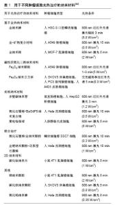
2.1 靶向纳米材料与肿瘤 纳米材料主要分为2大类:无机纳米材料和有机纳米材料,无机纳米材料主要包括半导体量子点、金属纳米材料、磁性纳米材料、金属氧化物、金材料和碳管纳米材料;有机纳米材料包括脂质体、人工合成纳米胶囊或树状聚合物[10]。靶向纳米材料则是利用抗原-抗体特异性结合原理,将不同的靶向分子承载在纳米材料表面所形成的,这些分子包括甘露糖、叶酸、多肽、蛋白或者抗体。靶向纳米材料可,通过表面修饰的靶向分子提高药物携载的效率和准确性,实现对特定部分肿瘤的成像或杀伤作用[11]。 肿瘤是指机体在各种致癌因子作用下,一类起源于正常组织的新生物,与正常组织不同,它具有不可控生长、细胞永生、侵袭临近组织和转移等特性[12]。由于解剖结构和生理功能与正常组织具有较大差异,肿瘤组织在病理结构上具有微环境低pH值、低氧、新生血管和淋巴管等特点[13]。基于以上的特点,肿瘤组织可被纳米材料特异性靶向,实现高效性、准确性的诊断和治疗[14]。 2.2 纳米材料在肿瘤诊断方面的应用 影像学的发展为各种疾病的诊断提供了便利,通过影像技术获得的图像可提供病变部位的信号甚至增强信号。同样,肿瘤的诊断也主要依靠与正常组织的信号对比来进行鉴别。目前,临床常用的影像学技术包括电子计算机断层扫描(CT)、正电子发射电子扫描技术(PET)、核磁共振成像(MRI)和拉曼成像等[10]。纳米材料在肿瘤诊断中的应用主要体现在其与影像学技术的结合,从而实现其对肿瘤的早期诊断,为患者赢得了宝贵的治疗时机。 2.2.1 纳米材料与CT CT是一种无创性的影像学检查技术,它利用X射线、γ射线或超声波对人体的某一区域进行断面扫描,通过不同细胞间的信号吸收差异达到鉴别诊断的效果[15]。金属纳米材料由于其高的X射线吸收率、高可塑性和易于表面修饰的特性,在CT技术中应用十分广泛[6]。Hu等[16]设计了一种可被肾脏代谢的铋-碳酸盐纳米管,发现铋-碳酸盐纳米管可高效地靶向肿瘤组织,CT成像显示高对比度,并且具有抑制肿瘤体积的作用。Wang等[17]发现表面包被二氧化硅的金纳米材料,可提高肝细胞癌的CT诊断特异性,其表面的抗癌药物分子亦可发挥治疗和增加放疗敏感性的作用;金纳米颗粒与丙烯酸和烯丙胺结合后,可用来诊断肝癌[18]。此外,脂质体等有机纳米材料在CT诊断中也被应用,Zhang等[19]将碳酸氢铵和金纳米管共同包裹进叶酸-脂质体中,既满足了CT成像中的高对比度要求,也实现了在肿瘤微环境中的高药物浓度。 显微CT,是一种新兴的3D成像技术,可在不破坏样本的情况下清楚地了解内部显微结构,相比于普通临床CT,其分辨率极高,可达到微米级别,检测出肿瘤新生血管,但敏感度较低。为了提高显微CT的敏感性,Liu等[20]设计了精氨酸-甘氨酸-天冬氨酸与磁性纳米材料的复合物,随后注射到H122肿瘤小鼠模型中,发现与未注射组相比,显微CT的敏感性大幅提高。 2.2.2 纳米材料和MRI MRI是断层成像的一种,在不破坏正常组织细胞的基础上,利用磁共振显像从人体中获得电磁信号并重建出人体信息,也是无创诊断的一种。虽然CT与MRI都可显示空间中的对比度分布,但MRI可得到任何方向的断层图像,甚至三维体图像。纳米材料与MRI技术结合,可提高MRI的敏感性和准确性,其中磁性纳米材料的研究最为广泛。Li等[21]将Fe3O4作为载体,其上结合透明质酸后形成复合物MPDA@ HA-MTX,可作为MRI对比剂,体内外实验均证实该应用可获得更高的诊断效率。Liu等[22]研究结果显示,使用金纳米冠层自组装对比剂可使MRI对比度更强,且加强了T2衰减效应。Chee等[23]通过将86种多钛富集于传统的超顺磁性氧化铁纳米粒子后,得到了更有效的T2加权像对比剂,可提高肝脏的对比度,增加肝癌检出率。胰腺癌作为一种早期无明显症状、诊断后已发展到晚期的癌症,早期诊断的意义便显得格外重要。Huang等[24]设计了一种钆-金的纳米团簇Gd-Au-NC-GPC-1,通过与GPC-1抗体结合,可特异性地与胰腺癌细胞表面的受体结合,体内实验证明MRI检测胰腺癌的水平明显提高,并且无明显生物毒性。 2.2.3 纳米材料和PET PET主要使用的物质是氟代脱氧葡萄糖,氟代脱氧葡萄糖作为生物生命代谢中所需要的物质,可进入检测的组织中,且根据代谢状态的不同,即在高代谢肿瘤组织中聚集较多,在低代谢的正常组织中聚集较少,进而将信号反映成图像表现出来,对病变进行诊断[25]。近年来,放射性同位素标记的金属材料被认为在PET成像中具有潜在的价值。Lee等[26]将金纳米材料聚乙二醇化并承载124I(PEG-124I-Au@ AuCBs),用体内实验验证其在PET诊断肿瘤的效果,结果显示其在不同的pH值、不同血清及各种模拟体内环境下都未对正常细胞产生毒性,并且在PET成像肿瘤中表现出高稳定性和灵敏性。考虑到PET的高生物相容性的要求,一些学者从最不会引起机体排斥且稳定性良好的二氧化硅材料入手,将二氧化硅的表面修饰后,连124I或者89Zr和靶向药物,成功地利用PET技术对肿瘤进行了诊断、检测和治疗。Chen等[27-28]以二氧化硅为基础,分别设计研发了124I/89Zr−cRGDY−PEG−Cy5−C′,利用M21和M21-L小鼠检测其生物相容性和靶向性,均获得了良好的结果,证明了靶向纳米材料在PET成像中的应用价值。 2.3 纳米材料在肿瘤治疗方面的应用 传统的肿瘤治疗方法,如放疗、化疗等在杀灭肿瘤细胞的同时,对正常组织细胞存在着较强的不良反应和低生物相容性等问题。靶向纳米材料由于其改良的药理和治疗功效,解决了传统治疗方法中的劣势。在过去几年里,纳米材料在研究人员手中历经了创新性的改造,包括聚合纳米颗粒、磁性纳米材料、非聚合纳米材料、纳米点和纳米金材料等,体内外实验中证明了这些材料在肿瘤治疗中的有效性和安全性[29]。 2.3.1 药物呈递 大多数关于纳米材料作为药物载体的研究,主要是应用于肿瘤的治疗[30],主要体现在纳米材料通过借助于其积极主动的靶向性,提高肿瘤组织携带的药物浓度,降低正常组织药物暴露浓度,达到准确、高效的治疗效果[31]。Zhai等[32]将表皮生长因子装配至脂质纳米材料,并连接紫杉醇形成复合纳米材料,体内实验证实相比于单纯紫杉醇治疗,使用这种复合纳米材料治疗的卵巢癌小鼠肿瘤体积明显减小,存活时间显著延长,表明靶向纳米材料可提高非可溶性化疗药物的功效。 药物靶向运输过程与成像技术结合,还可对药物的转运和释放进行实时监测。Kang等[33]将抗肿瘤药物阿霉素通过pH敏感的腙键连接到金纳米材料上,当阿霉素结合在金纳米材料上时,其表面增强了拉曼光谱,减弱了荧光信号;而当阿霉素脱离金纳米材料后,拉曼光谱被大幅度降低,取而代之的是荧光信号的增强。这不仅实现了靶向药物运输,也实现了在体外运用拉曼/荧光检测,并且在细胞水平上实现了对药物释放的实时定量检测。 近年来,DNA纳米技术越来越受人瞩目,作为天然有机纳米材料的一种,由于其可编辑性、可预测性和高生物相容性,被逐渐运用到生物医学当中[34]。此外,由于DNA分子排列方式的不同,通过纳米技术可根据不同需要将其排列成平面、3D、凝胶等不同结构,DNA组装成的纳米分子还可容纳抗癌药物分子,表现出卓越的肿瘤治疗效果[35-36]。Zhang等[37]设计了1个三角形的DNA纳米结构,在其中添加盐酸阿霉素,体内外实验均说明该设计对肿瘤具有明显的抑制效果,进一步通过细胞内成像发现,该材料在肿瘤细胞中能够大量聚集,具有良好的靶向特性。 2.3.2 光热治疗 近年来,以激光为首的光学技术在临床医学方面的应用越来越广泛[38]。在肿瘤治疗领域,光热治疗是其中研究较为广泛的一种。光热治疗的基础主要是:根据肿瘤局部紊乱的血管不易散热的特点,近红外光照射肿瘤部位后,探针可将光能转化为热能,通过局部高温直接杀灭肿瘤,或通过激活一些分子通路达到杀灭肿瘤细胞的目的,对正常组织损伤较少。光热治疗作为热疗的一种,相比其他超声和微波等物理方法的热疗更加高效[39]。 为了获得更好的治疗效果,光热治疗对探针的光学性质和靶向性有较高的要求,而纳米科技的崛起极大拓展了肿瘤光热治疗的应用范围。目前已研发出多种具备光吸收特性好、光热转化效能高的光热纳米材料,包括金纳米材料、重金属类、碳相关材料及有机纳米材料 等[40-42]。各类纳米材料探针都具有各自的优缺点,无机纳米材料探针的光热效率较高,但其会在体内蓄积,毒性作用周期长;有机小分子探针代谢较快,但容易和血浆蛋白非特异性结合,造成自身失活[43]。 近年来,光热治疗不单单作为一个独立的应用,往往与成像或抗肿瘤药物相结合,见表1[44]。Lu等[45]将西妥昔单抗装配进Fe3O4@Au的纳米结构中,通过对肿瘤组织的热刺激,可升高caspase-3、caspase-8、caspase-9的表达,促进肿瘤细胞的凋亡,与对照组或单独处理组相比,复合组明显抑制了肿瘤的生长。 2.3.3 光动力治疗 光动力治疗是光敏药物和激光活化治疗肿瘤疾病的一种方法,用特定波长照射肿瘤部位,激发光敏剂向肿瘤组织处释放氧分子,进而在组织内产生氧自由基、单线态氧(1O2)等活性氧簇,产生细胞毒性,达到杀灭低氧肿瘤组织的目的[46]。光动力治疗的优势在于可区域选择性地杀灭局部肿瘤组织,对正常组织的破坏极少[47]。目前临床应用的光动力疗法存在其局限性,即光源的穿透深度和在肿瘤部位光敏剂的低聚集浓度,并且肿瘤区域的低氧环境也为光动力治疗增加了困难。于是,光敏剂作为光动力疗法中的核心组件,需要更高的生物相容性和靶向性,以便增加其在肿瘤组织中的循环时间[48]。 纳米材料不仅可作为承载氧分子的优质载体,还可通过连接特定分子增加在肿瘤部位的浓度,增强光动力治疗的效果[49]。目前,多种形式纳米材料的光动力治疗有效性已被验证,包括有机纳米材料如脂质体[50]、凝 胶[51]、胶束聚合物等[52],无机纳米材料如金纳米材料[53]、二氧化硅[54]、碳纳米材料等[55]。为了提高光敏纳米材料释放氧的能力,Gao等[56]创新性地设计了一种双氧体系的特殊结构,在金属有机骨架具有良好气体吸附能力的基础上,使用锆材料作为承载氧分子的元件,连接吲哚菁绿,并且表面覆盖红细胞胞膜以增强其循环能力,在808 nm近红外光照射下,吲哚菁绿释放1O2裂解外层的红细胞胞膜,同时吲哚菁绿的光热转化能力,会使得锆金属有机骨架爆发性地释放氧分子,在缺氧的肿瘤组织中同样发挥了良好的光动力效应。 近期,一个新的2D纳米结构多层黑磷结构,在光动力治疗中显示出相比于其他纳米材料更加优越的性能,它具有更宽的吸光谱,更强的产氧能力和不可比拟的生物相容性[57]。Wang等[58]在2015年首先通过体内外实验证实了2D纳米结构多层黑磷结构可在光动力治疗中作为光敏剂使用,具有突出的1O2释放能力,还有广泛的吸收光谱。随后,Wang等[59]进一步改良2D纳米结构多层黑磷结构,制成超薄黑磷纳米片结构,再次增强了其光学转化和产生活性氧的能力。 2.4 放疗增敏 肿瘤放疗是利用放射线治疗肿瘤的一种局部治疗方法,随着影像学技术的不断发展,放疗在肿瘤治疗中的作用和地位日益突出。目前,约70%的癌症患者在治疗癌症的过程中需要采用放疗,其已成为治疗恶性肿瘤的主要手段之一[60]。然而,放疗也存在一定的弊端,如对放射不敏感的肿瘤无效、高频率高强度的放疗会对机体造成损害及具有合并症的患者禁忌放疗等[61]。随着靶向纳米材料的不断发展,研究人员发现相比传统放射增敏剂而言,靶向纳米材料改善的新型放射增敏剂具有更好的细胞穿透性、独特的光学性质、更小的毒副作用和易于表面修饰等优势,对临床提高肿瘤放疗疗效具有重要意义[62-63]。Ahn等[64]将Fe3O4/TaOx纳米材料作为增敏剂进行研究,CT、MRI检测发现其在体内具有良好的可吸收性,提高了放疗的增益比。 用于放疗增敏的靶向纳米材料,主要通过结合肿瘤细胞表面高表达受体的抗体或肿瘤必需的活性物质来达到定向、高剂量沉积的目的。Hazkani等[65]将淋巴瘤激酶偶联到金纳米材料上,表面包裹克唑替尼形成复合物ALK-GNPs,体内实验证实其对小鼠肿瘤的放疗作用增强。Amato等[66]通过将金纳米材料和葡萄糖相偶联,增加了肿瘤细胞对该复合物的摄取,使得金纳米材料更多的进入肿瘤细胞内,同时发现该复合物可通过对细胞周期中G2/M期的阻滞作用,增强肿瘤细胞对放疗的敏感性。 "

| [1] Jemal A,Bray F,Center M,et al.Global cancer statistics.CA Cancer J Clin.2011;61:69-90. [2] Siegel R,Miller K,Jemal A.Cancer statistics,2017.CA Cancer J Clin.2017;67:7-30. [3] de Planque MR,Aghdaei S,Roose T,et al.Electrophysiological characterization of membrane disruption by nanoparticles. ACS Nano.2011;5(5):3599-3606. [4] Aillon K,Xie Y,El-Gendy N,et al.Effects of nanomaterial physicochemical properties on in vivo toxicity.Adv Drug Deliv Rev.2009;61(6):457-466.[5] Bhattacharyya S,Kudgus RA,Bhattacharya R,et al.Inorganic nanoparticles in cancer therapy. Pharm Res. 2010;28(2): 237-259.[6] Nazir S,Hussain T,Ayub A,et al.Nanomaterials in combating cancer: therapeutic applications and developments. Nanomedicine.2014;10(1):19-34.[7] Moqharabi M,Abdollahi M,Faramarzi MA.Toxicity of nanomaterials; an undermined issue.Daru.2014; 22:59. [8] Yang Y,Xu L,Dekkers S,et al.Aggregation State of Metal-based Nanomaterials at the Pulmonary Surfactant Film Determines Biophysical Inhibition.Environ Sci Technol. 2018; 52(15):8920-8929.[9] Feng X,Chen A,Zhang Y,et al. Application of dental nanomaterials: potential toxicity to the central nervous system.Int J Nanomedicine.2015;10:3547-3565.[10] Janib SM,Moses AS,MacKay JA.Imaging and drug delivery using theranostic nanoparticles.Adv Drug Deliv Rev. 2010; 62(11):1052-1063. [11] Lu K,Aung T,Guo N,et al.Nanoscale Metal-Organic Frameworks for Therapeutic, Imaging, and Sensing Applications.Adv Mater.2018;30(37):e1707634.[12] Hanahan D,Weinberg RA.Hallmarks of cancer: the next generation. Cell. 2011; 144(5): 646-674.[13] Danquah MK, Zhang XA,Mahato RI. Extravasation of polymeric nanomedicines across tumor vasculature. Adv Drug Deliv Rev.2011;63(8):623-639. [14] Garbayo E,Estella-Hermoso de Mendoza A,Blanco-Prieto MJ.Diagnostic and therapeutic uses of nanomaterials in the brain.Curr Med Chem.2014;21(36):4100-4131.[15] Reuveni T,Motiei M,Romman Z,et al.Targeted gold nanoparticles enable molecular CT imaging of cancer: an in vivo study.Int J Nanomedicine.2011;6:2859-2864. [16] Hu X,Sun J,Li F,et al.Renal-Clearable Hollow Bismuth Subcarbonate Nanotubes for Tumor Targeted Computed Tomography Imaging and Chemoradiotherapy.Nano Lett. 2018;18(2):1196-1204. [17] Wang Z,Shao D,Chang Z,et al.Janus Gold Nanoplatform for Synergetic Chemoradiotherapy and Computed Tomography Imaging of Hepatocellular Carcinoma.ACS Nano. 2017;11(12): 12732-12741.[18] Rand D,Ortiz V,Liu Y,et al.Nanomaterials for X-ray imaging: gold nanoparticle enhancement of x-ray scatter imaging of hepatocellular carcinoma.Nano Lett.2011;11(7):2678-2683. [19] Zhang N, Li J, Hou R,et al.Bubble-generating nano-lipid carriers for ultrasound/CT imaging-guided efficient tumor therapy.Int J Pharm.2017;534(1-2):251-256. [20] Liu P,Li J,Zhang C,et al.Micro-CT molecular imaging of tumor angiogenesis using a magnetite nano-cluster probe.J Biomed Nanotechnol.2013;9(6):1041-1049.[21] Li Q,Chen Y,Zhou X,et al.Hyaluronic Acid-Methotrexate Conjugates Coated MagneticPolydopamine Nanoparticles for Multimodal Imaging-Guided Multistage Targeted Chemo- Photothermal Therapy. Mol Pharm. 2018;15(9):4049-4062. [22] Liu Y,Yang Z,Huang X,et al.Glutathione-Responsive Self-Assembled Magnetic Gold Nanowreath for Enhanced Tumor Imaging and Imaging-Guided Photothermal Therapy. ACS Nano.2018;12(8):8129-8137. [23] Chee HL,Gan CRR,Nq M,et al.Biocompatible Peptide-Coated Ultrasmall Superparamagnetic Iron Oxide Nanoparticles for In Vivo Contrast-Enhanced Magnetic Resonance Imaging.ACS Nano.2018;12(7): 6480-6491. [24] Huang X,Fan C,Zhu H,et al.Glypican-1-antibody-conjugated Gd-Au nanoclusters for FI/MRI dual-modal targeted detection of pancreatic cancer.Int J Nanomedicine.2018;13: 2585-2599. [25] Ni D,Jiang D,Ehlerding EB,et al.Radiolabeling Silica-Based Nanoparticles via Coordination Chemistry: Basic Principles, Strategies,and Applications.Acc Chem Res. 2018;51(3): 778-788. [26] Lee SB,Kumar D,Li Y,et al.PEGylated crushed gold shell-radiolabeled core nanoballs for in vivo tumor imaging with dual positron emission tomography and Cerenkov luminescent imaging.J Nanobiotechnology.2018;16(1):41.[27] Chen F,Ma K,Benezra M,et al.Cancer-Targeting Ultrasmall Silica Nanoparticles for Clinical Translation: Physicochemical Structure and Biological Property Correlations. Chem Mater. 2017; 29(20): 8766-8779. [28] Chen F,Ma K,Zhang L,et al.Target-or-Clear Zirconium-89 Labeled Silica Nanoparticles for Enhanced Cancer-Directed Uptake in Melanoma: A Comparison of Radiolabeling Strategies. Chem Mater.2017; 29(19): 8269-8281. [29] Wang Y,Liu K,Luo Z,et al.Preparation and tumor cell model based biobehavioral evaluation of the nanocarrier system using partially reduced graphene oxide functionalized by surfactant.Int J Nanomedicine.2015;10:4605-4620. [30] Park K.Controlled drug delivery systems: past forward and future back. J Control Release.2014;190:3-8. [31] Ghosh P,Han G,De M,et al.Gold nanoparticles in delivery applications.Adv Drug Deliv Rev.2008; 60(11):1307-1315. [32] Zhai J,Luwor RB,Ahmed N,et al.Paclitaxel-Loaded Self-Assembled Lipid Nanoparticles as Targeted Drug Delivery Systems for the Treatment of Aggressive Ovarian Cancer.ACS Appl Mater Interfaces. 2018;10(30): 25174-25185.[33] Kang B,Austin LA,El-Sayed MA.Observing real-time molecular event dynamics of apoptosis in living cancer cells using nuclear-targeted plasmonically enhanced Raman nanoprobes.ACS Nano.2014;8(5): 4883-4892. [34] Liu L,Xia D,Klausen LH,et al.The self-assembled behavior of DNA bases on the interface.Int J Mol Sci.2014;15(2): 1901-1914.[35] Powell JT,Akhuetie-Oni BO, Zhang Z, et al. DNA origami rotaxanes: Tailored synthesis and controlled structure switching.Angew Chem Int Engl.2016;55(38):11412-11416. [36] Zheng H, Xiao M, Yan Q,et al.Small circular DNA molecules act as rigid motifs to build DNA nanotubes. J Am Chem Soc. 2014;136(29):10194-10197.[37] Zhang Q,Jiang Q,Li N,et al.DNA origami as an in vivo drug delivery vehicle for cancer therapy. ACS Nano.2014;8(7): 6633-6643. [38] Chen Q,Wen J, Li H,et al.Recent advances in different modal imaging-guided photothermal therapy. Biomaterials. 2016; 106:144-166. [39] Chen YW,Su YL,Hu SH,et al.Functionalized graphene nanocomposites for enhancing photothermal therapy in tumor treatment.Adv Drug Deliv Rev.2016;105:190-204. [40] Liang X,Shang W, Chi C,et al.Dye-conjugated single-walled Carbon nanotubes induce photothermal therapy under the guidance of near-infrared imaging.Cancer Lett.2016;383(2): 243-249. [41] Liu T,Wang C,Gu X,et al.Drug delivery with PEGylated MoS2 nano-sheets for combined photothermal and chemotherapy of cancer.Adv Mater.2014;26(21):3433-3440. [42] Ahmad R, Fu J, He N,et al. Advanced Gold nanomaterials for photothermal therapy of cancer. J Nanosci Nanotechnol. 2016; 16(1):67-80. [43] Chen Q, Ke H,Dai Z,et al.Nanoscale theranostics for physical stimulus-responsive cancer therapies. Biomaterials.2015; 73:214-230. [44] Hussein EA,Zaqho MM,Nasrallah GK,et al.Recent advances in functional nanostructures as cancer photothermal therapy. Int J Nanomedicine.2018;13:2897-2906. [45] Lu Q,Dai X,Zhang P,et al.Fe3O4@Au composite magnetic nanoparticles modified with cetuximab for targeted magneto-photothermal therapy of glioma cells.Int J Nanomedicine.2018;13:2491-2505. [46] Abrajamse H,Kruger CA,Kadanyo S,et al.Nanoparticles for Advanced Photodynamic Therapy of Cancer. Photomed Laser Surg.2017;35(11):581-588. [47] Yuan A,Tang X,Qiu X,et al.Activatable photodynamic destruction of cancer cells by NIR dye/photosensitizer loaded liposomes.Chem Commun.2015;51(16):3340. [48] Guo J,Rahme K,He Y,et al.Gold nanoparticles enlighten the future of cancer theranostics. Int J Nanomedicine. 2017;12: 6131-6152. [49] Mesquita MQ,Dias CJ,Gamelas S,et al.An insight on the role of photosensitizer nanocarriers for Photodynamic Therapy.An Acad Bras Cienc.2018;90:1101-1130.[50] Sneider A,Jadia R,Piel B,et al. Engineering remotely triggered liposomes to target triple negative breast cancer. Oncomedicine. 2017;2:1-13. [51] Wang S,Yuan F,Chen K,et al.Synthesis of hemoglobin conjugated polymeric micelle: a ZnPc carrier with oxygen self-compensating ability for photodynamic therapy. Biomacromolecules.2015;16(9): 2693-2700.[52] Moretton MA,Hocht C,Taira C,et al.Rifampicin-loaded “flower-like” polymeric micelles for enhanced oral bioavailability in an extemporaneous liquid fixed-dose combination with isoniazid. Nanomedicine. 2014;9(11): 1635-1650. [53] Gao L, Liu R, Gao F,et al.Plasmon-mediated generation of reactive oxygen species from near-infrared light excited gold nanocages for photodynamic therapy in vitro.ACS Nano. 2014;8(7):7260-7271.[54] Ohulchanskyy TY,Roy I,Goswami LN,et al.Organically modified silica nanoparticles with covalently incorporated photosensitizer for photodynamic therapy of cancer. Nano Lett. 2007;7(9):2835-2842. [55] Zhang P,Huang H,Huang J,et al.Noncovalent ruthenium (Ⅱ) complexes-single-walled carbon nanotube composites for bimodal photothermal and photodynamic therapy with near-infrared irradiation. ACS Appl Mater Interfaces. 2015; 7(41):23278-23290.[56] Gao S, Zheng P,Li Z,et al.Biomimetic O2-Evolving metal-organic framework nanoplatform for highly efficient photodynamic therapy against hypoxic tumor.Biomaterials. 2018;178:83-94. [57] Qiu M,Ren WX,Jeong T,et al.Omnipotent phosphorene: a next-generation, two-dimensional nanoplatform for multidisciplinary biomedical applications.Chem Soc Rev. 2018;47(15):5588-5601. [58] Wang H,Yang X,Shao W,et al.Ultrathin Black Phosphorus Nanosheets for Efficient Singlet Oxygen Generation.J Am Chem Soc.2015;137(35):11376-11382.[59] Wang H,Jiang S,Shao W,et al.Optically Switchable Photocatalysis in Ultrathin Black Phosphorus Nanosheets.J Am Chem Soc.2018;140(9):3474-3480.[60] Doyen J,Falk AT,Floquet V,et al.Proton beams in cancer treatments: Clinical outcomes and dosimetric comparisons with photon therapy.Cancer Treat Rev.2016;43:104-112. [61] Ma J,Xu R,Sun J,et al.Nanoparticle surface and nanocore properties determine the effect on radiosensitivity of cancer cells upon ionizing radiation treatment.J Nanosci Nanotechanol.2013; 13(2):1472-1475. [62] Han L,Xia JM,Hai X,et al.Protein-stabilized gadolinium oxide-gold nanoclusters hybrid for multimodal imaging and drug delivery.ACS Appl Mater Interfaces. 2017;9(8): 6941-6949. [63] Shi M,Paquette B,Thippayamontri T,et al.Increased radiosensitivity of colorectal tumors with intra-tumoral injection of low dose of gold nanoparticles.Int J Nanomedicine. 2016;11:5323-5333. [64] Ahn SH,Lee N,Choi C,et al.Feasibility study of Fe3O4/TaOx nanoparticles as a radiosensitizer for proton therapy.Phys Med Biol.2018;64(11):114001. [65] Hazkani I,Motiei M,Betzer O,et al.Can molecular profiling enhance radiotherapy? Impact of personalized targeted gold nanoparticles on radiosensitivity and imaging of adenoid cystic carcinoma. Theranostics.2017;7(16):3962-3971. [66] Amato E,Italiano A,Leotta S,et al.Monte Carlo study of the dose enhancement effect of gold nanoparticles during X-ray therapies and evaluation of the anti-angiogenic effect on tumor capillary vessels.J Xray Sci Technol. 2013;21(2): 237-247. |
| [1] | Zhang Tongtong, Wang Zhonghua, Wen Jie, Song Yuxin, Liu Lin. Application of three-dimensional printing model in surgical resection and reconstruction of cervical tumor [J]. Chinese Journal of Tissue Engineering Research, 2021, 25(9): 1335-1339. |
| [2] | Zeng Yanhua, Hao Yanlei. In vitro culture and purification of Schwann cells: a systematic review [J]. Chinese Journal of Tissue Engineering Research, 2021, 25(7): 1135-1141. |
| [3] | Xu Dongzi, Zhang Ting, Ouyang Zhaolian. The global competitive situation of cardiac tissue engineering based on patent analysis [J]. Chinese Journal of Tissue Engineering Research, 2021, 25(5): 807-812. |
| [4] | Wu Zijian, Hu Zhaoduan, Xie Youqiong, Wang Feng, Li Jia, Li Bocun, Cai Guowei, Peng Rui. Three-dimensional printing technology and bone tissue engineering research: literature metrology and visual analysis of research hotspots [J]. Chinese Journal of Tissue Engineering Research, 2021, 25(4): 564-569. |
| [5] | Chang Wenliao, Zhao Jie, Sun Xiaoliang, Wang Kun, Wu Guofeng, Zhou Jian, Li Shuxiang, Sun Han. Material selection, theoretical design and biomimetic function of artificial periosteum [J]. Chinese Journal of Tissue Engineering Research, 2021, 25(4): 600-606. |
| [6] | Liu Fei, Cui Yutao, Liu He. Advantages and problems of local antibiotic delivery system in the treatment of osteomyelitis [J]. Chinese Journal of Tissue Engineering Research, 2021, 25(4): 614-620. |
| [7] | Li Xiaozhuang, Duan Hao, Wang Weizhou, Tang Zhihong, Wang Yanghao, He Fei. Application of bone tissue engineering materials in the treatment of bone defect diseases in vivo [J]. Chinese Journal of Tissue Engineering Research, 2021, 25(4): 626-631. |
| [8] | Zhang Zhenkun, Li Zhe, Li Ya, Wang Yingying, Wang Yaping, Zhou Xinkui, Ma Shanshan, Guan Fangxia. Application of alginate based hydrogels/dressings in wound healing: sustained, dynamic and sequential release [J]. Chinese Journal of Tissue Engineering Research, 2021, 25(4): 638-643. |
| [9] | Chen Jiana, Qiu Yanling, Nie Minhai, Liu Xuqian. Tissue engineering scaffolds in repairing oral and maxillofacial soft tissue defects [J]. Chinese Journal of Tissue Engineering Research, 2021, 25(4): 644-650. |
| [10] | Xing Hao, Zhang Yonghong, Wang Dong. Advantages and disadvantages of repairing large-segment bone defect [J]. Chinese Journal of Tissue Engineering Research, 2021, 25(3): 426-430. |
| [11] | Chen Siqi, Xian Debin, Xu Rongsheng, Qin Zhongjie, Zhang Lei, Xia Delin. Effects of bone marrow mesenchymal stem cells and human umbilical vein endothelial cells combined with hydroxyapatite-tricalcium phosphate scaffolds on early angiogenesis in skull defect repair in rats [J]. Chinese Journal of Tissue Engineering Research, 2021, 25(22): 3458-3465. |
| [12] | Wang Hao, Chen Mingxue, Li Junkang, Luo Xujiang, Peng Liqing, Li Huo, Huang Bo, Tian Guangzhao, Liu Shuyun, Sui Xiang, Huang Jingxiang, Guo Quanyi, Lu Xiaobo. Decellularized porcine skin matrix for tissue-engineered meniscus scaffold [J]. Chinese Journal of Tissue Engineering Research, 2021, 25(22): 3473-3478. |
| [13] | Mo Jianling, He Shaoru, Feng Bowen, Jian Minqiao, Zhang Xiaohui, Liu Caisheng, Liang Yijing, Liu Yumei, Chen Liang, Zhou Haiyu, Liu Yanhui. Forming prevascularized cell sheets and the expression of angiogenesis-related factors [J]. Chinese Journal of Tissue Engineering Research, 2021, 25(22): 3479-3486. |
| [14] | Liu Chang, Li Datong, Liu Yuan, Kong Lingbo, Guo Rui, Yang Lixue, Hao Dingjun, He Baorong. Poor efficacy after vertebral augmentation surgery of acute symptomatic thoracolumbar osteoporotic compression fracture: relationship with bone cement, bone mineral density, and adjacent fractures [J]. Chinese Journal of Tissue Engineering Research, 2021, 25(22): 3510-3516. |
| [15] | Liu Liyong, Zhou Lei. Research and development status and development trend of hydrogel in tissue engineering based on patent information [J]. Chinese Journal of Tissue Engineering Research, 2021, 25(22): 3527-3533. |
| Viewed | ||||||
|
Full text |
|
|||||
|
Abstract |
|
|||||