Chinese Journal of Tissue Engineering Research ›› 2025, Vol. 29 ›› Issue (22): 4653-4662.doi: 10.12307/2025.443
Previous Articles Next Articles
Chondrocyte proliferation and tissue formation enhanced by stromal cell derived factor-1 modified poly-L-lactic acid porous microspheres
Ma Yue, Tan Shiyu, Chu Feiyang, Chen Zhuoqi, Liu Siyu, Liu Wenshuai, Liu Xia
- Research Center of Plastic Surgery Hospital, Chinese Academy of Medical Sciences & Peking Union Medical College, Beijing 100144, China
-
Received:2024-03-07Accepted:2024-05-17Online:2025-08-08Published:2024-12-05 -
Contact:Liu Wenshuai, Assistant researcher, Research Center of Plastic Surgery Hospital, Chinese Academy of Medical Sciences & Peking Union Medical College, Beijing 100144, China Liu Xia, Researcher, Research Center of Plastic Surgery Hospital, Chinese Academy of Medical Sciences & Peking Union Medical College, Beijing 100144, China -
About author:Ma Yue, Master, Research Center of Plastic Surgery Hospital, Chinese Academy of Medical Sciences & Peking Union Medical College, Beijing 100144, China -
Supported by:National Natural Science Foundation of China (General Program), No. 81871575 (to LX); Chinese Academy of Medical Sciences Innovation Fund for Medical Sciences, No. 2021-I2M-1-052 (to LX, LWS)
CLC Number:
Cite this article
Ma Yue, Tan Shiyu, Chu Feiyang, Chen Zhuoqi, Liu Siyu, Liu Wenshuai, Liu Xia. Chondrocyte proliferation and tissue formation enhanced by stromal cell derived factor-1 modified poly-L-lactic acid porous microspheres[J]. Chinese Journal of Tissue Engineering Research, 2025, 29(22): 4653-4662.
share this article
Add to citation manager EndNote|Reference Manager|ProCite|BibTeX|RefWorks
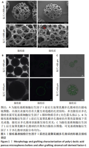
2.1 左旋聚乳酸多孔微球的形貌 扫描电镜下可见接枝与未接枝基质细胞衍生因子1左旋聚乳酸多孔微球内部及表面均存在大量互相连通的孔状结构,并且接枝基质细胞衍生因子1左旋聚乳酸表面有颗粒样物质存在(箭头所示),见图1A。未接枝基质细胞衍生因子1左旋聚乳酸多孔微球的孔隙率为90.63%,接枝基质细胞衍生因子1左旋聚乳酸多孔微球的孔隙率为89.83%。 2.2 基质细胞衍生因子1接枝表征 接枝基质细胞衍生因子1左旋聚乳酸多孔微球在不同层面扫描时均激发绿色荧光,见图1B。最终将荧光图像层层扫描后拟合输出,可见基质细胞衍生因子1均匀分布于微球,几乎遍布多孔微球表面,见图1C。利用基质细胞衍生因子1 ELISA试剂盒测量接枝完成后上清液中未成功接枝的因子量,通过间接法得到基质细胞衍生因子1 接枝率约为93.75%,与图1C中绿色荧光拟合图像相符,均验证了良好的接枝效果。"
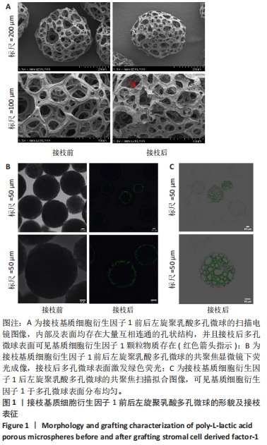
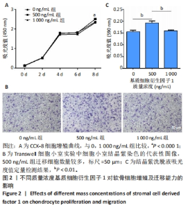
2.3 基质细胞衍生因子1对软骨细胞增殖、迁移、软骨表型维持的影响 CCK-8检测结果显示,随着培养时间的延长,各组细胞数量逐渐增加,培养第8天,500 ng/mL 基质细胞衍生因子1组细胞增殖吸光度值高于0,1 000 ng/mL 基质细胞衍生因子1组(P < 0.000 1),见图2A,说明添加500 ng/mL基质细胞衍生因子1对软骨细胞增殖具有促进作用。 Transwell细胞小室实验结果显示,500 ng/mL 基质细胞衍生因子1组细胞迁移能力强于0,1 000 ng/mL 基质细胞衍生因子1组(P < 0.01),见图2B,C,说明500 ng/mL基质细胞衍生因子1可促进软骨细胞的迁移。"
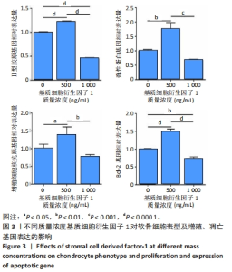
qRT-PCR检测结果显示,与0,1 000 ng/mL 基质细胞衍生因子1组比较,500 ng/mL基质细胞衍生因子1组软骨细胞内Ⅱ型胶原、弹性蛋白、增殖细胞核抗原、Bcl-2 mRNA表达均升高(P < 0.05,P < 0.01,P < 0.001,P < 0.000 1),见图3,说明500 ng/mL基质细胞衍生因子1可进软骨细胞的胶原蛋白合成能力,促进软骨细胞增殖并抑制其凋亡。"

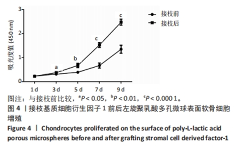
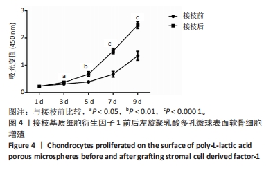
2.4 左旋聚乳酸多孔微球对软骨细胞增殖与黏附的影响 CCK-8检测结果显示,从培养第3天起,接枝基质细胞衍生因子1左旋聚乳酸多孔微球组软骨细胞吸光度值显著高于未接枝基质细胞衍生因子1左旋聚乳酸多孔微球组(P < 0.05,P < 0.01,P < 0.000 1),见图4,提示基质细胞衍生因子1有助于提高左旋聚乳酸多孔微球的细胞相容性。"
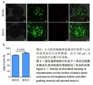
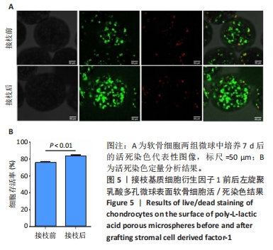
两组微球活死染色结果如图5A所示,可见细胞分布均匀,以绿色活细胞为主。活死染色定量分析结果显示,接枝基质细胞衍生因子1左旋聚乳酸多孔微球组细胞存活率高于未接枝基质细胞衍生因子1左旋聚乳酸多孔微球组(P < 0.01),如图5B所示,说明在左旋聚乳酸多孔微球表面接枝基质细胞衍生因子1有利于软骨细胞的黏附和存活。"

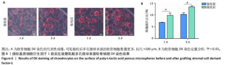
两组微球Dil免疫荧光染色如图6A所示,可见接枝基质细胞衍生因子1左旋聚乳酸多孔微球组细胞量更多、增殖速度更快;定量分析结果显示,接枝基质细胞衍生因子1左旋聚乳酸多孔微球组细胞面积占比高于未接枝基质细胞衍生因子1左旋聚乳酸多孔微球组(P < 0.01),如图6B所示,说明基质细胞衍生因子1有利于软骨细胞增殖。"
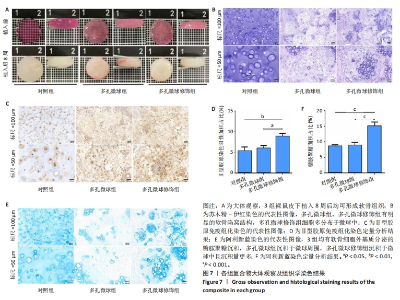
2.5 左旋聚乳酸多孔微球植入裸鼠皮下后体内成软骨检测 2.5.1 实验动物数量分析 12只裸鼠全部进入结果分析。 2.5.2 各组复合物大体观察 对照组、多孔微球组、多孔微球修饰组的组织块大小及厚度无明显差别,形态维持良好,3组组织块硬度手感无明显差异,均可生成乳白色软骨样组织,见图7A。 2.5.3 各组复合物组织学染色结果 苏木精-伊红染色结果显示,对照组软骨细胞散在分布于甲基丙烯酰胺基明胶中,相比之下,多孔微球组及多孔微球修饰组软骨细胞更倾向于聚集分布,多孔微球修饰组中有更多的细胞黏附、生长于多孔微球中,见图7B。Ⅱ型胶原免疫组化染色结果显示,多孔微球修饰组软骨细胞Ⅱ型胶原分泌多于对照组(P < 0.01)、多孔微球组(P < 0.05),见图7C,D。阿利新蓝染色结果显示,多孔微球修饰组软骨细胞的细胞外基质糖胺聚糖沉积多于对照组、多孔微球组(P均< 0.001),见图7E,F。"


2.5.4 qRT-PCR检测结果 多孔微球修饰组软骨相关基因Ⅱ型胶原、弹性蛋白的mRNA 表达明显高于对照组、多孔微球组(P < 0.000 1),说明在接枝基质细胞衍生因子1左旋聚乳酸多孔微球可更好地维持软骨细胞表型;多孔微球修饰组增殖相关基因增殖细胞核抗原和凋亡抑制基因Bcl-2 mRNA表达明显高于对照组和多孔微球组(P < 0.001,P < 0.000 1),见图8,说明接枝基质细胞衍生因子1左旋聚乳酸多孔微球能够促进软骨细胞的增殖并抑制其凋亡。这些结果与体外实验中基质细胞衍生因子1对软骨细胞的作用是一致的。"

| [1] STAMPOULTZIS T, KARAMI P, PIOLETTI DP. Thoughts on cartilage tissue engineering: A 21st century perspective. Curr Res Transl Med. 2021;69(3):103299. [2] BALUCH N, NAGATA S, PARK C, et al. Auricular reconstruction for microtia: A review of available methods. Plast Surg (Oakv). 2014; 22(1):39-43. [3] CHEN FM, LIU X. Advancing biomaterials of human origin for tissue engineering. Prog Polym Sci. 2016;53:86-168. [4] RASTOGI P, KANDASUBRAMANIAN B. Review of alginate-based hydrogel bioprinting for application in tissue engineering. Biofabrication. 2019;11(4):42001. [5] SALEHI S, KOECK K, SCHEIBEL T. Spider Silk for Tissue Engineering Applications. Molecules. 2020;25(3):737. [6] CATOIRA MC, FUSARO L, DI FRANCESCO D, et al. Overview of natural hydrogels for regenerative medicine applications. J Mater Sci Mater Med. 2019;30(10):115. [7] WU DT, MUNGUIA-LOPEZ JG, CHO YW, et al. Polymeric Scaffolds for Dental, Oral, and Craniofacial Regenerative Medicine. Molecules. 2021;26(22):7043. [8] LEE HI, HEO Y, BAEK SW, et al. Multifunctional Biodegradable Vascular PLLA Scaffold with Improved X-ray Opacity, Anti-Inflammation, and Re-Endothelization. Polymers (Basel). 2021;13(12):1979. [9] REDDY M, PONNAMMA D, CHOUDHARY R, et al. A Comparative Review of Natural and Synthetic Biopolymer Composite Scaffolds. Polymers (Basel). 2021;13(7):1105. [10] XIAO G, YIN H, XU W, et al. Modification and cytocompatibility of biocomposited porous PLLA/HA-microspheres scaffolds. J Biomater Sci Polym Ed. 2016;27(14):1462-1475. [11] RAVI M, PARAMESH V, KAVIYA SR, et al. 3D cell culture systems: advantages and applications. J Cell Physiol. 2015;230(1):16-26. [12] KANG SW, SEO SW, CHOI CY, et al. Porous poly(lactic-co-glycolic acid) microsphere as cell culture substrate and cell transplantation vehicle for adipose tissue engineering. Tissue Eng Part C Methods. 2008;14(1):25-34. [13] LIN A, LIU S, XIAO L, et al. Controllable preparation of bioactive open porous microspheres for tissue engineering. J Mater Chem B. 2022;10(34):6464-6471. [14] ZENG Y, LI X, LIU X, et al. PLLA Porous Microsphere-Reinforced Silk-Based Scaffolds for Auricular Cartilage Regeneration. ACS Omega. 2021;6(4):3372-3383. [15] LIU S, WANG YN, MA B, et al. Gingipain-Responsive Thermosensitive Hydrogel Loaded with SDF-1 Facilitates In Situ Periodontal Tissue Regeneration. ACS Appl Mater Interfaces. 2021;13(31):36880-36893. [16] WANG Y, SUN X, LV J, et al. Stromal Cell-Derived Factor-1 Accelerates Cartilage Defect Repairing by Recruiting Bone Marrow Mesenchymal Stem Cells and Promoting Chondrogenic Differentiation<sup/>. Tissue Eng Part A. 2017;23(19-20):1160-1168. [17] LI J, CHEN H, ZHANG D, et al. The role of stromal cell-derived factor 1 on cartilage development and disease. Osteoarthritis Cartilage. 2021;29(3):313-322. [18] YUE K, TRUJILLO-DE SG, ALVAREZ MM, et al. Synthesis, properties, and biomedical applications of gelatin methacryloyl (GelMA) hydrogels. Biomaterials. 2015;73:254-271. [19] LU W, ZENG M, LIU W, et al. Human urine-derived stem cell exosomes delivered via injectable GelMA templated hydrogel accelerate bone regeneration. Mater Today Bio. 2023;19:100569. [20] JIN C, XU G. Study on the Promotion of hADSCs Migration and Chemotaxis by SDF-1. Asia Pac J Ophthalmol (Phila). 2023;12(3):303-309. [21] CHIESA-ESTOMBA CM, HERNAEZ-MOYA R, RODINO C, et al. Ex Vivo Maturation of 3D-Printed, Chondrocyte-Laden, Polycaprolactone-Based Scaffolds Prior to Transplantation Improves Engineered Cartilage Substitute Properties and Integration. Cartilage. 2022;13(4):105-118. [22] LI X, LI X, YANG J, et al. Living and Injectable Porous Hydrogel Microsphere with Paracrine Activity for Cartilage Regeneration. Small. 2023;19(17):e2207211. [23] TAN YJ, TAN X, YEONG WY, et al. Hybrid microscaffold-based 3D bioprinting of multi-cellular constructs with high compressive strength: A new biofabrication strategy. Sci Rep. 2016;6:39140. [24] BIJU TS, PRIYA VV, FRANCIS AP. Role of three-dimensional cell culture in therapeutics and diagnostics: an updated review. Drug Deliv Transl Res. 2023;13(9):2239-2253. [25] JIN GZ, KIM HW. Porous microcarrier-enabled three-dimensional culture of chondrocytes for cartilage engineering: A feasibility study. Tissue Eng Regen Med. 2016;13(3):235-241. [26] GUPTA V, KHAN Y, BERKLAND CJ, et al. Microsphere-Based Scaffolds in Regenerative Engineering. Annu Rev Biomed En. 2017;19:135-161. [27] YUAN X, YANG W, FU Y, et al. Four-Arm Polymer-Guided Formation of Curcumin-Loaded Flower-Like Porous Microspheres as Injectable Cell Carriers for Diabetic Wound Healing. Adv Healthc Mater. 2023; 12(30):e2301486. [28] QIAO Z, ZHANG W, JIANG H, et al. 3D-printed composite scaffold with anti-infection and osteogenesis potential against infected bone defects. RSC Adv. 2022;12(18):11008-11020. [29] PAN Q, SU W, YAO Y. Progress in microsphere-based scaffolds in bone/cartilage tissue engineering. Biomed Mater. 2023;18(6). doi: 10.1088/1748-605X/acfd78. [30] FINKLEA FB, TIAN Y, KERSCHER P, et al. Engineered cardiac tissue microsphere production through direct differentiation of hydrogel-encapsulated human pluripotent stem cells. Biomaterials. 2021;274: 120818. [31] DENG S, ZHAO X, ZHU Y, et al. Efficient hepatic differentiation of hydrogel microsphere-encapsulated human pluripotent stem cells for engineering prevascularized liver tissue. Biofabrication. 2022;15(1). doi: 10.1088/1758-5090/aca79b. [32] GHUMAN H, MATTA R, TOMPKINS A, et al. ECM hydrogel improves the delivery of PEG microsphere-encapsulated neural stem cells and endothelial cells into tissue cavities caused by stroke. Brain Res Bull. 2021;168:120-137. [33] YANG Y, YAO X, LI X, et al. Non-mulberry silk fiber-based scaffolds reinforced by PLLA porous microspheres for auricular cartilage: An in vitro study. Int J Biol Macromol. 2021;182:1704-1712. [34] HUANG L, XIAO L, JUNG PA, et al. Porous chitosan microspheres as microcarriers for 3D cell culture. Carbohydr Polym. 2018;202:611-620. [35] WANG SQ, XIA J, CHEN J, et al. Influence of biological scaffold regulation on the proliferation of chondrocytes and the repair of articular cartilage. Am J Transl Res. 2016;8(11):4564-4573. [36] ZHANG Y, CHOI SW, XIA Y. Modifying the pores of an inverse opal scaffold with chitosan microstructures for truly three-dimensional cell culture. Macromol Rapid Commun. 2012;33(4):296-301. [37] LI Y, XIAO Y, LIU C. The Horizon of Materiobiology: A Perspective on Material-Guided Cell Behaviors and Tissue Engineering. Chem Rev. 2017;117(5):4376-4421. [38] LUO Y, XIAO M, ALMAQRAMI BS, et al. Regenerated silk fibroin based on small aperture scaffolds and marginal sealing hydrogel for osteochondral defect repair. Biomater Res. 2023;27(1):50. [39] 黄元亮,穆琳,林燕娴,等.丝素蛋白-左旋聚乳酸微载体扩增脂肪来源干细胞的实验研究[J].中国修复重建外科杂志,2021,35(5):611-619. [40] LI Q, CHANG B, DONG H, et al. Functional microspheres for tissue regeneration. Bioact Mater. 2023;25:485-499. [41] BAI L, HAN Q, HAN Z, et al. Stem Cells Expansion Vector via Bioadhesive Porous Microspheres for Accelerating Articular Cartilage Regeneration. Adv Healthc Mater. 2024;13(3):e2302327. [42] THAKUR G, RODRIGUES FC, SINGH K. Crosslinking Biopolymers for Advanced Drug Delivery and Tissue Engineering Applications. Adv Exp Med Biol. 2018;1078:213-231. [43] NAIR M, JOHAL RK, HAMAIA SW, et al. Tunable bioactivity and mechanics of collagen-based tissue engineering constructs: A comparison of EDC-NHS, genipin and TG2 crosslinkers. Biomaterials. 2020;254:120109. [44] GRABSKA-ZIELINSKA S, SIONKOWSKA A, CARVALHO A, et al. Biomaterials with Potential Use in Bone Tissue Regeneration-Collagen/Chitosan/Silk Fibroin Scaffolds Cross-Linked by EDC/NHS. Materials (Basel). 2021;14(5):1105. [45] LI L X, ZHANG XF, BAI X, et al. SDF-1 promotes ox-LDL induced vascular smooth muscle cell proliferation. Cell Biol Int. 2013;37(9):988-994. [46] SADRI F, REZAEI Z, FEREIDOUNI M. The significance of the SDF-1/CXCR4 signaling pathway in the normal development. Mol Biol Rep. 2022;49(4):3307-3320. |
| [1] | Zhao Jiyu, Wang Shaowei. Forkhead box transcription factor O1 signaling pathway in bone metabolism [J]. Chinese Journal of Tissue Engineering Research, 2025, 29(9): 1923-1930. |
| [2] | Yin Lu, Jiang Chuanfeng, Chen Junjie, Yi Ming, Wang Zihe, Shi Houyin, Wang Guoyou, Shen Huarui. Effect of Complanatoside A on the apoptosis of articular chondrocytes [J]. Chinese Journal of Tissue Engineering Research, 2025, 29(8): 1541-1547. |
| [3] | Liu Qi, Li Linzhen, Li Yusheng, Jiao Hongzhuo, Yang Cheng, Zhang Juntao. Icariin-containing serum promotes chondrocyte proliferation and chondrogenic differentiation of stem cells in the co-culture system of three kinds of cells [J]. Chinese Journal of Tissue Engineering Research, 2025, 29(7): 1371-1379. |
| [4] | Yang Zhihang, Sun Zuyan, Huang Wenliang, Wan Yu, Chen Shida, Deng Jiang. Nerve growth factor promotes chondrogenic differentiation and inhibits hypertrophic differentiation of rabbit bone marrow mesenchymal stem cells [J]. Chinese Journal of Tissue Engineering Research, 2025, 29(7): 1336-1342. |
| [5] | Xiang Pan, Che Yanjun, Luo Zongping. Compressive stress induces degeneration of cartilaginous endplate cells through the SOST/Wnt/beta-catenin pathway [J]. Chinese Journal of Tissue Engineering Research, 2025, 29(5): 951-957. |
| [6] | Yu Shuangqi, Ding Fan, Wan Song, Chen Wei, Zhang Xuejun, Chen Dong, Li Qiang, Lin Zuoli. Effects of polylactic acid-glycolic acid copolymer/lysine-grafted graphene oxide nanoparticle composite scaffolds on osteogenic differentiation of MC3T3 cells [J]. Chinese Journal of Tissue Engineering Research, 2025, 29(4): 707-712. |
| [7] | Ma Weibang, Xu Zhe, Yu Qiao, Ouyang Dong, Zhang Ruguo, Luo Wei, Xie Yangjiang, Liu Chen. Screening and cytological validation of cartilage degeneration-related genes in exosomes from osteoarthritis synovial fluid [J]. Chinese Journal of Tissue Engineering Research, 2025, 29(36): 7783-7789. |
| [8] | Sima Xinli, Liu Danping, Qi Hui. Effect and mechanism of metformin-modified bone marrow mesenchymal stem cell exosomes on regulating chondrocytes [J]. Chinese Journal of Tissue Engineering Research, 2025, 29(36): 7728-7734. |
| [9] | Yin Hang, Song Kui. Effect of crocin hydrogel on chondrocytes and MC3T3-E1 cells [J]. Chinese Journal of Tissue Engineering Research, 2025, 29(34): 7293-7300. |
| [10] | Yu Qinghe, Cai Ziming, Wu Jintao, Ma Pengfei, Zhang Xin, Zhou Longqian, Wang Yakun, Lin Xiaoqin, Lin Wenping. Vanillic acid inhibits inflammatory response and extracellular matrix degradation of endplate chondrocytes [J]. Chinese Journal of Tissue Engineering Research, 2025, 29(30): 6391-9397. |
| [11] | An Xingqi, Li Wenjin. Mechanism of Notch signaling pathway regulating condylar cartilage development and temporomandibular joint inflammation [J]. Chinese Journal of Tissue Engineering Research, 2025, 29(26): 5673-5679. |
| [12] | Liang Zhifeng, Yang Yingcai, Cheng Qiangang, Jia Yongxing, Wang Bo . Effect of stromal cell-derived factor-1 in cartilage and subchondral bone homeostasis [J]. Chinese Journal of Tissue Engineering Research, 2025, 29(25): 5422-5433. |
| [13] | Zheng Li, Ding Yiheng, Li Xinhao, Wen Zekai, Jiang Bingzheng, Lin Xuexia. Role of neuropilin 1 in promoting angiogenesis-osteogenesis coupling during fracture healing [J]. Chinese Journal of Tissue Engineering Research, 2025, 29(21): 4576-4583. |
| [14] | Yang Dujuan, Cheng Mengke, Liu Jia. Osteogenic/odontogenic differentiation ability of human dental pulp stem cells under photocrosslinked composite hydrogel scaffold [J]. Chinese Journal of Tissue Engineering Research, 2025, 29(19): 4022-4028. |
| [15] | Wang Zhe, Qi Yansong, Xu Yongsheng. Diagnosis and treatment of osteoarthritis with exosomes derived from different stem cells and carrying non-coding RNA [J]. Chinese Journal of Tissue Engineering Research, 2025, 29(19): 4122-4131. |
| Viewed | ||||||
|
Full text |
|
|||||
|
Abstract |
|
|||||