Chinese Journal of Tissue Engineering Research ›› 2025, Vol. 29 ›› Issue (22): 4748-4760.doi: 10.12307/2025.432
Previous Articles Next Articles
Vascularization characteristics of tissue-engineered oral mucosa equivalents
Shi Lijuan1, 2, 3, Wei Jian1, 2, 3, Zhang Xuan1, 2, 3, He Lingxiao1, 2, 3, Jiang Xiaoxi1, 2, 3, Nie Minhai1, 2, 3, Chen Jiana1, 2, 3, Liu Xuqian1, 2, 3
- 1Department of Periodontal Mucosal Diseases, Affiliated Stomatological Hospital of Southwest Medical University, Luzhou 646000, Sichuan Province, China; 2Luzhou Key Laboratory of Oral & Maxillofacial Reconstruction and Regeneration, Luzhou 646000, Sichuan Province, China; 3Institute of Stomatology, Southwest Medical University, Luzhou 646000, Sichuan Province, China
-
Received:2024-03-21Accepted:2024-05-06Online:2025-08-08Published:2024-12-06 -
Contact:Liu Xuqian, MD, Associate professor, Master’s supervisor, Department of Periodontal Mucosal Diseases, Affiliated Stomatological Hospital of Southwest Medical University, Luzhou 646000, Sichuan Province, China; Luzhou Key Laboratory of Oral & Maxillofacial Reconstruction and Regeneration, Luzhou 646000, Sichuan Province, China; Institute of Stomatology, Southwest Medical University, Luzhou 646000, Sichuan Province, China -
About author:Shi Lijuan, Master candidate, Department of Periodontal Mucosal Diseases, Affiliated Stomatological Hospital of Southwest Medical University, Luzhou 646000, Sichuan Province, China; Luzhou Key Laboratory of Oral & Maxillofacial Reconstruction and Regeneration, Luzhou 646000, Sichuan Province, China; Institute of Stomatology, Southwest Medical University, Luzhou 646000, Sichuan Province, China -
Supported by:1Department of Periodontal Mucosal Diseases, Affiliated Stomatological Hospital of Southwest Medical University, Luzhou 646000, Sichuan Province, China; 2Luzhou Key Laboratory of Oral & Maxillofacial Reconstruction and Regeneration, Luzhou 646000, Sichuan Province, China; 3Institute of Stomatology, Southwest Medical University, Luzhou 646000, Sichuan Province, China
CLC Number:
Cite this article
Shi Lijuan, Wei Jian, Zhang Xuan, He Lingxiao, Jiang Xiaoxi, Nie Minhai, Chen Jiana, Liu Xuqian. Vascularization characteristics of tissue-engineered oral mucosa equivalents[J]. Chinese Journal of Tissue Engineering Research, 2025, 29(22): 4748-4760.
share this article
Add to citation manager EndNote|Reference Manager|ProCite|BibTeX|RefWorks
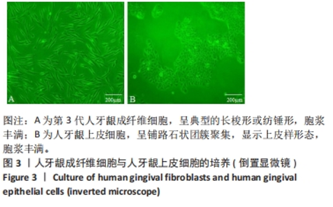
2.1 HGFs、HGECs细胞培养 倒置显微镜下观察第3代HGFs呈典型的长梭形或纺锤形,胞浆丰满;第3代HGECs呈铺路石状团簇聚集,细胞呈上皮样形态,胞浆丰满,呈立方形,细胞表面无突起,见图3。"

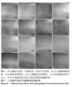
2.2 人牙龈间充质干细胞单克隆培养结果 单个人牙龈间充质干细胞呈梭形,轮廓清楚,似HGFs,每日可见人牙龈间充质干细胞扩增,初始生长缓慢,5 d左右细胞增殖速度加快,细胞呈长梭形或纺锤形,胞浆丰满;6-12 d细胞呈束状排列,12 d后细胞弥漫成片生长;经16 d连续培养后镜下观察到密集细胞团块,见图4。"
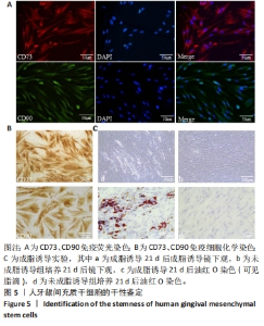
2.3 人牙龈间充质干细胞的干性鉴定结果 免疫荧光染色结果显示,CD73标记的人牙龈间充质干细胞胞浆表达红色荧光,CD90标记的人牙龈间充质干细胞胞浆表达绿色荧光,DAPI标记的细胞核表达蓝色荧光,位于胞浆中央。免疫细胞染色结果显示,CD73、CD90在人牙龈间充质干细胞胞浆中呈阳性表达。成脂诱导实验结果显示,诱导组镜下观察可见人牙龈间充质干细胞胞浆中富含空白的脂肪液滴,油红O染色后见深红色脂滴分布于淡蓝色胞核周围,对照组镜下观及油红O染色均无明显改变,见图5,提示人牙龈间充质干细胞具有成脂分化的潜力。"


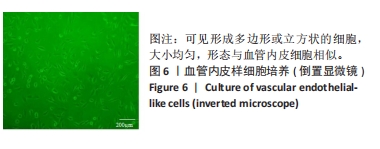
2.4 VELCs细胞培养 倒置显微镜下观察人牙龈间充质干细胞经血管内皮生长因子165诱导分化形成多边形或立方状的细胞,大小均匀,细胞形态与血管内皮细胞相似,见图6。"
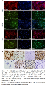
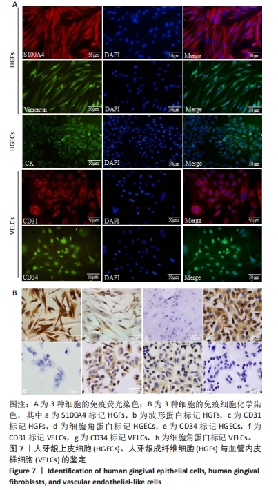
2.5 HGFs、HGECs、VELCs的鉴定 免疫荧光染色结果显示:S100A4标记的HGFs胞浆表达红色荧光,波形蛋白标记的HGFs胞浆表达绿色荧光,DAPI标记的细胞核表达蓝色荧光,位于胞浆中央;细胞角蛋白标记的HGECs胞浆表达绿色荧光,DAPI标记的细胞核表达蓝色荧光,位于胞浆中央;CD31标记的VELCs胞浆表达红色荧光,CD34标记的VELCs胞浆表达绿色荧光,DAPI标记的细胞核表达蓝色荧光,位于胞浆中央,见图7A。免疫细胞染色结果显示:S100A4、波形蛋白在HGFs胞浆中呈阳性表达,CD31在胞浆中阴性表达;细胞角蛋白在HGECs胞浆中呈阳性表达,CD34在胞浆中阴性表达;CD31、CD34在VELCs胞浆中呈阳性表达,细胞角蛋白在胞浆中阴性表达,见图7B。"
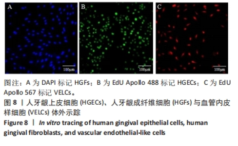
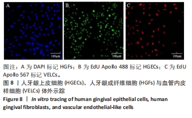
2.6 EdU Apollo体外示踪标记种子细胞 DAPI标记HGFs,被标记的胞核均呈蓝色荧光表达;HGECs标记组中,90%的HGECs被EdU Apollo 488标记,胞核呈绿色荧光表达;VELCs标记组中,90%的VELCs被EdU Apollo 567标记,胞核呈红色荧光表达,见图8。"

2.7 经典动物模型生物反应器体内动态强化实验 实验组皮下植入HGECs-HGFs-VELCs-脱细胞基质-0.25%类人Ⅰ型胶原复合体,未见明显瘢痕组织形成,4周后进行组织取材;对照组皮下植入脱细胞基质-0.25%类人Ⅰ型胶原支架,4周后进行组织取材,见图9。"
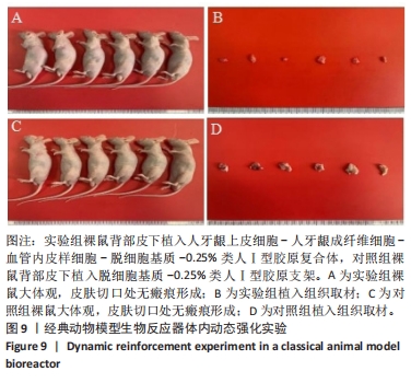
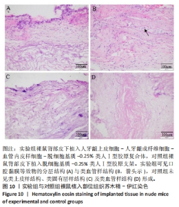
2.7.1 实验动物数量分析 12只裸鼠全部进入结果分析。 2.7.2 形态学观察结果 实验组类似于正常口腔黏膜组织结构,即存在类上皮样结构、类固有层样结构、类血管腔样结构,细胞层次清晰,类血管腔样结构内存在散在红细胞;对照组未见类上皮样结构、类固有层样结构、类血管腔样结构形成,见图10。"
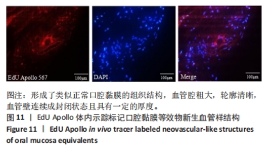
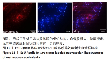
2.7.3 EdU Apollo体内示踪标记口腔黏膜等效物新生血管样结构 HGECs-HGFs-VELCs-脱细胞基质-0.25%类人Ⅰ型胶原复合体在生物反应器体内形成了类似正常口腔黏膜的组织结构,其中EdU Apollo 567标记的VELCs增殖、分化并形成了类血管样结构,呈红色荧光表达,血管腔粗大,轮廓清晰,血管壁连续成封闭状态,且具有一定的厚度;DAPI标记的HGFs形成了类固有层样结构,呈蓝色荧光表达,分布在红色荧光标记的新生血管样结构周围,见图11。"
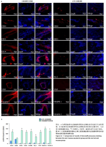
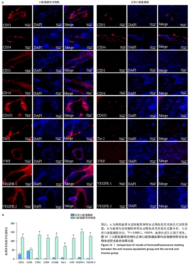
2.7.4 免疫荧光标记口腔黏膜等效物中的新生血管样结构 对动物体内构建的口腔黏膜等效物组织和口腔黏膜组织分别采用较为全面的血管内皮细胞特异性标志物表达谱进行免疫荧光染色,结果显示:CD31、CD34、CD51、CD54、CD105、Tie-2、VWF、血管内皮生长因子受体1、血管内皮生长因子受体2均表达于口腔黏膜等效物的新生血管样结构,VELCs在HGECs-HGFs-VELCs-脱细胞基质-0.25%类人Ⅰ型胶原复合体上形成连续封闭的新生血管样结构,血管腔粗大,血管壁具有一定的厚度,呈不规则扁圆形分布于HGFs形成的类固有层样结构中,表达红色荧光;广谱血管内皮细胞标志物CD31、CD34表达于正常口腔黏膜组织中的血管结构,血管腔细小,表达红色荧光,呈不规则扁圆形分布于蓝色荧光标记的固有层结构中,而对新生血管具有特异性标志的CD51、CD54、CD105、Tie-2、VWF、血管内皮生长因子受体1、血管内皮生长因子受体2在正常口腔黏膜组中未见明显表达,仅见DAPI标记的固有层组织结构。与正常口腔黏膜血管结构相比,口腔黏膜等效物血管样结构中的CD31、CD51、CD54、CD105、Tie-2、VWF、血管内皮生长因子受体1、V血管内皮生长因子受体2表达升高(P < 0.000 1),两者CD34表达比较差异无显著性意义(P > 0.05),见图12。"
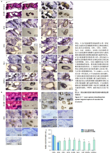
2.7.5 激光捕获显微切割系统靶向捕获血管样结构 苏木精-伊红染色:口腔黏膜等效物中新生血管样结构清晰,血管腔粗大,呈扁圆形、连续封闭状,血管腔内可见一个到数个不等的红细胞分布,血管壁轮廓清晰,具有一定的厚度,新生血管样结构分布于类固有层样结构中;正常口腔黏膜组中毛细血管结构清晰,呈不规则扁圆形分布于上皮组织下的固有层结构中,血管腔细小,血管壁不如口腔黏膜等效物中的管壁厚,腔内见红细胞分布。 免疫组化染色:CD31、CD34、CD51、CD54、CD105、Tie-2、VWF、血管内皮生长因子受体1、血管内皮生长因子受体2均功能性表达于口腔黏膜等效物的新生血管样结构中,呈不同程度特异性着色,标志物仅表达于血管壁的内皮细胞,呈棕褐色标记新生血管样结构的形态和位置,血管腔较粗大,形态与苏木精-伊红染色中一致;血管内皮细胞广谱标志物CD31、CD34表达于正常口腔黏膜组织中的毛细血管结构,呈棕褐色表达于正常口腔黏膜血管组织内皮细胞,清晰标记出血管形态和位置,血管壁较口腔黏膜等效物新生血管管壁薄,管腔也较小,而血管内皮细胞特异性标志物CD51、CD54、CD105、Tie-2、VWF、血管内皮生长因子受体1、血管内皮生长因子受体2在正常口腔黏膜组织中几乎不表达,组织中的血管结构未见特异性着色,血管结构及位置不能被标记显示。与正常口腔黏膜的血管结构相比,口腔黏膜等效物的血管样结构中CD51、CD54、CD105、Tie-2、VWF、血管内皮生长因子受体1、血管内皮生长因子受体2表达升高(P < 0.000 1),两者CD31、CD34表达比较差异无显著性意义(P > 0.05),见图13。"

| [1] CHIANG TY, HUANG CH, KUAN CH, et al. Resuming Oral Feeding in Patients With Oral Squamous Cell Carcinoma With Free Anterolateral Thigh Flap Reconstruction. Ann Plast Surg. 2021;86(1):S108-S112. [2] NIKOLOUDAKI G, CREBER K, HAMILTON DW. Wound healing and fibrosis: a contrasting role for periostin in skin and the oral mucosa. Am J Physiol Cell Physiol. 2020;318(6):C1065-C1077. [3] IZUMI K, YORTCHAN W, AIZAWA Y, et al. Recent trends and perspectives in reconstruction and regeneration of intra/extra-oral wounds using tissue-engineered oral mucosa equivalents. Jpn Dent Sci Re. 2023;59: 365-374. [4] MASCHARAK S, DESJARDINS-PARK HE, DAVITT MF, et al. Preventing Engrailed-1 activation in fibroblasts yields wound regeneration without scarring. Science. 2021;372(6540):eaba2374. [5] EELEN G, TREPS L, LI X, et al. Basic and Therapeutic Aspects of Angiogenesis Updated. Circ Res. 2020;127(2):310-329. [6] GRIFFIOEN AW, DUDLEY AC. Angiogenesis: a year in review. Angiogenesis. 2021;24(2):195-196. [7] HOSSEINI M, KOEHLER KR, SHAFIEE A. Biofabrication of human skin with its appendages. Adv Healthc Mater. 2022;11(22):2201626. [8] LEE HJ, HONG YJ, KIM M. Angiogenesis in Chronic Inflammatory Skin Disorders. Int J Mol Sci. 2021;22(21):12035. [9] 李光照,陈锐,贾洪林,等.组织工程血管化基因治疗的研究进展[J].中国组织工程研究,2022,26(28):4569-4574. [10] ZHOU MY, CHEN X, QIU YL, et al. Study of tissue engineered vascularised oral mucosa-like structures based on ACVM-0.25%HLC-I scaffold in vitro and in vivo. Artif Cells Nanomed Biotechnol. 2020; 48(1):1167-1177. [11] LIU XQ, WANG J, DONG FS, et al. Study of composite vascular scaffold combining with differentiated VSMC- and VEC-like cells in vitro and in vivo. J Biomater Appl. 2017;32(2):219-229. [12] QIU YL, CHEN X, HOU YL, et al. Characterization of different biodegradable scaffolds in tissue engineering. Mol Med Rep. 2019; 19(5):4043-4056. [13] MASSON DS, BERTASSONI LE, TAYEBI L. Oral mucosa equivalents, prevascularization approaches, and potential applications. Connect Tissue Res. 2022;63(5):514-529. [14] CUCCI LM, SATRIANO C, MARZO T, et al. Angiogenin and Copper Crossing in Wound Healing. Int J Mol Sci. 2021; 22(19):10704. [15] CUI X, LI Z, YUAN Y. Knockdown of FOXO4 protects against OGD/R-induced cerebral microvascular endothelial cell injury and regulates the AMPK/Nrf2/HO-1 pathway through transcriptional activation of CTRP6. Exp Ther Med. 2024;27(3):94. [16] TOYODA M, FUKUDA T, FUJIMOTO R, et al. Scaffold-free bone-like 3D structure established through osteogenic differentiation from human gingiva-derived stem cells. Biochem Biophys Rep. 2024;38:101656. [17] CALIGIURI G. CD31 as a Therapeutic Target in Atherosclerosis. Circ Res. 2020;126(9):1178-1189. [18] NOGUCHI Y, MAEDA A, WANG HT, et al. Human CD31 on Swine Endothelial Cells Induces SHP-1 Phosphorylation in Macrophages. Transplant Proc. 2020;52(6):1913-1915. [19] WU H, YIN G, PU X, et al. Inhibitory Effects of Combined Bone Morphogenetic Protein 2, Vascular Endothelial Growth Factor, and Basic Fibroblast Growth Factor on Osteoclast Differentiation and Activity. Tissue Eng Part A. 2021;27(21-22):1387-1398. [20] 刘洁.VEGFR1下调对Aβ1-42诱导人脑微血管内皮细胞衰老及PI3K信号通路的影响[J].河北医药,2021,43(7):990-993+999. [21] YANG M, LI CJ, XIAO Y, et al. Ophiopogonin D promotes bone regeneration by stimulating CD31hi EMCNhi vessel formation. Cell Prolif. 2020;53(3):e12784. [22] NAITO H, IBA T, TAKAKURA N. Mechanisms of new blood-vessel formation and proliferative heterogeneity of endothelial cells. Int Immunol. 2020;32(5):295-305. [23] WANG C, DAI X, WU S, et al. FUNDC1-dependent mitochondria-associated endoplasmic reticulum membranes are involved in angiogenesis and neoangiogenesis. Nat Commun. 2021;12(1):2616. [24] MODENA DAO, SOARES CD, CANDIDO EC, et al. Effect of extracorporeal shock waves on inflammation and angiogenesis of integumentary tissue in obese individuals: stimulating repair and regeneration. Lasers Med Sci. 2022;37(2):1289-1297. [25] 时华,杨中,李颖.扶正祛毒方抑制人乳腺癌MCF-7细胞增殖及对PI3KAkt信号通路和血管内皮生长因子C、血管内皮细胞生长因子受体3表达的影响[J].中国医院用药评价与分析,2020,20(12): 1437-1441. [26] CHEN B, ZHANG Y, CHEN S, et al. The role of vascular endothelial growth factor in ischemic stroke. Pharmazie. 2021;76(4):127-131. [27] CECI C, ATZORI MG, LACAL PM, et al. Role of VEGFs/VEGFR-1 Signaling and its Inhibition in Modulating Tumor Invasion: Experimental Evidence in Different Metastatic Cancer Models. Int J Mol Sci. 2020;21(4):1388. [28] OLLAURI-IBÁÑEZ C, NÚÑEZ-GÓMEZ E, EGIDO-TURRIÓN C, et al. Continuous endoglin (CD105) overexpression disrupts angiogenesis and facilitates tumor cell metastasis. Angiogenesis. 2020;23(2):231-247. [29] MARCIANÒ A, IENI A, MAUCERI R, et al. CD34 and CD105 Microvessels in Resected Bone Specimen May Implicate Wound Healing in MRONJ. Int J Environ Res Public Health. 2021;18(21):11362. [30] RAO BH, SOUČEK P, HLAVÁČ V. Laser Capture Microdissection: A Gear for Pancreatic Cancer Research. Int J Mol Sci. 2022;23(23):14566. [31] LLOSA N. Interrogating the Tumor Microenvironment by Deep Tissue Immunoprofiling. Methods Mol Biol. 2022;2435:139-156. [32] ZHOU T, WANG J. Laser Capture Microdissection of Vascular Endothelial Cells from Frozen Heart Tissues. Methods Mol Biol. 2021;2319:105-110. [33] PANDEY S, TUMA Z, SMRHOVA T, et al. Laser Capture Microdissection Coupled Capillary Immunoassay to Study the Expression of PCK-2 on Spatially-Resolved Islets of Rat Langerhans. Pharmaceutics. 2021; 13(6):883. [34] CARRARO C, BONAGURO L, SRINIVASA R, et al. Chromatin accessibility profiling of targeted cell populations with laser capture microdissection coupled to ATAC-seq. Cell Reports Methods. 2023;3(10):100598. |
| [1] | Li Zikai, Zhang Chengcheng, Xiong Jiaying, Yang Xirui, Yang Jing, Shi Haishan. Potential effects of ornidazole on intracanal vascularization in endodontic regeneration [J]. Chinese Journal of Tissue Engineering Research, 2025, 29(在线): 1-7. |
| [2] | Xiong Bohan, Wang Guoliang, Yu Yang, Xue Wenqiang, Yu Hong, Liu Jinrui, Ruan Zhaohui, Li Yajuan, Liu Haolong, Dong Kaiyan, Long Dan, Chen Zhao. Internal tension relieving technique assisted anterior cruciate ligament reconstruction to promote ligamentization of Achilles tendon grafts in small ear pigs in southern Yunnan province [J]. Chinese Journal of Tissue Engineering Research, 2025, 29(4): 713-720. |
| [3] | Wang Kaigang, Hao Dongsheng, Ma Pei, Zhou Shuo, Li Ruimin. Comparison of efficacy of different biological scaffolds for pulp regeneration therapy in immature permanent teeth: a Bayesian network meta-analysis [J]. Chinese Journal of Tissue Engineering Research, 2025, 29(34): 7447-7460. |
| [4] | Li Zikai, Zhang Chengcheng, Xiong Jiaying, Yang Xirui, Yang Jing, Shi Haishan. Potential effects of ornidazole on intracanal vascularization in endodontic regeneration [J]. Chinese Journal of Tissue Engineering Research, 2025, 29(14): 2892-2898. |
| [5] | Liu Mingyu, Fan Wenjuan. Construction strategy for vascularization of organoids [J]. Chinese Journal of Tissue Engineering Research, 2025, 29(13): 2774-2783. |
| [6] | Shen Ziqing, Xia Tian, Shan Yibo, Zhu Ruijun, Wan Haoxin, Ding Hao, Pan Shu, Zhao Jun. Vascularized tracheal substitutes constructed by exosome-load hydrogel-modified 3D printed scaffolds [J]. Chinese Journal of Tissue Engineering Research, 2024, 28(5): 697-705. |
| [7] | Liu Junpeng, Yao Xingchen, Zhao Hui, Xu Ziyu, Wu Yue, Pei Fuchun, Zhang Lin, Du Xinru. Antibiotic-loaded bone cement enhances ability of tibial cortex transverse transport for treating infected wounds [J]. Chinese Journal of Tissue Engineering Research, 2024, 28(29): 4599-4604. |
| [8] | Gao Li, Liu Liu, Ren Wenyan, Liu Xue, Wang Yiyu. Influential mechanism of graphene and its derivatives on angiogenesis and vascularized bone [J]. Chinese Journal of Tissue Engineering Research, 2024, 28(17): 2716-2722. |
| [9] | Zhao Lixin, Tang Qingxi, Zhang Kezhong. Influence of adipose tissue and its derivative on wound repair and vascularization [J]. Chinese Journal of Tissue Engineering Research, 2024, 28(13): 2120-2125. |
| [10] | Xu Yan, Li Ping, Lai Chunhua, Zhu Peijun, Yang Shuo, Xu Shulan. Piezoelectric materials for vascularized bone regeneration [J]. Chinese Journal of Tissue Engineering Research, 2023, 27(7): 1126-1132. |
| [11] | Cao Congcong, Ling Gengfei, Yang Chunhua. Local injection of ginsenoside Rg1 nanoparticles in the treatment of myocardial infarction in rats [J]. Chinese Journal of Tissue Engineering Research, 2023, 27(25): 3977-3983. |
| [12] | Cao Jin, Wang Ansu, Huang Nijiao, Wu Fujun, Chen Ping, Li Chengmei, Wang Xin. Role and mechanism of polyphosphate in bone tissue regeneration [J]. Chinese Journal of Tissue Engineering Research, 2023, 27(21): 3375-3381. |
| [13] | Zhou Lanxi, Shao Lu, Dong Shiwu, Yu Zhengwen. Molecular mechanism of angiogenesis promoted by medical metal materials [J]. Chinese Journal of Tissue Engineering Research, 2023, 27(16): 2616-2624. |
| [14] | He Ruya, Liu Yunling, Nie Minhai, Liu Xuqian. Repairing equivalent injury of oral mucosa with concentrated growth factor fibrin membrane combined with recombinant human epidermal growth factor active protein polypeptide complex [J]. Chinese Journal of Tissue Engineering Research, 2023, 27(12): 1848-1855. |
| [15] | Lu Yangyang, Yan Ruihong, Qiu Zhongpeng, Dai Yi, Wang Zixin, Wang Weishan, Shi Chenhui, Du Xinhui, Li Gang. Intervention with platelet-rich fibrin in rabbit models after microvascular anastomosis surgery of the femoral artery: changes in vascular structure and soft tissue [J]. Chinese Journal of Tissue Engineering Research, 2023, 27(11): 1659-1668. |
| Viewed | ||||||
|
Full text |
|
|||||
|
Abstract |
|
|||||