Chinese Journal of Tissue Engineering Research ›› 2025, Vol. 29 ›› Issue (2): 262-268.doi: 10.12307/2025.241
Previous Articles Next Articles
Effects and mechanisms of swimming for inhibiting traumatic joint contracture in a rat model
Shui Xiaoping1, 2, Li Chunying3, Zhang xin4, Li Bin2, Feng Chao2, Zhou Hongyu1, Chen Ke4, Liao Yingying4
- 1Sichaun College of Traditional Chinese Medicine, Mianyang 621000, Sichuan Province, China; 2Mianyang Orthopedic Hospital, Mianyang 621000, Sichuan Province, China; 3Mianyang Hospital of Traditional Chinese Medicine, Mianyang 621000, Sichuan Province, China; 4Sichuan Province Orthopedic Hospital, Chengdu 610041, Sichuan Province, China
-
Received:2024-01-09Accepted:2024-02-26Online:2025-01-18Published:2024-05-24 -
Contact:Shui Xiaoping, Sichaun College of Traditional Chinese Medicine, Mianyang 621000, Sichuan Province, China; Mianyang Orthopedic Hospital, Mianyang 621000, Sichuan Province, China -
About author:Shui Xiaoping, PhD, Associate professor, Sichaun College of Traditional Chinese Medicine, Mianyang 621000, Sichuan Province, China; Mianyang Orthopedic Hospital, Mianyang 621000, Sichuan Province, China -
Supported by:Science and Technology Development Cultivation Project of the Chinese Association of Rehabilitation Medicine (Cultivation Project 14) (to SXP); Scientific Research and Innovation Team of Sichaun College of Traditional Chinese Medicine, No. TD-2022-04 (to SXP); Scientific Research and Innovation Team of Mianyang Orthopaedic Hospital and Mianyang Municipal Higher Education "Three-Shift Project" (Advantageous and Characteristic Specialties - Rehabilitation Therapy Technology Specialty Construction) Project, No. 202055 (to SXP)
CLC Number:
Cite this article
Shui Xiaoping, Li Chunying, Zhang xin, Li Bin, Feng Chao, Zhou Hongyu, Chen Ke, Liao Yingying. Effects and mechanisms of swimming for inhibiting traumatic joint contracture in a rat model [J]. Chinese Journal of Tissue Engineering Research, 2025, 29(2): 262-268.
share this article
Add to citation manager EndNote|Reference Manager|ProCite|BibTeX|RefWorks
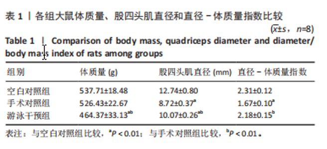
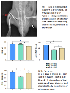
2.1 实验动物数量分析 16只大鼠造模均成功,每组8只大鼠进入结果分析,所有大鼠均无脱落。 2.2 大鼠体质量、股四头肌直径和直径-体质量指数 表1和图2显示,游泳干预组大鼠体质量明显低于空白对照组和手术对照组(P < 0.01)。与空白对照组相比,手术对照组大鼠股四头肌直径和直径-体质量指数明显降低(P < 0.01),游泳干预组大鼠股四头肌直径降低(P < 0.01);与手术对照组比较,游泳干预组大鼠股四头肌直径和直径-体质量指数增加(P < 0.01)。"


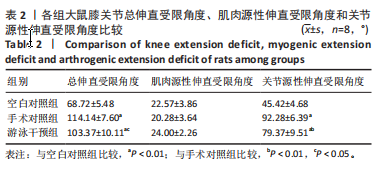
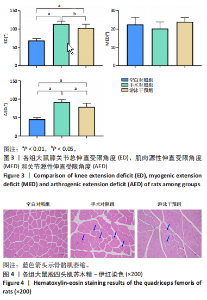
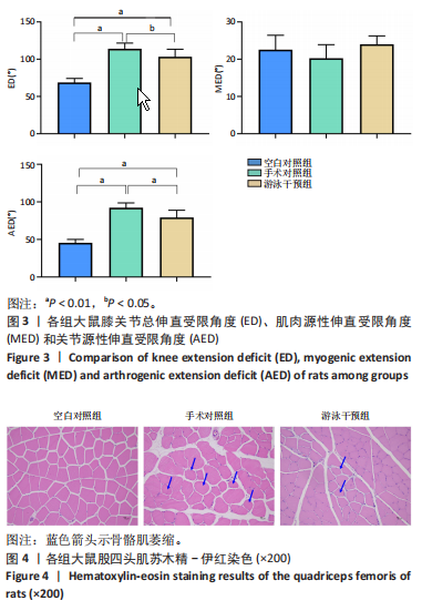
2.3 膝关节活动度 表2和图3显示,与空白对照组相比,手术对照组和游泳干预组大鼠膝关节总伸直受限角度和关节源性伸直受限角度均明显增加(P < 0.01)。与手术对照组比较,游泳干预组大鼠总伸直受限角度和关节源性伸直受限角度均降低(P < 0.05,P < 0.01)。"
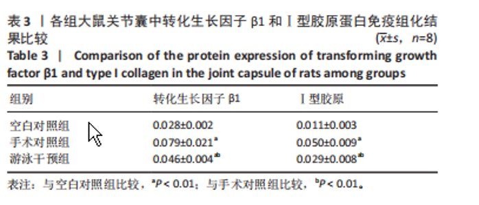
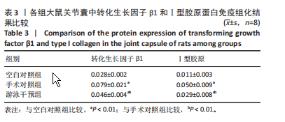
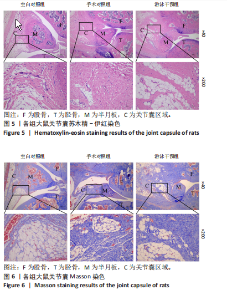
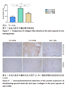
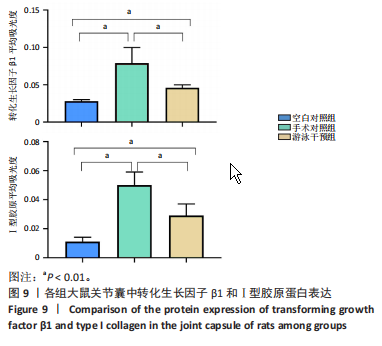
2.4 股四头肌苏木精-伊红染色结果 图4显示,空白对照组大鼠股四头肌细胞呈不规则的多边形,细胞核多且在边缘,细胞间隙均匀;与空白对照组比较,手术对照组大鼠股四头肌细胞明显萎缩,表现为细胞核减少,细胞固缩,形态圆润,相邻细胞间隙增宽;而游泳干预组细胞萎缩程度明显减轻。 2.5 关节囊苏木精-伊红染色结果 图 5显示,空白对照组大鼠关节囊中有少量形态规则的胶原纤维和成纤维细胞,周边有较多排列规则的脂肪细胞。手术对照组关节囊纤维化病变明显,可见大量不规则团块状胶原纤维且排列紊乱,关节囊中脂肪细胞减少。游泳干预组大鼠关节囊纤维化病变程度减轻,胶原纤维减少,成纤维细胞、胶原纤维和脂肪细胞排列较规则。 2.6 关节囊Masson染色结果 图6显示,空白对照组大鼠关节囊胶原纤维数量少,呈波浪状规则排列;手术对照组大鼠关节囊纤维明显增加,表现为致密的团块状,且排列紊乱;游泳干预组大鼠关节囊胶原减少,且部分区域胶原纤维排列规则整齐。图7显示,与空白对照组比较,手术对照组与游泳干预组大鼠关节囊胶原密度均显著增加(P < 0.01);与手术对照组比较,游泳干预组大鼠关节囊胶胶原密度降低(P < 0.01)。 2.7 各组大鼠关节囊转化生长因子β1和Ⅰ型胶原蛋白免疫组化结果 表3,图8,9显示,与空白对照组比较,手术对照组和游泳干预组大鼠关节囊中转化生长因子β1和Ⅰ型胶原蛋白表达增加(P < 0.01);与手术对照组比较,游泳干预组大鼠关节囊中转化生长因子β1和Ⅰ型胶原蛋白表达减少(P < 0.01)。"
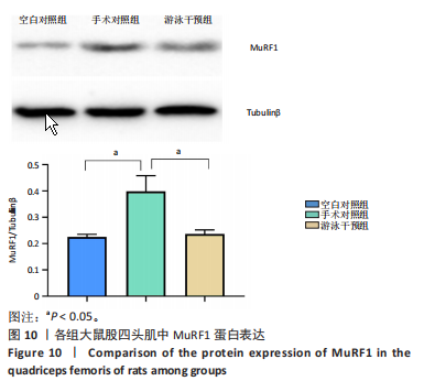
2.8 各组大鼠股四头肌MuRF1蛋白表达 图10显示,与空白对照组比较,手术对照组和游泳干预组大鼠股四头肌中MuRF1蛋白表达明显增加(P < 0.05);与手术对照组比较,游泳干预组大鼠股四头肌中MuRF1蛋白表达减少 (P < 0.05)。"

"
"

"


"

"
| [1] ITAYA N, YABE Y, HAGIWARA Y, et al. Effects of Low-Intensity Pulsed Ultrasound for Preventing Joint Stiffness in Immobilized Knee Model in Rats. Ultrasound Med Biol. 2018;44(6):1244-1256. [2] LEPLEY LK, DAVI SM, BURLAND JP, et al. Muscle Atrophy After ACL Injury: Implications for Clinical Practice. Sports Health. 2020;12(6):579-586. [3] TOKUDA K, YAMANAKA Y, MANO Y, et al. Effect of metformin treatment and its time of administration on joint capsular fibrosis induced by mouse knee immobilization. Sci Rep. 2021;11(1):17978. [4] ZHOU H, TRUDEL G, GOUDREAU L, et al. Knee joint stiffness following immobilization and remobilization: A study in the rat model. J Biomech. 2020;99:109471. [5] KANEGUCHI A, OZAWA J. Inflammation and Fibrosis Induced by Joint Remobilization, and Relevance to Progression of Arthrogenic Joint Contracture: A Narrative Review. Physiol Res. 2022;71(4):447-488. [6] KANEGUCHI A, OZAWA J, MINAMIMOTO K, et al. Active exercise on immobilization-induced contractured rat knees develops arthrogenic joint contracture with pathological changes. J Appl Physiol (1985). 2018;124(2):291-301. [7] REITER AJ, CASTILE RM, SCHOTT HR, et al. Investigating the Effects of Physical Therapy Timing, Intensity and Duration on Post-Traumatic Joint Contracture in a Rat Elbow Model. Muscles Ligaments Tendons J. 2021;11(3):547-553. [8] BECKER BE. Aquatic therapy: scientific foundations and clinical rehabilitation applications. PM R. 2009;1(9):859-872. [9] QU XM, LI G, LI ZM, et al. Effects of low-intensity swimming on radiation-induced leg contracture in mice. Chin J Dent Res. 2011;14(1):41-45. [10] SHUI XP, YE F, LI CY, et al. Effects of millimeter-wave for preventing joint stiffness in the immobilized knee rat model. Knee. 2023;42:236-245. [11] 蔡任,钱佳佳,许奇,等.游泳运动对膝骨关节炎小鼠软骨退变和NF-κB激活的影响[J].山东体育学院学报,2021,37(6):25-33. [12] ZHANG Y, WANG Z, ZONG C, et al. Platelet-rich plasma attenuates the severity of joint capsule fibrosis following post-traumatic joint contracture in rats. Front Bioeng Biotechnol. 2023;10:1078527. [13] 廖莹莹,张鑫,温呈洪,等.膝关节僵硬动物模型的造模方法及特点[J].中国组织工程研究,2022,26(15):2439-2445. [14] TRUDEL G, LANEUVILLE O, COLETTA E, et al. Quantitative and temporal differential recovery of articular and muscular limitations of knee joint contractures; results in a rat model. J Appl Physiol (1985). 2014;117(7): 730-737. [15] WEGNER E, SLOTINA E, MICKAN T, et al. Pleiotropic Long-Term Effects of Atorvastatin on Posttraumatic Joint Contracture in a Rat Model. Pharmaceutics. 2022;14(3):523. [16] DUNHAM CL, CASTILE RM, HAVLIOGLU N, et al. Persistent motion loss after free joint mobilization in a rat model of post-traumatic elbow contracture. J Shoulder Elbow Surg. 2017;26(4):611-618. [17] MATTYASOVSZKY SG, WOLLSTÄDTER J, MARTIN A, et al. Inhibition of Contractile Function in Human Joint Capsule Myofibroblasts by Targeting the TGF-β1 and PDGF Pathways. PLoS One. 2016;11(1): e0145948. [18] MONUMENT MJ, HART DA, BEFUS AD, et al. The mast cell stabilizer ketotifen reduces joint capsule fibrosis in a rabbit model of post-traumatic joint contractures. Inflamm Res. 2012;61(4):285-292. [19] HAGIWARA Y, CHIMOTO E, TAKAHASHI I, et al. Expression of transforming growth factor-beta1 and connective tissue growth factor in the capsule in a rat immobilized knee model. Ups J Med Sci. 2008; 113(2):221-234. [20] SASABE R, SAKAMOTO J, GOTO K, et al. Effects of joint immobilization on changes in myofibroblasts and collagen in the rat knee contracture model. J Orthop Res. 2017;35(9):1998-2006. [21] DUAN YC, SHI L, JIN Z, et al. Swimming Exercise Ameliorates Hypertension-Induced Kidney Dysfunction via Alleviating Renal Interstitial Fibrosis and Apoptosis. Kidney Blood Press Res. 2021;46(2):219-228. [22] FAKHRAEI S, REZA ALMASI M, PEERI M, et al. The effect of 4-Week rehabilitation by aerobic exercise on hippocampus BDNF and TGF-β1 gene expressions inAβ 1-42-induced rat model of Alzheimer’s disease. J Clin Neurosci. 2022;95:106-111. [23] LIN S, JIN S, ZHOU F, et al. Effects of endurance exercise on serum inflammatory cytokine level and kidney structure in a rat diabetes model. Exp Ther Med. 2021;22(4):1125. [24] KANEGUCHI A, TAKAHASHI A, SHIMOE A, et al. The combined effects of treadmill exercise and steroid administration on anterior cruciate ligament reconstruction-induced joint contracture and muscle atrophy in rats. Steroids. 2023;192:109183. [25] WANG X, WANG Z, TANG D. Aerobic Exercise Alleviates Inflammation, Oxidative Stress, and Apoptosis in Mice with Chronic Obstructive Pulmonary Disease. Int J Chron Obstruct Pulmon Dis. 2021;16:1369-1379. [26] LUBIS AM, LUBIS VK. Matrix metalloproteinase, tissue inhibitor of metalloproteinase and transforming growth factor-beta 1 in frozen shoulder, and their changes as response to intensive stretching and supervised neglect exercise. J Orthop Sci. 2013;18(4):519-527. [27] SCHILATY ND, MCPHERSON AL, NAGAI T, et al. Arthrogenic muscle inhibition manifests in thigh musculature motor unit characteristics after anterior cruciate ligament injury. Eur J Sport Sci. 2023;23(5):840-850. [28] YIN L, LI N, JIA W, et al. Skeletal muscle atrophy: From mechanisms to treatments. Pharmacol Res. 2021;172:105807. [29] HU SL, YANG WC, ZUO LY, et al. Effects of different types of exercise on contractile properties and expressions of MuRF1, PGC-1α /FNDC5 in soleus muscle of unloaded rats. Zhongguo Ying Yong Sheng Li Xue Za Zhi. 2022;38(4):356-360. [30] CIEMINSKI K, FLIS DJ, DZIK K, et al. Swim training affects Akt signaling and ameliorates loss of skeletal muscle mass in a mouse model of amyotrophic lateral sclerosis. Sci Rep. 2021;11(1):20899. |
| [1] | Yu Shuai, Liu Jiawei, Zhu Bin, Pan Tan, Li Xinglong, Sun Guangfeng, Yu Haiyang, Ding Ya, Wang Hongliang. Hot issues and application prospects of small molecule drugs in treatment of osteoarthritis [J]. Chinese Journal of Tissue Engineering Research, 2025, 29(9): 1913-1922. |
| [2] | Sun Yundi, Cheng Lulu, Wan Haili, Chang Ying, Xiong Wenjuan, Xia Yuan. Effect of neuromuscular exercise for knee osteoarthritis pain and function: a meta-analysis [J]. Chinese Journal of Tissue Engineering Research, 2025, 29(9): 1945-1952. |
| [3] | Miao Jiahang, Ma Sheng, Li Qupeng, Yu Huilin, Hu Tianyu, Gao Xiao, Feng Hu. Cervical lordosis ratio can be used as a decision-making indicator for selection of posterior surgical approach for multi-level cervical spondylotic myelopathy [J]. Chinese Journal of Tissue Engineering Research, 2025, 29(9): 1796-1802. |
| [4] | Liu Lin, Liu Shixuan, Lu Xinyue, Wang Kan. Metabolomic analysis of urine in a rat model of chronic myofascial trigger points [J]. Chinese Journal of Tissue Engineering Research, 2025, 29(8): 1585-1592. |
| [5] | Su Xiaoyang, Chen Wenting, Fu Yidan, Zhao Yan, Lan Danfeng, Yang Qiuping. Correlation between Mer receptor tyrosine kinase and diabetic peripheral neuropathy in Sprague-Dawley rats [J]. Chinese Journal of Tissue Engineering Research, 2025, 29(8): 1593-1599. |
| [6] | Li Jun, Gong Jingjing, Sun Guobin, Guo Rui, Ding Yang, Qiang Lijuan, Zhang Xiaoli, Fang Zhanhai . miR-27a-3p promotes the proliferation of human hypertrophic scar fibroblasts by regulating mitogen-activated protein kinase signaling pathway [J]. Chinese Journal of Tissue Engineering Research, 2025, 29(8): 1609-1617. |
| [7] | Jing Ruyi, Chen Yingxin, Cao Lei . Prognosis of deep lamellar keratoplasty versus penetrating keratoplasty in the treatment of stromal corneal dystrophy [J]. Chinese Journal of Tissue Engineering Research, 2025, 29(8): 1626-1633. |
| [8] | Wang Qiuyue, Jin Pan, Pu Rui . Exercise intervention and the role of pyroptosis in osteoarthritis [J]. Chinese Journal of Tissue Engineering Research, 2025, 29(8): 1667-1675. |
| [9] | Wang Yida, Liu Jun, Wang Xiaoling, Wang Liyan, Yang Chengru, Zhang Xuexiao. Effects of wearable electronic device-based interventions on physical activity and sedentary behavior in healthy adolescents: a meta-analysis [J]. Chinese Journal of Tissue Engineering Research, 2025, 29(8): 1693-1704. |
| [10] | Wang Juan, Wang Guanglan, Zuo Huiwu. Efficacy of exercise therapy in the treatment of anterior cruciate ligament reconstruction patients: #br# a network meta-analysis #br# [J]. Chinese Journal of Tissue Engineering Research, 2025, 29(8): 1714-1726. |
| [11] | Zheng Huakun, Yin Mingyue, Liu Qian. Effects of interval and continuous training on the quality of life in physically inactive adults: a meta-analysis [J]. Chinese Journal of Tissue Engineering Research, 2025, 29(8): 1727-1740. |
| [12] | Cai Yaohao, Lang Lyu, Li Hong. Assessing the bone mass of the residual alveolar ridge in the first molar for implant placement by cone-beam computed tomography [J]. Chinese Journal of Tissue Engineering Research, 2025, 29(8): 1572-1577. |
| [13] | Hu Taotao, Liu Bing, Chen Cheng, Yin Zongyin, Kan Daohong, Ni Jie, Ye Lingxiao, Zheng Xiangbing, Yan Min, Zou Yong. Human amniotic mesenchymal stem cells overexpressing neuregulin-1 promote skin wound healing in mice [J]. Chinese Journal of Tissue Engineering Research, 2025, 29(7): 1343-1349. |
| [14] | Jin Kai, Tang Ting, Li Meile, Xie Yuan. Effects of conditioned medium and exosomes of human umbilical cord mesenchymal stem cells on proliferation, migration, invasion, and apoptosis of hepatocellular carcinoma cells [J]. Chinese Journal of Tissue Engineering Research, 2025, 29(7): 1350-1355. |
| [15] | Lou Guo, Zhang Min, Fu Changxi. Exercise preconditioning for eight weeks enhances therapeutic effect of adipose-derived stem cells in rats with myocardial infarction [J]. Chinese Journal of Tissue Engineering Research, 2025, 29(7): 1363-1370. |
| Viewed | ||||||
|
Full text |
|
|||||
|
Abstract |
|
|||||