Chinese Journal of Tissue Engineering Research ›› 2025, Vol. 29 ›› Issue (19): 3961-3967.doi: 10.12307/2025.075
Programmed death receptor 1 inhibits osteogenic differentiation of rat bone marrow mesenchymal stem cells in a high glucose environment
Han Nianrong, Huang Yifei, Akram · Osman, Liu Yanlu, Hu Wei
- Hospital of Traditional Chinese Medicine Affiliated to Xinjiang Medical University, Urumqi 830000, Xinjiang Uygur Autonomous Region, China
-
Received:2024-02-08Accepted:2024-05-08Online:2025-07-08Published:2024-09-12 -
Contact:Hu Wei, Chief physician, Hospital of Traditional Chinese Medicine Affiliated to Xinjiang Medical University, Urumqi 830000, Xinjiang Uygur Autonomous Region, China -
About author:Han Nianrong, Doctoral candidate, Attending physician, Hospital of Traditional Chinese Medicine Affiliated to Xinjiang Medical University, Urumqi 830000, Xinjiang Uygur Autonomous Region, China -
Supported by:Natural Science Foundation of Xinjiang Uygur Autonomous Region - Outstanding Youth Science Foundation, No. 2022D01E29 (to HW)
CLC Number:
Cite this article
Han Nianrong, Huang Yifei, Akram · Osman, Liu Yanlu, Hu Wei . Programmed death receptor 1 inhibits osteogenic differentiation of rat bone marrow mesenchymal stem cells in a high glucose environment[J]. Chinese Journal of Tissue Engineering Research, 2025, 29(19): 3961-3967.
share this article
Add to citation manager EndNote|Reference Manager|ProCite|BibTeX|RefWorks
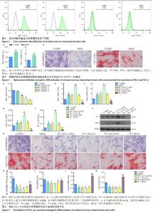
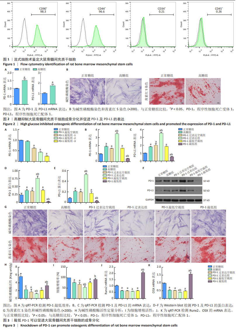
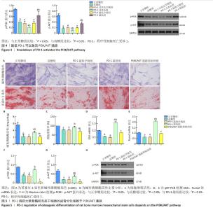
2.1 骨髓间充质干细胞鉴定结果 流式细胞术鉴定结果显示表面抗原CD90和CD44阳性,CD34与CD45阴性,见图1。 2.2 高糖抑制大鼠骨髓间充质干细胞成骨分化并促进PD-1及PD-L1的表达 采用qPCR检测高糖组与正常糖组PD-1、PD-L1的mRNA表达,结果显示,高糖组PD-1及PD-L1的mRNA表达水平显著高于正常糖组(P < 0.05),见图2A。成骨诱导分化21 d后,与正常糖组相比,高糖组碱性磷酸酶染色强度和形成矿化结节的能力降低,见图2B。结果表明,高糖环境会抑制大鼠骨髓间充质干细胞的成骨能力,提高大鼠骨髓间充质干细胞成骨分化过程中PD-1及PD-L1的表达水平。 2.3 敲低PD-1可以促进大鼠骨髓间充质干细胞的成骨分化 为了进一步探索PD-1对大鼠骨髓间充质干细胞成骨分化的调控功能,选择了1个敲低效率最好的siRNA(siRNA3)构建PD-1敲低慢病毒(sh-PD-1),见图3A。结果显示,PD-1过表达组PD-1和PD-L1的mRNA与蛋白表达水平较高糖组显著升高,差异均有显著性意义(P < 0.05);PD-1敲低组PD-1和PD-L1的mRNA与蛋白表达水平较高糖组显著降低,差异均有显著性意义(P < 0.05),见图3B-F;茜素红S染色和碱性磷酸酶染色显示PD-1敲低组细胞成骨分化能力较高糖组增加,而PD-1过表达组细胞成骨分化能力较高糖组降低,见图3G,H;PD-1敲低组细胞增殖活性较高糖组增加,PD-1过表达组细胞增殖活性较高糖组降低,差异均有显著性意义(P < 0.05),见图3I。同时,PD-1敲低组成骨标志物Runx2、OSX mRNA表达水平较高糖组增加(P < 0.05);而PD-1过表达组成骨标志物OSX mRNA表达水平较高糖组降低(P < 0.05),见图3J,K。"
2.4 降低PD-1可以激活PI3K/AKT通路 如图4所示,PD-1敲低组p-PI3K和p-AKT表达水平较高糖组显著升高(P < 0.05);而PD-1过表达组p-PI3K和p-AKT表达水平较高糖组显著降低(P < 0.05)。结果表明PD-1可以调控PI3K/AKT信号通路影响大鼠骨髓间充质干细胞的成骨分化。 2.5 PD-1调控大鼠骨髓间充质干细胞的成骨分化依赖于PI3K/AKT通路 为了进一步了解PD-1对大鼠骨髓间充质干细胞成骨分化的调控机制依赖于PI3K/AKT通路,加入PI3K/AKT通路抑制剂LY294002,茜素红S染色和碱性磷酸酶染色显示成骨细胞分化能力较PD-1敲低组降低,与高糖组相似,见图5A,B;PI3K/AKT通路抑制剂组的细胞增殖活性较PD-1敲低组降低(P < 0.05),与高糖组相比无明显差异,见图5C。同时,PI3K/AKT通路抑制剂组的成骨标志物Runx2、OSX mRNA表达水平较PD-1敲低组显著降低(P < 0.05),与高糖组相比无明显差异,见图5D,E。PI3K/AKT通路抑制剂组PI3K和AKT信号通路的蛋白磷酸化水平较PD-1敲低组均显著降低(P < 0.05),见图5F-H。 "

| [1] ALA M, JAFARI RM, DEHPOUR AR. Diabetes Mellitus and Osteoporosis Correlation: Challenges and Hopes. Curr Diabetes Rev. 2020;16(9): 984-1001. [2] 刘长路,马丽波,刘晓民,等.高糖环境LIF介导STAT3/SOCS3信号通路参与成骨细胞分化机制研究[J].中国骨质疏松杂志,2020,26(2): 191-197. [3] WU B, FU Z, WANG X, et al. A narrative review of diabetic bone disease: Characteristics, pathogenesis, and treatment. Front Endocrinol (Lausanne). 2022;13:1052592. [4] ALMUTLAQ N, NEYMAN A, DIMEGLIO LA. Are diabetes microvascular complications risk factors for fragility fracture? Curr Opin Endocrinol Diabetes Obes. 2021;28(4):354-359. [5] SCHWARTZ AV. Efficacy of Osteoporosis Therapies in Diabetic Patients. Calcif Tissue Int. 2017;100(2):165-173. [6] BONE HG, HOSKING D, DEVOGELAER JP, et al. Ten years’ experience with alendronate for osteoporosis in postmenopausal women. N Engl J Med. 2004;350(12):1189-1199. [7] LIU Q, ZHANG X, JIAO Y, et al. In vitro cell behaviors of bone mesenchymal stem cells derived from normal and postmenopausal osteoporotic rats. Int J Mol Med. 2018;41(2):669-678. [8] WANG C, MENG H, WANG X, et al. Differentiation of Bone Marrow Mesenchymal Stem Cells in Osteoblasts and Adipocytes and its Role in Treatment of Osteoporosis. Med Sci Monit. 2016;22:226-233. [9] LUO M, ZHAO Z, YI J. Osteogenesis of bone marrow mesenchymal stem cell in hyperglycemia. Front Endocrinol (Lausanne). 2023;14:1150068. [10] KHOSLA S, SAMAKKARNTHAI P, MONROE DG, et al. Update on the pathogenesis and treatment of skeletal fragility in type 2 diabetes mellitus. Nat Rev Endocrinol. 2021;17(11):685-697. [11] KAWADA-HORITANI E, KITA S, OKITA T, et al. Human adipose-derived mesenchymal stem cells prevent type 1 diabetes induced by immune checkpoint blockade. Diabetologia. 2022;65(7):1185-1197. [12] FALCONE M, FOUSTERI G. Role of the PD-1/PD-L1 Dyad in the Maintenance of Pancreatic Immune Tolerance for Prevention of Type 1 Diabetes. Front Endocrinol (Lausanne). 2020;11:569. [13] XIN GLL, KHEE YP, YING TY, et al. Current Status on Immunological Therapies for Type 1 Diabetes Mellitus. Curr Diab Rep. 2019;19(5):22. [14] ZUO H, WAN Y. Inhibition of myeloid PD-L1 suppresses osteoclastogenesis and cancer bone metastasis. Cancer Gene Ther. 2022;29(10):1342-1354. [15] LEE SC, SHIN MK, JANG BY, et al. Immunomodulatory Effect and Bone Homeostasis Regulation in Osteoblasts Differentiated from hADMSCs via the PD-1/PD-L1 Axis. Cells. 2022;11(19):3152. [16] WANG K, GU Y, LIAO Y, et al. PD-1 blockade inhibits osteoclast formation and murine bone cancer pain. J Clin Invest. 2020;130(7):3603-3620. [17] LIN Z, XIONG Y, MENG W, et al. Exosomal PD-L1 induces osteogenic differentiation and promotes fracture healing by acting as an immunosuppressant. Bioact Mater. 2021;13:300-311. [18] LI N, LI Z, FU L, et al. PD-1 Suppresses the Osteogenic and Odontogenic Differentiation of Stem Cells from Dental Apical Papilla via Targeting SHP2/NF-κB Axis. Stem Cells. 2022;40(8):763-777. [19] WANG Z, WANG L, JIANG R, et al. Ginsenoside Rg1 prevents bone marrow mesenchymal stem cell senescence via NRF2 and PI3K/Akt signaling. Free Radic Biol Med. 2021;174:182-194. [20] MAI L, HE G, CHEN J, et al. Proteomic Analysis of Hypoxia-Induced Senescence of Human Bone Marrow Mesenchymal Stem Cells. Stem Cells Int. 2021;2021:5555590. [21] MA Y, RAN D, ZHAO H, et al. Cadmium exposure triggers osteoporosis in duck via P2X7/PI3K/AKT-mediated osteoblast and osteoclast differentiation. Sci Total Environ. 2021;750:141638. [22] 武永利,刘娣,王铎,等.温针灸调控膝骨关节炎模型兔关节软骨中PI3K/Akt信号通路的变化[J].中国组织工程研究,2022,26(35): 5596-5601. [23] 黄燕霞,梅思.糖尿病性骨质疏松的研究进展[J].临床与病理杂志, 2020,40(1):182-187. [24] FERRARI SL, ABRAHAMSEN B, NAPOLI N, et al. Diagnosis and management of bone fragility in diabetes: an emerging challenge. Osteoporos Int. 2018;29(12):2585-2596. [25] CHEN SC, SHEPHERD S, MCMILLAN M, et al. Skeletal Fragility and Its Clinical Determinants in Children With Type 1 Diabetes. J Clin Endocrinol Metab. 2019;104(8):3585-3594. [26] XU Y, WU Q. Trends in osteoporosis and mean bone density among type 2 diabetes patients in the US from 2005 to 2014. Sci Rep. 2021; 11(1):3693. [27] WANG X, YANG X, ZHANG C, et al. Tumor cell-intrinsic PD-1 receptor is a tumor suppressor and mediates resistance to PD-1 blockade therapy. Proc Natl Acad Sci U S A. 2020;117(12):6640-6650. [28] 孔伟浩,张剑. PD-1/PD-L1信号通路在不同组织来源间充质干细胞中免疫调节作用的研究进展[J].器官移植,2017,8(6):483-485. [29] KAVIARASAN V, DEKA D, BALAJI D, et al. Signaling Pathways in Trans-differentiation of Mesenchymal Stem Cells: Recent Advances. Methods Mol Biol. 2024;2736:207-223. [30] LI R, ZOU X, HUANG H, et al. HMGB1/PI3K/Akt/mTOR Signaling Participates in the Pathological Process of Acute Lung Injury by Regulating the Maturation and Function of Dendritic Cells. Front Immunol. 2020;11:1104. [31] LI Y, SUN D, SUN W, et al. Retracted: Ras-PI3K-AKT signaling promotes the occurrence and development of uveal melanoma by downregulating H3K56ac expression. J Cell Physiol. 2019;234(9): 16032-16042. [32] ALZAHRANI AS. PI3K/Akt/mTOR inhibitors in cancer: At the bench and bedside. Semin Cancer Biol. 2019;59:125-132. [33] YAN Y, ZHANG H, LIU L, et al. Periostin reverses high glucose-inhibited osteogenesis of periodontal ligament stem cells via AKT pathway. Life Sci. 2020;242:117184. [34] XING Q, FENG J, ZHANG X. Semaphorin3B Promotes Proliferation and Osteogenic Differentiation of Bone Marrow Mesenchymal Stem Cells in a High-Glucose Microenvironment. Stem Cells Int. 2021;2021:6637176. [35] DAI X, HENG BC, BAI Y, et al. Restoration of electrical microenvironment enhances bone regeneration under diabetic conditions by modulating macrophage polarization. Bioact Mater. 2020;6(7):2029-2038. [36] ZHANG J, TANG Z, GUO X, et al. Synergistic effects of nab-PTX and anti-PD-1 antibody combination against lung cancer by regulating the Pi3K/AKT pathway through the Serpinc1 gene. Front Oncol. 2022;12:933646. [37] WANG F, YANG L, XIAO M, et al. PD-L1 regulates cell proliferation and apoptosis in acute myeloid leukemia by activating PI3K-AKT signaling pathway. Sci Rep. 2022;12(1):11444. [38] TANG X, YANG J, SHI A, et al. CD155 Cooperates with PD-1/PD-L1 to Promote Proliferation of Esophageal Squamous Cancer Cells via PI3K/Akt and MAPK Signaling Pathways. Cancers (Basel). 2022;14(22):5610. [39] SONG L, LIU S, ZHAO S. Everolimus (RAD001) combined with programmed death-1 (PD-1) blockade enhances radiosensitivity of cervical cancer and programmed death-ligand 1 (PD-L1) expression by blocking the phosphoinositide 3-kinase (PI3K)/protein kinase B (AKT)/mammalian target of rapamycin (mTOR)/S6 kinase 1 (S6K1) pathway. Bioengineered. 2022;13(4):11240-11257. [40] QUAN Z, YANG Y, ZHENG H, et al. Clinical implications of the interaction between PD-1/PD-L1 and PI3K/AKT/mTOR pathway in progression and treatment of non-small cell lung cancer. J Cancer. 2022;13(13): 3434-3443. [41] PENG M, FAN S, LI J, et al. Programmed death-ligand 1 signaling and expression are reversible by lycopene via PI3K/AKT and Raf/MEK/ERK pathways in tongue squamous cell carcinoma. Genes Nutr. 2022; 17(1):3. |
| [1] | Liu Qi, Li Linzhen, Li Yusheng, Jiao Hongzhuo, Yang Cheng, Zhang Juntao. Icariin-containing serum promotes chondrocyte proliferation and chondrogenic differentiation of stem cells in the co-culture system of three kinds of cells [J]. Chinese Journal of Tissue Engineering Research, 2025, 29(7): 1371-1379. |
| [2] | Aikepaer · Aierken, Chen Xiaotao, Wufanbieke · Baheti. Osteogenesis-induced exosomes derived from human periodontal ligament stem cells promote osteogenic differentiation of human periodontal ligament stem cells in an inflammatory microenvironment [J]. Chinese Journal of Tissue Engineering Research, 2025, 29(7): 1388-1394. |
| [3] | Zhang Zhenyu, Liang Qiujian, Yang Jun, Wei Xiangyu, Jiang Jie, Huang Linke, Tan Zhen. Target of neohesperidin in treatment of osteoporosis and its effect on osteogenic differentiation of bone marrow mesenchymal stem cells [J]. Chinese Journal of Tissue Engineering Research, 2025, 29(7): 1437-1447. |
| [4] | Sun Yuting, Wu Jiayuan, Zhang Jian. Physical factors and action mechanisms affecting osteogenic/odontogenic differentiation of dental pulp stem cells [J]. Chinese Journal of Tissue Engineering Research, 2025, 29(7): 1531-1540. |
| [5] | Yang Zhihang, Sun Zuyan, Huang Wenliang, Wan Yu, Chen Shida, Deng Jiang. Nerve growth factor promotes chondrogenic differentiation and inhibits hypertrophic differentiation of rabbit bone marrow mesenchymal stem cells [J]. Chinese Journal of Tissue Engineering Research, 2025, 29(7): 1336-1342. |
| [6] | Zhao Ruihua, Chen Sixian, Guo Yang, Shi Lei, Wu Chengjie, Wu Mao, Yang Guanglu, Zhang Haoheng, Ma Yong. Wen-Shen-Tong-Du Decoction promoting spinal cord injury repair in mice [J]. Chinese Journal of Tissue Engineering Research, 2025, 29(6): 1118-1126. |
| [7] | Xu Tianjie, Fan Jiaxin, Guo Xiaoling, Jia Xiang, Zhao Xingwang, Liu kainan, Wang Qian. Metformin exerts a protective effect on articular cartilage in osteoarthritis rats by inhibiting the PI3K/AKT/mTOR pathway [J]. Chinese Journal of Tissue Engineering Research, 2025, 29(5): 1003-1012. |
| [8] | Sun Xianjuan, Wang Qiuhua, Zhang Jinyi, Yang Yangyang, Wang Wenshuang, Zhang Xiaoqing. Adhesion, proliferation, and vascular smooth muscle differentiation of bone marrow mesenchymal stem cells on different electrospinning membranes [J]. Chinese Journal of Tissue Engineering Research, 2025, 29(4): 661-669. |
| [9] | Ge Xiao, Zhao Zhuangzhuang, Guo Shuyu, Xu Rongyao. HOXA10 gene-modified bone marrow mesenchymal stem cells promote bone regeneration [J]. Chinese Journal of Tissue Engineering Research, 2025, 29(36): 7701-7708. |
| [10] | Zhang Xiongjinfu, Chen Yida, Cheng Xinyi, Liu Daihui, Shi Qin . Exosomes derived from bone marrow mesenchymal stem cells of young rats to reverse senescence in aged rat bone marrow mesenchymal stem cells [J]. Chinese Journal of Tissue Engineering Research, 2025, 29(36): 7709-7718. |
| [11] | Sima Xinli, Liu Danping, Qi Hui. Effect and mechanism of metformin-modified bone marrow mesenchymal stem cell exosomes on regulating chondrocytes [J]. Chinese Journal of Tissue Engineering Research, 2025, 29(36): 7728-7734. |
| [12] | Li Tingyue, Guo Qian, He Wenxi, Wu Jiayuan. Long noncoding RNA TP53TG1 promotes odontogenic and osteogenic differentiation of stem cells from the apical papilla [J]. Chinese Journal of Tissue Engineering Research, 2025, 29(36): 7776-7782. |
| [13] |
Li Yunzhe, Niu Zefan, Wang Zirou, Ai Chongyi, Chen Gang, Wang Xinxing.
Asperosaponin VI promotes osteogenic differentiation of MC3T3-E1 cells under hypoxia environment #br#
#br#
[J]. Chinese Journal of Tissue Engineering Research, 2025, 29(35): 7481-7489.
|
| [14] | Liu Chengyuan, Guo Qianping. Differential effects of kartogenin on chondrogenic and osteogenic differentiation of rat and rabbit bone marrow mesenchymal stem cells [J]. Chinese Journal of Tissue Engineering Research, 2025, 29(35): 7490-7498. |
| [15] | Tang Haoxu, Liang Yingjie, Li Ce, Ding Penglin, Qian Minlong, Yuan Lingli. Deferoxamine alleviates the inhibitory effect of glucocorticoids on osteogenic differentiation [J]. Chinese Journal of Tissue Engineering Research, 2025, 29(32): 6821-6827. |
| Viewed | ||||||
|
Full text |
|
|||||
|
Abstract |
|
|||||