Chinese Journal of Tissue Engineering Research ›› 2024, Vol. 28 ›› Issue (13): 2030-2035.doi: 10.12307/2024.147
Previous Articles Next Articles
Silencing of activating transcription factor 4 inhibits neuronal necroptosis after intracerebral hemorrhage in vitro
Feng Dengfeng, Wei Min, Zhang Hengzhu
- Clinical Medical College, Yangzhou University, Yangzhou 225001, Jiangsu Province, China
-
Received:2023-03-24Accepted:2023-05-05Online:2024-05-08Published:2023-08-28 -
Contact:Zhang Hengzhu, MD, Professor, Chief physician, Doctoral supervisor, Clinical Medical College, Yangzhou University, Yangzhou 225001, Jiangsu Province, China -
About author:Feng Dengfeng, Master candidate, Clinical Medical College, Yangzhou University, Yangzhou 225001, Jiangsu Province, China -
Supported by:Jiangsu Province “333 High-Level Talents” Training Project, No. BRA2019026 (to ZHZ); Jiangsu Provincial Health Commission, No. H2018064 (to ZHZ)
CLC Number:
Cite this article
Feng Dengfeng, Wei Min, Zhang Hengzhu. Silencing of activating transcription factor 4 inhibits neuronal necroptosis after intracerebral hemorrhage in vitro[J]. Chinese Journal of Tissue Engineering Research, 2024, 28(13): 2030-2035.
share this article
Add to citation manager EndNote|Reference Manager|ProCite|BibTeX|RefWorks

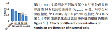
2.1 氯化血红素对神经元增殖的影响 为了探索构建脑出血体外模型所用到氯化血红素的有效浓度,实验选用不同浓度(0,10,25,50,100 μmol/L) 氯化血红素处理共培养细胞24 h,MTT实验结果显示,细胞活力随着氯化血红素浓度上升呈下降的趋势,与空白对照组相比,50 μmol/L氯化血红素对神经细胞增殖活力有明显的抑制作用,差异有显著性意义(P < 0.001),见图1。与100 μmol/L氯化血红素组相比,50 μmol/L氯化血红素作为安全范围内的最低浓度,对神经细胞增殖活力的抑制更有效,差异有显著性意义(P < 0.05),见图1。"
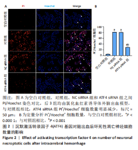
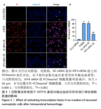
2.2 沉默ATF4对脑出血后神经元坏死性凋亡数量的影响 与空白对照组相比,其余3组神经细胞坏死性凋亡数量(PI+/Hoechst+)明显增多(P < 0.000 1);与对照组相比,NC siRNA组PI+/Hoechst+细胞数量无明显差异;与对照组相比,ATF4 siRNA组神经细胞坏死性凋亡数量明显减少(P < 0.001),见图2。"

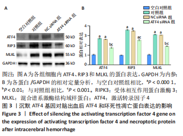
2.3 沉默ATF4对脑出血后神经细胞坏死性凋亡蛋白表达的影响 与空白对照组相比,用氯化血红素构建脑出血体外模型24 h后,ATF4蛋白表达明显升高(P < 0.000 1);与对照组相比,ATF4 siRNA组ATF4蛋白表达被明显抑制(P < 0.001),见图3。为了进一步明确沉默ATF4,对脑出血后神经细胞坏死性凋亡蛋白表达的影响,由Western blot结果可以看出,与空白对照组相比,对照组、NC siRNA组和ATF4 siRNA组的神经细胞坏死性凋亡蛋白(RIP3、MLKL)表达明显上升(P < 0.000 1);与对照组相比,ATF4 siRNA组的坏死性凋亡蛋白表达明显被抑制(P < 0.001),见图3。"
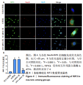
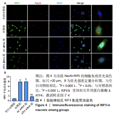
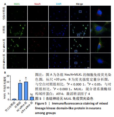
2.4 沉默ATF4对脑出血后神经元坏死性凋亡的调控作用 为了将ATF4对脑出血后神经细胞坏死性凋亡的调控作用进一步定位于神经元,而不是对小胶质细胞调控而产生的效应,使用双重免疫荧光染色(NeuN+坏死性凋亡蛋白,NeuN是神经元的标志性抗体)。各组NeuN、RIP3染色图片见图4A,首先通过NeuN阳性细胞定位为神经元,再对各组之间RIP3荧光密度进行对比,脑出血后神经元坏死性凋亡较空白对照组明显升高(P < 0.000 1),而ATF4 siRNA组的神经元坏死性凋亡较对照组明显降低(P < 0.000 1),见图4B;MLKL的免疫荧光染色结果同RIP3,见图5A,B。上述结果进一步证实了ATF4对脑出血后神经元坏死性凋亡具有调控作用。"

"
| [1] SHETH KN. Spontaneous Intracerebral Hemorrhage. N Engl J Med. 2022;387(17):1589-1596. [2] KASE CS, HANLEY DF. Intracerebral Hemorrhage: Advances in Emergency Care. Neurol Clin. 2021;39(2):405-418. [3] KIRSHNER H, SCHRAG M. Management of Intracerebral Hemorrhage: Update and Future Therapies. Curr Neurol Neurosci Rep. 2021; 21(10):57. [4] XUE M, YONG VW. Neuroinflammation in intracerebral haemorrhage: immunotherapies with potential for translation. Lancet Neurol. 2020; 19(12):1023-1032. [5] FRANK D, VINCE JE. Pyroptosis versus necroptosis: similarities, differences, and crosstalk. Cell Death Differ. 2019;26(1):99-114. [6] NAYAK D, ROTH TL, MCGAVERN DB. Microglia development and function. Annu Rev Immunol. 2014;32:367-402. [7] LI X, ZHANG M, HUANG X, et al. Ubiquitination of RIPK1 regulates its activation mediated by TNFR1 and TLRs signaling in distinct manners. Nat Commun. 2020;11(1):6364. [8] YUAN J, AMIN P, OFENGEIM D. Necroptosis and RIPK1-mediated neuroinflammation in CNS diseases. Nat Rev Neurosci. 2019;20(1):19-33. [9] CHEN AQ, FANG Z, CHEN XL, et al. Microglia-derived TNF-α mediates endothelial necroptosis aggravating blood brain-barrier disruption after ischemic stroke. Cell Death Dis. 2019;10(7):487. [10] DENG XX, LI SS, SUN FY. Necrostatin-1 Prevents Necroptosis in Brains after Ischemic Stroke via Inhibition of RIPK1-Mediated RIPK3/MLKL Signaling. Aging Dis. 2019;10(4):807-817. [11] LI J, ZHANG J, ZHANG Y, et al. TRAF2 protects against cerebral ischemia-induced brain injury by suppressing necroptosis. Cell Death Dis. 2019; 10(5):328. [12] KRUKOWSKI K, NOLAN A, FRIAS ES, et al. Small molecule cognitive enhancer reverses age-related memory decline in mice. Elife. 2020;9: e62048. [13] YANG T, ZHANG Y, CHEN L, et al. The potential roles of ATF family in the treatment of Alzheimer’s disease. Biomed Pharmacother. 2023; 161:114544. [14] NAKAGAWA T, OHTA K. Quercetin Regulates the Integrated Stress Response to Improve Memory. Int J Mol Sci. 2019;20(11):2761. [15] KEERTHIGA R, PEI DS, FU A. Mitochondrial dysfunction, UPRmt signaling, and targeted therapy in metastasis tumor. Cell Biosci. 2021; 11(1):186. [16] WU X, LUO J, LIU H, et al. Recombinant adiponectin peptide promotes neuronal survival after intracerebral haemorrhage by suppressing mitochondrial and ATF4-CHOP apoptosis pathways in diabetic mice via Smad3 signalling inhibition. Cell Prolif. 2020;53(2):e12759. [17] KARUPPAGOUNDER SS, ALIM I, KHIM SJ, et al. Therapeutic targeting of oxygen-sensing prolyl hydroxylases abrogates ATF4-dependent neuronal death and improves outcomes after brain hemorrhage in several rodent models. Sci Transl Med. 2016;8(328):328ra29. [18] ZHANG J, ZHANG P, MENG C, et al. The PERK Pathway Plays a Neuroprotective Role During the Early Phase of Secondary Brain Injury Induced by Experimental Intracerebral Hemorrhage. Acta Neurochir Suppl. 2020;127:105-119. [19] JHA RM, KOCHANEK PM, SIMARD JM. Pathophysiology and treatment of cerebral edema in traumatic brain injury. Neuropharmacology. 2019;145(Pt B):230-246. [20] CHEN S, PENG J, SHERCHAN P, et al. TREM2 activation attenuates neuroinflammation and neuronal apoptosis via PI3K/Akt pathway after intracerebral hemorrhage in mice. J Neuroinflammation. 2020; 17(1):168. [21] YIN K, LU H, ZHANG Y, et al. Secondary brain injury after polystyrene microplastic-induced intracerebral hemorrhage is associated with inflammation and pyroptosis. Chem Biol Interact. 2022;367:110180. [22] PENG C, FU X, WANG K, et al. Dauricine alleviated secondary brain injury after intracerebral hemorrhage by upregulating GPX4 expression and inhibiting ferroptosis of nerve cells. Eur J Pharmacol. 2022;914:174461. [23] BOBINGER T, BURKARDT P, B HUTTNER H, et al. Programmed Cell Death after Intracerebral Hemorrhage. Curr Neuropharmacol. 2018; 16(9):1267-1281. [24] ZHANG Y, KHAN S, LIU Y, et al. Modes of Brain Cell Death Following Intracerebral Hemorrhage. Front Cell Neurosci. 2022;16:799753. [25] ROBINSON N, GANESAN R, HEGEDŰS C, et al. Programmed necrotic cell death of macrophages: Focus on pyroptosis, necroptosis, and parthanatos. Redox Biol. 2019;26:101239. [26] TONG X, TANG R, XIAO M, et al. Targeting cell death pathways for cancer therapy: recent developments in necroptosis, pyroptosis, ferroptosis, and cuproptosis research. J Hematol Oncol. 2022;15(1):174. [27] HUAN Y, WU XQ, CHEN T, et al. Necroptosis plays a crucial role in the exacerbation of retinal injury after blunt ocular trauma. Neural Regen Res. 2023;18(4):922-928. [28] HSU SK, LI CY, LIN IL, et al. Inflammation-related pyroptosis, a novel programmed cell death pathway, and its crosstalk with immune therapy in cancer treatment. Theranostics. 2021;11(18):8813-8835. [29] COLONNA M, BUTOVSKY O. Microglia Function in the Central Nervous System During Health and Neurodegeneration. Annu Rev Immunol. 2017;35:441-468. [30] SUBHRAMANYAM CS, WANG C, HU Q, et al. Microglia-mediated neuroinflammation in neurodegenerative diseases. Semin Cell Dev Biol. 2019;94:112-120. [31] JIN X, LIU MY, ZHANG DF, et al. Baicalin mitigates cognitive impairment and protects neurons from microglia-mediated neuroinflammation via suppressing NLRP3 inflammasomes and TLR4/NF-κB signaling pathway. CNS Neurosci Ther. 2019;25(5):575-590. [32] CORNELL J, SALINAS S, HUANG HY, et al. Microglia regulation of synaptic plasticity and learning and memory. Neural Regen Res. 2022;17(4): 705-716. [33] UMPIERRE AD, WU LJ. How microglia sense and regulate neuronal activity. Glia. 2021;69(7):1637-1653. [34] ALMEIDA LM, PINHO BR, DUCHEN MR, et al. The PERKs of mitochondria protection during stress: insights for PERK modulation in neurodegenerative and metabolic diseases. Biol Rev Camb Philos Soc. 2022;97(5):1737-1748. [35] MENG C, ZHANG J, DANG B, et al. PERK Pathway Activation Promotes Intracerebral Hemorrhage Induced Secondary Brain Injury by Inducing Neuronal Apoptosis Both in Vivo and in Vitro. Front Neurosci. 2018; 12:111. [36] FONT-BELMONTE E, UGIDOS IF, SANTOS-GALDIANO M, et al. Post-ischemic salubrinal administration reduces necroptosis in a rat model of global cerebral ischemia. J Neurochem. 2019;151(6):777-794. [37] ESTORNES Y, AGUILETA MA, DUBUISSON C, et al. RIPK1 promotes death receptor-independent caspase-8-mediated apoptosis under unresolved ER stress conditions. Cell Death Dis. 2015;6(6):e1798. [38] JIANG H, WANG C, ZHANG A, et al. ATF4 protects against sorafenib-induced cardiotoxicity by suppressing ferroptosis. Biomed Pharmacother. 2022;153:113280. [39] WANG X, ZHANG G, DASGUPTA S, et al. ATF4 Protects the Heart From Failure by Antagonizing Oxidative Stress. Circ Res. 2022;131(1):91-105. [40] SZEWCZYK MM, LUCIANI GM, VU V, et al. PRMT5 regulates ATF4 transcript splicing and oxidative stress response. Redox Biol. 2022;51: 102282. |
| [1] | Yue Yun, Wang Peipei, Yuan Zhaohe, He Shengcun, Jia Xusheng, Liu Qian, Li Zhantao, Fu Huiling, Song Fei, Jia Menghui. Effects of croton cream on JNK/p38 MAPK signaling pathway and neuronal apoptosis in cerebral ischemia-reperfusion injury rats [J]. Chinese Journal of Tissue Engineering Research, 2024, 28(8): 1186-1192. |
| [2] | Li Longyang, Zhang Songjiang, Zhao Xianmin, Zhou Chunguang, Gao Jianfeng. Electroacupuncture intervention on the proliferation and differentiation of hippocampal neurons and oligodendrocytes in Alzheimer’s disease model mice [J]. Chinese Journal of Tissue Engineering Research, 2024, 28(7): 1029-1035. |
| [3] | Chen Zepeng, Hou Yonghui, Chen Shudong, Hou Yu, Lin Dingkun. Tauroursodeoxycholic acid treats spinal cord injury by reducing apoptosis of spinal cord neurons under glucose and oxygen deprivation [J]. Chinese Journal of Tissue Engineering Research, 2024, 28(4): 528-534. |
| [4] | Chen Simin, Hu Yingjun, Yan Wenrui, Ji Le, Shao Mengli, Sun Ze, Zheng Hongxing, Qi Shanshan. Establishment and evaluation of a streptozotocin-induced diabetic encephalopathy rat model [J]. Chinese Journal of Tissue Engineering Research, 2024, 28(2): 237-241. |
| [5] | Li Jiahui, Qi Xue, Zhu Yuanfeng, Yu Lu, Liu Lifeng, Wang Peng. Protective effect of C2 ceramide on dopaminergic neurons in a mouse model of Parkinson’s disease [J]. Chinese Journal of Tissue Engineering Research, 2024, 28(11): 1653-1659. |
| [6] | Liao Yidong, Ming Jiang, Song Wenxue, Wang Zili, Zhang Yu, Liao Yifei, Xu Kaya, Yang Hua. An experimental method for simultaneously culturing primary cortical and hippocampal neurons [J]. Chinese Journal of Tissue Engineering Research, 2023, 27(6): 897-902. |
| [7] | Hao Liufang, Duan Hongmei, Wang Zijue, Hao Fei, Hao Peng, Zhao Wen, Gao Yudan, Yang Zhaoyang, Li Xiaoguang. Spatiotemporal dynamic changes of ependymal cells after spinal cord injury in transgenic mice [J]. Chinese Journal of Tissue Engineering Research, 2023, 27(6): 883-889. |
| [8] | Yuan Changshen, Guan Yanbing, Li Zhe, Rong Weiming, Liao Shuning, Chen Lewei, Mei Qijie, Duan Kan. Screening and verification of key genes of necroptosis in osteoarthritis [J]. Chinese Journal of Tissue Engineering Research, 2023, 27(5): 695-700. |
| [9] | Zhao Siqi, Du Juan, Qu Haifeng, Li Jianmin, Zhang Yuxin, Liu Junjie. Effects of enriched environment combined with melatonin on learning and memory function and brain neuron apoptosis in SAMP8 mice [J]. Chinese Journal of Tissue Engineering Research, 2023, 27(5): 701-706. |
| [10] | Chen Guodong, Zheng Meiyan, Zhang Peng, Wang Zhenchao, Jin Lixin. Changes in sensory neurons and astrocytes and the expression of interleukin 1beta and glial fibrillary acidic protein in the rat spinal cord after selective dorsal rhizotomy [J]. Chinese Journal of Tissue Engineering Research, 2023, 27(5): 726-731. |
| [11] | Guo Yongjuan, Zhang Li, Lu Huamei. Effects of sevoflurane combined with propofol on pain mediators and neuronal activity in rats with spinal cord fracture [J]. Chinese Journal of Tissue Engineering Research, 2023, 27(36): 5850-5855. |
| [12] | Luo Zhangrong, Cao Liang, Zhang Yi, Pi Wenjun, Li Qing. Characteristics of neural stem cells of cerebrospinal fluid-contacting neurons verified by multimodal imaging molecules in vitro [J]. Chinese Journal of Tissue Engineering Research, 2023, 27(34): 5505-5509. |
| [13] | Zhang Huiyu, Yu Jingwen, Bai Zhenjun, Li Liang, Mu Bingtao, Zhang Jinfeng, Xie Jiawei. Triptolide protects damaged neurons by regulating microglial polarization [J]. Chinese Journal of Tissue Engineering Research, 2023, 27(33): 5342-5347. |
| [14] | Xu Luchun, Yang Yongdong, Zhao He, Zhong Wenqing, Ma Yukun, Yu Xing. Effect and mechanism of non-coding RNA in regulating neuronal apoptosis after spinal cord injury [J]. Chinese Journal of Tissue Engineering Research, 2023, 27(33): 5404-5412. |
| [15] | Lu Xiaojun, Xiong Bohan, Yang Tengyun, Wang Xu, Zhang Yaozhang, Zhong Ruiying, Li Yanlin. Novel programmed cell death of chondrocytes in osteoarthritis [J]. Chinese Journal of Tissue Engineering Research, 2023, 27(28): 4571-4576. |
| Viewed | ||||||
|
Full text |
|
|||||
|
Abstract |
|
|||||