[1] WANG W, YEUNG KWK. Bone grafts and biomaterials substitutes for bone defect repair: A review. Bioact Mater. 2017;2(4):224-247.
[2] 杨思敏,王新卫.自体骨移植修复骨缺损的临床研究进展[J].中国疗养医学,2019,28(90):945-948.
[3] STARCH-JENSEN T, AHMAD M, BRUUN NH, et al. Patient’s perception of recovery after maxillary sinus floor augmentation with autogenous bone graft compared with composite grafts: a single-blinded randomized controlled trial. Int J Implant Dent. 2021;7(1):99.
[4] 黄涛,孟志斌,金大地,等.生物珊瑚人工骨支架材料生物相容性检测[J].中国现代医学杂志,2012,22(11):36-41.
[5] 谭海涛,孟志斌,李俊,等.β磷酸三钙和α半水硫酸钙复合人工骨生物相容性及在脊柱融合模型中的应用[J].中国组织工程研究, 2017,21(26):4119-4124
[6] 王挺锐,孟志斌,李俊.人工珊瑚骨治疗脊柱结核临床观察[J].中国热带医学,2008,8(9):1526-1547.
[7] NOVACK DV, TEITELBAUM SL. The osteoclast: friend or foe?. Annu Rev Pathol. 2008;3:457-484.
[8] GUILLEMIN G, PATAT JL, FOURNIE J, et al. The use of coral as a bone graft substitute. J Biomed Mater Res. 1987;21(5):557-567.
[9] 李超,张贤,邵家豪.骨代谢过程中钙离子通道TRPV5、TRPV6的作用[J].中国组织工程研究,2022,26(12):1950-1955.
[10] SONG T, LIN T, MA J, et al. Regulation of TRPV5 transcription and expression by E2/ERα signalling contributes to inhibition of osteoclastogenesis. J Cell Mol Med. 2018;22(10):4738-4750.
[11] CHAMOUX E, BISSON M, PAYET MD, et al. TRPV-5 mediates a receptor activator of NF-kappaB (RANK) ligand-induced increase in cytosolic Ca2+ in human osteoclasts and down-regulates bone resorption. J Biol Chem. 2010;285(33):25354-25362.
[12] VAN DER EERDEN BC, HOENDEROP JG, DE VRIES TJ, et al. The epithelial Ca2+ channel TRPV5 is essential for proper osteoclastic bone resorption. Proc Natl Acad Sci U S A. 2005;102(48):17507-17512.
[13] LIEDTKE W, CHOE Y, MARTÍ-RENOM MA, et al. Vanilloid receptor-related osmotically activated channel (VR-OAC), a candidate vertebrate osmoreceptor. Cell. 2000;103(3):525-535.
[14] ROSCHER A, HASEGAWA T, DOHNKE S, et al. The F-actin modulator SWAP-70 controls podosome patterning in osteoclasts. Bone Rep. 2016;5:214-221.
[15] WHITE RA, WEBER JN, WHITE EW. Replamineform: a new process for preparing porous ceramic, metal, and polymer prosthetic materials. Science. 1972;176(4037):922-924.
[16] SARTORIS DJ, HOLMES RE, BUCHOLZ RW, et al. Coralline hydroxyapatite bone-graft substitutes in a canine diaphyseal defect model. Radiographic-histometric correlation. Invest Radiol. 1987;22(7):590-596.
[17] KOËTER S, TIGCHELAAR SJ, FARLA P, et al. Coralline hydroxyapatite is a suitable bone graft substitute in an intra-articular goat defect model. J Biomed Mater Res B Appl Biomater. 2009;90(1):116-122.
[18] DAY AGE, FRANCIS WR, FU K, et al. Osteogenic Potential of Human Umbilical Cord Mesenchymal Stem Cells on Coralline Hydroxyapatite/Calcium Carbonate Microparticles. Stem Cells Int. 2018;2018:4258613.
[19] BEGLEY CT, DOHERTY MJ, HANKEY DP, et al. The culture of human osteoblasts upon bone graft substitutes. Bone. 1993;14(4):661-666.
[20] HAUGEN HJ, LYNGSTADAAS SP, ROSSI F, et al. Bone grafts: which is the ideal biomaterial? J Clin Periodontol. 2019;46 Suppl 21:92-102.
[21] ZHAO R, YANG R, COOPER PR, et al. Bone Grafts and Substitutes in Dentistry: A Review of Current Trends and Developments. Molecules. 2021;26(10):3007.
[22] XIA YJ, WANG W, XIA H, et al. Preparation of Coralline Hydroxyapatite Implant with Recombinant Human Bone Morphogenetic Protein-2-Loaded Chitosan Nanospheres and Its Osteogenic Efficacy. Orthop Surg. 2020;12(6):1947-1953.
[23] 付 昆,孟志斌,李 俊,等.珊瑚羟基磷灰石植入修复良性骨肿瘤缺损[J].中南大学学报(医学版),2008,33(5):421-424.
[24] 李 俊,孟志斌,周健强.一期前路手术内固定珊瑚人工骨植入治疗脊柱结核[J].颈腰痛杂志,2007,28(3):177-178.
[25] KHAN MUA, RAZAK SIA, ANSARI MNM, et al. Development of Biodegradable Bio-Based Composite for Bone Tissue Engineering: Synthesis, Characterization and In Vitro Biocompatible Evaluation. Polymers (Basel). 2021;13(21):3611.
[26] KITAURA H, MARAHLEH A, OHORI F, et al. Osteocyte-Related Cytokines Regulate Osteoclast Formation and Bone Resorption. Int J Mol Sci. 2020;21(14):5169.
[27] KONG L, SMITH W, HAO D. Overview of RAW264.7 for osteoclastogensis study: Phenotype and stimuli. J Cell Mol Med. 2019;23(5):3077-3087.
[28] SONG C, YANG X, LEI Y, et al. Evaluation of efficacy on RANKL induced osteoclast from RAW264.7 cells. J Cell Physiol. 2019;234(7):11969-11975.
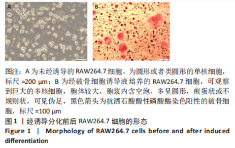
[29] 许丹, 律颖, 朱小语.小鼠单核巨噬细胞白血病细胞(RAW264.7)的培养及其在诱导破骨细胞中的应用[J]. 中国骨质疏松杂志,2016, 22(10):1355-1360.
[30] WIDBILLER M, ROTHMAIER C, SALITER D, et al. Histology of human teeth: Standard and specific staining methods revisited. Arch Oral Biol. 2021;127:105136.
[31] WU L, LUO Z, LIU Y, et al. Aspirin inhibits RANKL-induced osteoclast differentiation in dendritic cells by suppressing NF-κB and NFATc1 activation. Stem Cell Res Ther. 2019;10(1):375.
[32] KAJIYA H. Calcium signaling in osteoclast differentiation and bone resorption. Adv Exp Med Biol. 2012;740:917-932.
[33] UDAGAWA N, KOIDE M, NAKAMURA M, et al. Osteoclast differentiation by RANKL and OPG signaling pathways. J Bone Miner Metab. 2021; 39(1):19-26.
[34] RYU S, MCDONNELL K, CHOI H, et al. Suppression of miRNA-708 by polycomb group promotes metastases by calcium-induced cell migration. Cancer Cell. 2013;23(1):63-76.
[35] XU J, ZEUG A, RIEDERER B, et al. Calcium-sensing receptor regulates intestinal dipeptide absorption via Ca2+ signaling and IKCa activation. Physiol Rep. 2020;8(1):e14337.
[36] HUGHES TET, LODOWSKI DT, HUYNH KW, et al. Structural basis of TRPV5 channel inhibition by econazole revealed by cryo-EM. Nat Struct Mol Biol. 2018;25(1):53-60.
[37] TANG D, LIU X, CHEN K, et al. Cytoplasmic PCNA is located in the actin belt and involved in osteoclast differentiation. Aging (Albany NY). 2020; 12(13):13297-13317.
[38] VAN DE GRAAF SF, CHANG Q, MENSENKAMP AR, Et al. Direct interaction with Rab11a targets the epithelial Ca2+ channels TRPV5 and TRPV6 to the plasma membrane. Mol Cell Biol. 2006;26(1):303-312.
[39] KASPER D, PLANELLS-CASES R, FUHRMANN JC, et al. Loss of the chloride channel ClC-7 leads to lysosomal storage disease and neurodegeneration. EMBO J. 2005;24(5):1079-1091.
[40] NIJENHUIS T, VAN DER EERDEN BC, HOENDEROP JG, et al. Bone resorption inhibitor alendronate normalizes the reduced bone thickness of TRPV5(-/-) mice. J Bone Miner Res. 2008;23(11):1815-1824. |