Chinese Journal of Tissue Engineering Research ›› 2023, Vol. 27 ›› Issue (20): 3117-3122.doi: 10.12307/2023.148
Digital morphology of foramen magnum and occipital condyle: an observation based on three-dimensional reconstruction
Wang Jian1, Wang Xing1, 2, Li Kun1, 2, Gao Shang1, Wang Chaoqun3, Zhang Shaojie1, 2, Li Zhijun1, 2
- 1Human Anatomy Teaching and Research Section, 2Digital Medicine Center, 3Imaging Center of Affiliated Hospital, Inner Mongolia Medical University, Hohhot 010110, Inner Mongolia Autonomous Region, China
-
Received:2021-12-08Accepted:2022-05-21Online:2023-07-18Published:2022-11-18 -
Contact:Li Zhijun, Professor, Doctoral supervisor, Human Anatomy Teaching and Research Section, Inner Mongolia Medical University, Hohhot 010110, Inner Mongolia Autonomous Region, China; Digital Medicine Center, Inner Mongolia Medical University, Hohhot 010110, Inner Mongolia Autonomous Region, China Zhang Shaojie, Professor, Master’s supervisor, Human Anatomy Teaching and Research Section, Inner Mongolia Medical University, Hohhot 010110, Inner Mongolia Autonomous Region, China; Digital Medicine Center, Inner Mongolia Medical University, Hohhot 010110, Inner Mongolia Autonomous Region, China -
About author:Wang Jian, Master candidate, Human Anatomy Teaching and Research Section, Inner Mongolia Medical University, Hohhot 010110, Inner Mongolia Autonomous Region, China Wang Xing, MD, Human Anatomy Teaching and Research Section, Inner Mongolia Medical University, Hohhot 010110, Inner Mongolia Autonomous Region, China; Digital Medicine Center, Inner Mongolia Medical University, Hohhot 010110, Inner Mongolia Autonomous Region, China -
Supported by:the National Natural Science Foundation of China, Nos. 81860383 (to LZJ) and 81860382 (to WX); the Natural Science Foundation of Inner Mongolia Autonomous Region, Nos. 2020LH08021 (to LZJ), 2020MS03061 (to WX), and 2019MS08017 (to ZSJ); the College Youth Science and Technology Talent Support Program in Inner Mongolia Autonomous Region, No. NJYT22009 (to WX); Inner Mongolia Autonomous Region Science and Technology Plan Project, No. 2019GG158 (to WX); Inner Mongolia Medical University Key Research Project, No. YKD2021ZD011 (to WX); Medical and Health Science and Technology Plan Project of Inner Mongolia Autonomous Region Health Commission, No. 202201217 (to WX); Inner Mongolia Medical University Youth Fund Project, No. YKD2020QNCX055 (to LK)
CLC Number:
Cite this article
Wang Jian, Wang Xing, Li Kun, Gao Shang, Wang Chaoqun, Zhang Shaojie, Li Zhijun. Digital morphology of foramen magnum and occipital condyle: an observation based on three-dimensional reconstruction[J]. Chinese Journal of Tissue Engineering Research, 2023, 27(20): 3117-3122.
share this article
Add to citation manager EndNote|Reference Manager|ProCite|BibTeX|RefWorks
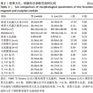
2.1 参与者数量分析 符合纳入标准的样本共673例,其中男448例,女225例,男女各分为5个年龄组。 2.2 枕骨大孔形态参数整体结果 通过分析发现,枕骨大孔指数整体男性小于女性,且性别间无差异(P > 0.05);枕骨大孔最大长度、枕骨大孔最大宽度、FMAR、FMAT均为男性大于女性,且性别间差异有显著性意义(P < 0.05);在男、女中枕骨大孔最大长度>枕骨大孔最大宽度,即枕骨大孔长大于宽;且FMAT在男、女中均大于 FMAR,见表2。"
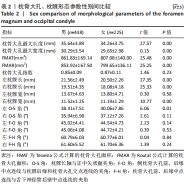
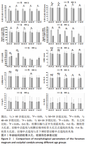
2.3 枕髁形态参数整体结果 参数分析发现男性左枕髁宽整体小于女性,但性别间无统计学差异(P > 0.05);男性左枕髁长、右枕髁长、右枕髁宽指标均大于女性,且性别间差异有显著性意义(P < 0.05);男性左O-S角、右O-S角、右F-H角整体均小于女性,但仅左O-S角在性别间差异有显著性意义(P < 0.05);男性左F-O角、右F-O角、左F-H角均大于女性,但均无性别间的差异(P > 0.05)。通过枕髁指标在左、右侧别间的分析发现,枕髁长、枕髁宽、O-S角、F-O角在侧别间差异均有显著性意义(P < 0.05),且左枕髁长、左枕髁宽、左O-S角均大于右侧,左F-O角、左F-H角均小于右侧,见表2。 2.4 枕骨大孔形态参数年龄分组结果 枕骨大孔形态学参数的年龄组分析发现枕骨大孔最大长度总体随年龄的增长呈波浪状递增趋势,最大值在50-59岁组,最小值在20-29岁组;枕骨大孔最大宽度、FMAT、FMAR、枕骨大孔指数总体随年龄的增长呈波浪状递减趋势,最大值在30-39岁组,最小值在≥60岁组。男性枕骨大孔最大长度、FMAT、FMAR在所有年龄组都大于女性,枕骨大孔最大长度在20-29岁组、30-39岁组、50-59岁组内有男女性别间的差异(P < 0.05),枕骨大孔最大宽度、FMAT、FMAR在20-29岁组、30-39岁组、40-49岁组、50-59岁组内有男女性别间差异(P < 0.05);枕骨大孔指数在所有年龄组中都无性别间的差异(P > 0.05),见图2。"
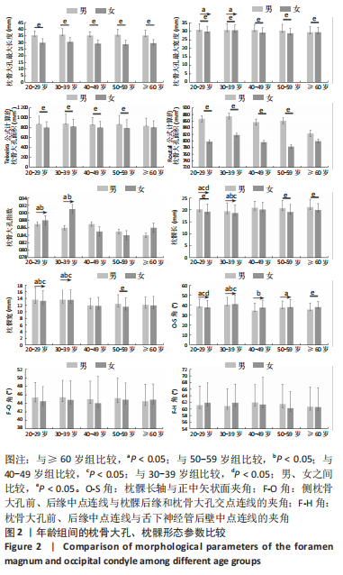

2.5 枕髁形态参数年龄分组结果 枕髁形态学参数的年龄组分析发现,枕髁长总体随年龄的增长呈波浪状递增趋势,最大值在≥60岁组,最小值在30-39岁组,枕髁长总体在20-29岁组、50-59岁组、≥60岁组内均有性别间差异(P < 0.05);枕髁宽总体随年龄的增长呈波浪状递减趋势,最大值在30-39岁组,最小值在40-49岁组,枕髁宽总体只在50-59岁组内有性别间差异(P < 0.05)。 左枕髁长、右枕髁宽总体随年龄的增长变化不明显,在各年龄组间总体值接近;右枕髁长总体随年龄的增长呈波浪状递增趋势,最大值在40-49岁组,最小值在30-39岁组;左枕髁宽总体随年龄的增长呈波浪状递减趋势,最大值在30-39岁组,最小值在40-49岁组。男性枕髁长在所有年龄组都大于女性,左枕髁长在20-29岁组、30-39岁组、50-59岁组、≥60岁组内均有性别间差异(P < 0.05);右枕髁长在50-59岁组、≥60岁组内均有性别间差异(P < 0.05);左枕髁宽只在50-59岁组有性别间差异(P < 0.05);右枕髁宽在40-49岁组、50-59岁组、≥60岁组内均有性别间差异(P < 0.05)。 O-S角总体、F-O角总体随年龄的增长变化呈平缓递减趋势,最大值在30-39岁组,最小值在40-49岁组;F-H角总体随年龄的增长呈波浪状递减趋势,最大值在30-39岁组,最小值在≥60岁组。O-S角总体只在≥60岁组内有性别间差异(P < 0.05)。F-O角总体、F-H角总体在任何年龄组内均无性别间差异(P > 0.05)。 左O-S角只在40-49岁组内有性别间差异(P < 0.05);右O-S角只在60岁以上组内有性别间差异(P < 0.05);枕骨大孔最大宽度、枕骨大孔指数、右枕髁长、枕髁宽、O-S角、F-H角在不同年龄组间比较差异均有显著性意义(P < 0.05) ,见图2。"

| [1] VASQUEZ C, YANG A, YOUSSEF AS. Foramen Magnum Meningioma: Far Lateral Approach. J Neurol Surg B Skull Base. 2019;80(Suppl 4): S363-S364. [2] Beer-Furlan A, Vellutini EA, Balsalobre L, et al. Endoscopic Endonasal Approach to Ventral Posterior Fossa Meningiomas: From Case Selection to Surgical Management. Neurosurg Clin N Am. 2015; 26(3):413-426. [3] AMELOT A, TERRIER LM, LOT G. Craniovertebral Junction Transoral Approach: Predictive Factors of Complications. World Neurosurg. 2018; 110:568-574. [4] Heros RC. Lateral suboccipital approach for vertebral and vertebrobasilar artery lesions. J Neurosurg. 1986;64(4):559-562. [5] NANDA A, VINCENT DA, VANNEMREDDY PS, et al. Far-lateral approach to intradural lesions of the foramen magnum without resection of the occipital condyle. J Neurosurg. 2002;96(2):302-309. [6] BASSIOUNI H , NTOUKAS V, ASGARI S, et al. Foramen magnum meningiomas: clinical outcome after microsurgical resection via a posterolateral suboccipital retrocondylar approach. Neurosurgery. 2006;59(6):1177-1185. [7] BOULTON MR , CUSIMANO MD. Foramen magnum meningiomas: concepts, classifications, and nuances. Neurosurg Focus. 2003;14(6): e10. [8] BOLZ S, GAPERT R, HARTWIG S, et al. Evaluation of foramen magnum sexual dimorphism in a modern documented German population using post-mortem computed tomography. Forensic Imaging. 2020; 21:200352. [9] TONEVA D, NIKOLOVA S, HARIZANOV S, et al. Sex estimation by size and shape of foramen magnum based on CT imaging. Leg Med (Tokyo). 2018;35:50-60. [10] TEIXEIRA WR. Sex identification utilizing the size of the foramen magnum. Am J Forensic Med Pathol. 1982;3(3):203-206. [11] ROUTAL RR, PAL GP, BHAGAWAT SS, et al. Metrical studies with sexual dimorphism in foramen magnum of human crania. J Anat Soc India. 1984;2(33):85-89. [12] GARGI V, RAVI PRAKASH SM, SANGEETA MALIK S, et al. Sexual Dimorphism of Foramen Magnum between Two Different Groups of Indian Population: A CrossSectional ConeBeam Computed Tomography Study. J Forens Sci Med. 2021;4(3):150-155. [13] AGARWAL HKS, SETIA PS, PANDEY S. Virtual Determination of Sex: Estimating Cut off Value of Digital Metric Traits of Foramen Magnum on Threedimensional Computed Tomography with Receiver Operating Characteristic and Logistic Regression Analysis. J Forens Sci Med. 2021; 1(7):1-8. [14] ZDILLA MJ, RUSSELL ML, BLISS KN, et al. The size and shape of the foramen magnum in man. J Craniovertebr Junction Spine. 2017;8(3): 205-221. [15] GONZÁLEZ-COLMENARES G, SANABRIA MEDINA C, ROJAS-SÁNCHEZ MP, et al. Sex estimation from skull base radiographs in a contemporary Colombian population. J Forensic Leg Med. 2019;62:77-81. [16] GONZÁLEZ-COLMENARES G, MEDINA CS, ROJAS-SÁNCHEZ MP. Sexual Dimorphism in the Foramen Magnum of Nigerian Adult. Int J Biol Med Res. 2011;2(4):878-881. [17] 朱祥清.利用枕骨大孔和乳突进行性别判别的研究[D].昆明:昆明医科大学,2017. [18] HEND MH, ABDEL-RAHMAN RH, EL-HAWARY G, et al. Sexual dimorphism of foramen magnum: An Egyptian study. Egypt J Forensic Sci. 2020;10(3):1715-1722. [19] GAPERT R, BLACK S, LAST J. Sex determination from the foramen magnum: discriminant function analysis in an eighteenth and nineteenth century British sample. Int J Legal Med. 2009;123(1):25-33. [20] LUCENA JD, SANDERS J, BRITO HD, et al. Morphometric Analysis of the Foramen Magnum in Dry Human Skulls in Northeastern Brazil. J Morphol Sci. 2019;36:97-104. [21] UTHMAN AT, AL-RAWI NH, AL-TIMIMI JF. Evaluation of foramen magnum in gender determination using helical CT scanning. Dentomaxillofac Radiol. 2012;41(3):197-202. [22] MOODLEY M, RENNIE C, LAZARUS L, et al. The Morphometry and Morphology of the Foramen Magnum In Age And Sex Determination Within The South African Black Population Utilizing Computer Tomography (CT) Scans. Int J Morphol. 2019;37(1):251-257. [23] KUMAR NP, PRIYANKA EN, KOLAPPAN R, et al. Morphometric analysis of foramen magnum using helical computed tomogrophy for gender determination. World J Adv Res Rev. 2020;8(3):1-6. [24] MADADIN M, MENEZES RG, AL SAIF HS, et al. Morphometric evaluation of the foramen magnum for sex determination: A study from Saudi Arabia. J Forensic Leg Med. 2017;46:66-71. [25] LE TV, DAKWAR E, HAN S, et al. Computed tomography-based morphometric analysis of the human occipital condyle for occipital condyle-cervical fusion. J Neurosurg Spine. 2011;15(3):328-331. [26] WANIBUCHI M, FUKUSHIMA T, ZENGA F, et al. Simple identification of the third segment of the extracranial vertebral artery by extreme lateral inferior transcondylar-transtubercular exposure (ELITE). Acta Neurochir (Wien). 2009;151(11):1499-1503. [27] 胡海建.远外侧入路中枕髁相关解剖学参数的CT影像研究[D].新乡:新乡医学院,2020. [28] REHAB I, ABDEL-KARIM, HOUSSEINI AM, et al. Adult sex estimation using Three Dimensional Volume Rendering Multislice Computed Tomography of the foramen magnum and occipital condyles: A study in Egyptian population. Int J Adv Res. 2015;3(5):1212-1215. [29] CHOVALOPOULOU ME, BERTSATOS A, CARDOSO H. Estimating Sex of Modern Greeks Based on the Foramen Magnum Region. J Anthropology. 2017;2017:1-7. [30] ALJARRAH K, PACKIRISAMY V, AL ANAZI N, et al. Morphometric analysis of foramen magnum and occipital condyle using CT images for sex determination in a Saudi Arabian population. Morphologie. 2021;11: S1286-0115(21)192-192. [31] KALTHUR SG, PADMASHALI S, GUPTA C, et al. Anatomic study of the occipital condyle and its surgical implications in transcondylar approach. J Craniovertebr Junction Spine. 2014;5(2):71-77. [32] ZHOU J, ESPINOZA ORÍAS AA, KANG X, et al. CT-based morphometric analysis of the occipital condyle: focus on occipital condyle screw insertion. J Neurosurg Spine. 2016;25(5):572-579. [33] GUMUSSOY I, DUMAN SB. Morphometric analysis of occipital condyles using alternative imaging technique. Surg Radiol Anat. 2020;42(2): 161-169. [34] KIRNAZ S, GERGES MM, RUMALLA K, et al. Occipital Condyle Screw Placement in Patients with Chiari Malformation: A Radiographic Feasibility Analysis and Cadaveric Demonstration. World Neurosurg. 2020;136:470-478. [35] 周晓平,岳志建,胡小吴,等. 枕下远外侧入路切除枕大孔前方及外侧肿瘤[J]. 中华神经外科疾病研究杂志,2003,2(3):238-241. [36] SPEKTOR S, ANDERSON GJ, MCMENOMEY SO, et al. Quantitative description of the far-lateral transcondylar transtubercular approach to the foramen magnum and clivus. J Neurosurg. 2000;92(5):824-831. |
| [1] | Cai Zhihao, Xie Zhaoyong. Femoral neck anteversion measurement assessment: how to establish a unified method and standard [J]. Chinese Journal of Tissue Engineering Research, 2023, 27(9): 1448-1454. |
| [2] | Wu Tong, Yin Caiyun, Zhao Mingzhe, Zhu Yishen. Application of functional peptides for biomedical diagnosis [J]. Chinese Journal of Tissue Engineering Research, 2023, 27(3): 478-485. |
| [3] | Li Daen, Wu Yafei, Qiu Shang, Wang Gang, Gao Xuren, Chen Xiangyang. Factors associated with subscapularis injury and predictive efficacy based on three-dimensional CT reconstruction [J]. Chinese Journal of Tissue Engineering Research, 2023, 27(27): 4351-4356. |
| [4] | Xu Biao, Lu Tan, Yang Junliang, Jiang Yaqiong, Yin Yujiao. Three-dimensional finite element analysis of the stress of the patellofemoral joint after medial patellofemoral ligament reconstruction by different femoral reconstruction insertion points [J]. Chinese Journal of Tissue Engineering Research, 2023, 27(22): 3463-3468. |
| [5] | Jiang Daixiang, Lu Hui, Ma Ling, Zhang Hua, Liu Dingxi, Sheng Zhaoyou, Wu Qimei, Liu Rong. Study on the distribution of calcaneal fracture line based on three-dimensional fracture map technology [J]. Chinese Journal of Tissue Engineering Research, 2023, 27(18): 2842-2847. |
| [6] | Liu Changzhen, Sun Ning, Zhu Kai, Liu Xin, Dou Yongfeng, Wang Jianye, Bi Jingwei, Zhu Tengyue, Sun Zhaozhong. Safety of interbody fusion with one-hole split endoscope for L4/5 spondylolisthesis evaluated by three-dimensional CT [J]. Chinese Journal of Tissue Engineering Research, 2023, 27(18): 2884-2891. |
| [7] | Zhan Ying, Wu Xiaodan, Hao Shanhu. Establishment of an animal model of radioactive 131I-induced hypothyroidism in rats [J]. Chinese Journal of Tissue Engineering Research, 2023, 27(17): 2631-2636. |
| [8] | Wei Guoqiang, Li Yunfeng, Wang Yi, Niu Xiaofen, Che Lifang, Wang Haiyan, Li Zhijun, Shi Guopeng, Bai Ling, Mo Kai, Zhang Chenchen, Xu Yangyang, Li Xiaohe. Biomechanical analysis of non-uniform material femur under different loads [J]. Chinese Journal of Tissue Engineering Research, 2022, 26(9): 1318-1322. |
| [9] | Yi Xinrong, Jia Fuquan, He Xin, Zhang Shaojie, Ren Xiaoyan, Li Zhijun. Establishment of cervical bone age equation for male adolescents aged 8-16 years old in Hohhot based on thin-slice CT [J]. Chinese Journal of Tissue Engineering Research, 2022, 26(6): 954-958. |
| [10] | Yuan Jing, Sun Xiaohu, Chen Hui, Qiao Yongjie, Wang Lixin. Digital measurement and analysis of the distal femur in adults with secondary knee valgus deformity [J]. Chinese Journal of Tissue Engineering Research, 2022, 26(6): 881-885. |
| [11] | Zhang Qiang, Wu Zongde, Liu Liang, Wei Guohua, Peng Liang. Finite element analysis of medial and lateral locking plates for fixation of externally rotated spiral fractures of the lower tibia [J]. Chinese Journal of Tissue Engineering Research, 2022, 26(36): 5750-5754. |
| [12] | Bi Jingwei, Ren Jiabin, Liu Xin, Sun Ning, Li Rui, Li Yuefei, Sun Zhaozhong. 3D-CT-guided unilateral biportal endoscopic localization of L5 and S1 nerve roots and intervertebral space [J]. Chinese Journal of Tissue Engineering Research, 2022, 26(33): 5283-5289. |
| [13] | Li Xiaojun, Li Jia, Gao Feng, Li Lang, Li Qin, Ouyang Xiangyu. Functional differentiation and visualization of nerve fibers in the human sciatic nerve [J]. Chinese Journal of Tissue Engineering Research, 2022, 26(32): 5148-5154. |
| [14] | Deng Zhaoyang, Yang Zhaohui. Three-dimensional model analysis of tibial plateau fracture lines [J]. Chinese Journal of Tissue Engineering Research, 2022, 26(3): 334-339. |
| [15] | Yang Ruijuan, Li Yangyang, Cai Ruiyan, Liu Huibin, Guo Chun. Interleukin-1 alpha induces osteoclast activation and bone loss [J]. Chinese Journal of Tissue Engineering Research, 2022, 26(23): 3691-3699. |
| Viewed | ||||||
|
Full text |
|
|||||
|
Abstract |
|
|||||