Chinese Journal of Tissue Engineering Research ›› 2022, Vol. 26 ›› Issue (23): 3762-3767.doi: 10.12307/2022.680
Previous Articles Next Articles
Moist exposed burn ointment intervenes with wound healing and expression of alpha-smooth muscle actin in burn model rats
Yang Shukai, Wa Qingbiao, Yuan Xiaoyan
- Department of Plastic Surgery, Chengdu Second People’s Hospital, Chengdu 610000, Sichuan Province, China
-
Received:2021-05-19Accepted:2021-08-04Online:2022-08-18Published:2022-02-15 -
About author:Yang Shukai, Master, Attending physician, Department of Plastic Surgery, Chengdu Second People’s Hospital, Chengdu 610000, Sichuan Province, China
CLC Number:
Cite this article
Yang Shukai, Wa Qingbiao, Yuan Xiaoyan. Moist exposed burn ointment intervenes with wound healing and expression of alpha-smooth muscle actin in burn model rats[J]. Chinese Journal of Tissue Engineering Research, 2022, 26(23): 3762-3767.
share this article
Add to citation manager EndNote|Reference Manager|ProCite|BibTeX|RefWorks
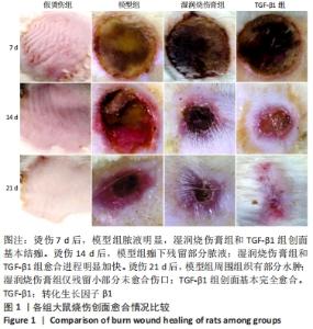
2.1 实验动物数量分析 实验选用大鼠80只,分为4组,实验过程无脱失,全部进入结果分析。 2.2 各组大鼠烧伤创面愈合情况比较 各组大鼠创面均无明显感染迹象,随着时间的推移,伤口逐渐愈合。烫伤7 d后,各组大鼠创面出现愈合,模型组脓液明显,湿润烧伤膏组和TGF-β1组创面基本结痂。烫伤14 d后,各组大鼠创面愈合明显,模型组痂下残留部分脓液;湿润烧伤膏组和TGF-β1组愈合进程明显加快。烫伤21 d后,模型组大鼠创面颜色较深,周围组织有部分水肿,创面逐步愈合;湿润烧伤膏组创面明显缩小,仅残留小部分未愈合伤口;TGF-β1组创面基本完全愈合,见图1。"
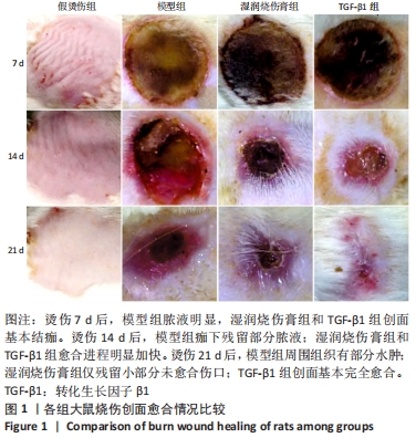
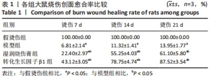
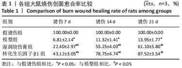
与假烫伤组相比,模型组创面愈合缓慢;与模型组相比,湿润烧伤膏组和TGF-β1组创面愈合率明显升高(P < 0.05),见表1。"
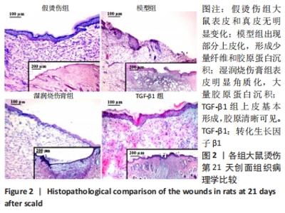
2.3 各组大鼠创面组织病理学比较 苏木精-伊红染色结果显示,假烫伤组大鼠表皮和真皮无明显变化;烫伤21 d,模型组大鼠出现部分上皮化,形成少量纤维和胶原蛋白沉积,存在丰富的肉芽组织和严重的炎症反应;湿润烧伤膏组大鼠表面覆盖坚固的表皮,表皮衬里存在明显的重构和角质化,少量淋巴细胞出现,纤维蛋白渗出减少,大量胶原蛋白沉积;TGF-β1组大鼠上皮基本形成,成纤维细胞肥大且密集分布,胶原组织清晰可见,见图2。"

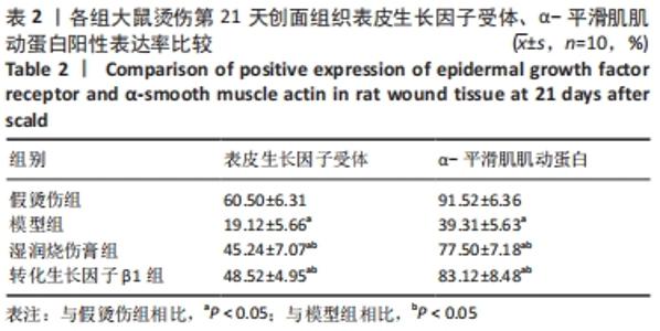
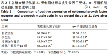
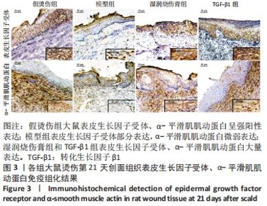
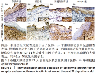
2.4 各组大鼠创面组织表皮生长因子受体、α-平滑肌肌动蛋白表达水平比较 免疫组化结果显示,表皮生长因子受体定位于上皮细胞中,α-平滑肌肌动蛋白定位于真皮中,阳性染色为棕黄色。假烫伤组大鼠表皮生长因子受体、α-平滑肌肌动蛋白呈强阳性表达;烫伤21 d,模型组大鼠表皮生长因子受体部分表达,α-平滑肌肌动蛋白微弱表达;湿润烧伤膏组和TGF-β1组在基底层和基底上层均发现表皮生长因子受体大量表达,真皮中α-平滑肌肌动蛋白阳性率显著增加。与假烫伤组相比,模型组大鼠表皮生长因子受体、α-平滑肌肌动蛋白表达水平显著降低;与模型组相比,湿润烧伤膏组和TGF-β1组表皮生长因子受体、α-平滑肌肌动蛋白表达水平显著升高(P < 0.05),见表2及图3。"
"
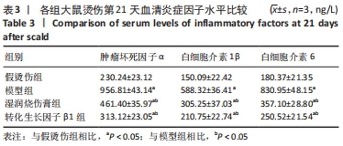
2.5 各组大鼠血清炎症因子肿瘤坏死因子α、白细胞介素1β、白细胞介素6水平比较 ELISA结果显示,与假烫伤组相比,模型组大鼠肿瘤坏死因子α、白细胞介素1β、白细胞介素6表达水平显著升高;与模型组相比,湿润烧伤膏组和TGF-β1组大鼠血清炎症因子肿瘤坏死因子α、白细胞介素1β、白细胞介素6水平显著下降(P < 0.05),见表3。"

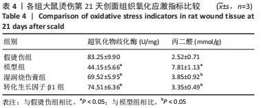
2.6 各组大鼠创面组织氧化应激指标超氧化物歧化酶、丙二醛水平比较 检测结果显示,与假烫伤组相比,模型组大鼠超氧化物歧化酶水平显著降低,丙二醛水平显著升高;与模型组相比,湿润烧伤膏组和TGF-β1组大鼠超氧化物歧化酶水平显著升高,丙二醛水平显著下降(P < 0.05);而湿润烧伤膏组和TGF-β1组超氧化物歧化酶、丙二醛水平差异无显著性意义(P > 0.05),见表4。"
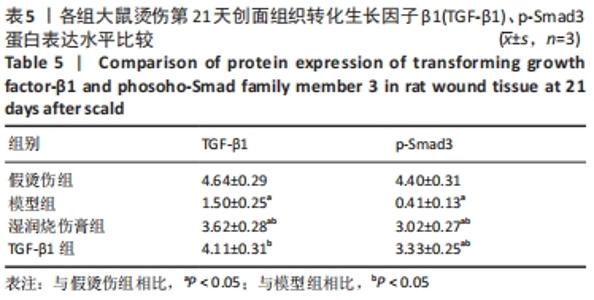
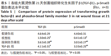
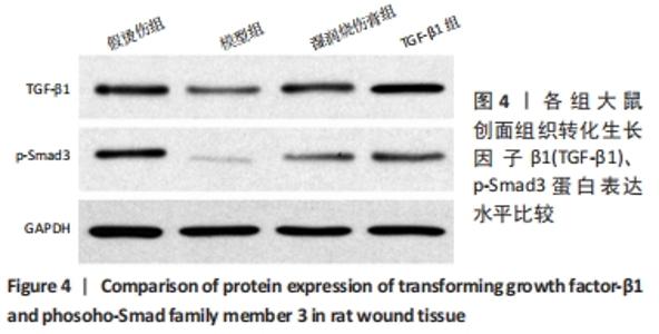
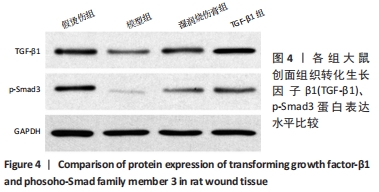
2.7 各组大鼠创面组织TGF-β1、p-Smad3蛋白表达水平比较 Western blot结果显示,与假烫伤组相比,模型组大鼠TGF-β1、p-Smad3水平显著降低;与模型组相比,湿润烧伤膏组和TGF-β1组大鼠TGF-β1、p-Smad3蛋白水平显著升高(P < 0.05);而湿润烧伤膏组和TGF-β1组的TGF-β1、p-Smad3水平相比差异无显著性意义(P > 0.05),见表5及图4。"
"
| [1] JESCHKE MG, PECK MD. Burn care of the elderly. J Burn Care Res. 2017;38(3): e625-e628. [2] 李蓉, 宋志芳. 心理弹性的心理支持方案用于面部深度烧伤美容患者的临床研究[J].中国健康心理学杂志,2018,26(9):1337-1340. [3] SON B, LEE S, KIM H, et al. Low dose radiation attenuates inflammation and promotes wound healing in a mouse burn model. J Dermatol Sci. 2019;96(2):81-89. [4] LEILA R, JAFAR SR, RAZIYEH K, et al. Skin Burns: Review of Molecular Mechanisms and Therapeutic Approaches. Wounds. 2019;31(12):308-315. [5] DU HY, ZHOU YW, LIANG X, et al. CCN1 accelerates re-epithelialization by promoting keratinocyte migration and proliferation during cutaneous wound healing. Biochem Biophys Res Commun. 2018;505(4):966-972. [6] 冯志芳. 湿润烧伤膏治疗烧伤创面患者的效果分析[J]. 中国医学创新,2019, 16(20):105-108. [7] 杨新蕾, 许建允, 孟红阳. 湿润烧伤膏治疗供皮区残余创面的疗效观察[J]. 当代医学,2021,27(12):121-122. [8] 梁吉星. 湿润烧伤膏治疗深Ⅱ度烧伤效果及对血清炎症因子的影响[J]. 中医药临床杂志,2019,31(8):1571-1573. [9] 张丽艳. 湿润烧伤膏(MEBO)对糖尿病皮肤溃疡小鼠抗氧化作用及机制研究[D]. 沈阳:辽宁中医药大学,2019. [10] JIANG TC, WANG ZY, SUN J. Human bone marrow mesenchymal stem cell-derived exosomes stimulate cutaneous wound healing mediates through TGF-beta/Smad signaling pathway. Stem Cell Res Ther. 2020;11(1):198-207. [11] JI YY, ZHANG AJ, CHEN XB, et al. Sodium humate accelerates cutaneous wound healing by activating TGF-beta/Smads signaling pathway in rats. Acta Pharm Sin B. 2016;6(2):132-140. [12] 张萍, 蒋仕秋, 陈雪莲, 等. 基于TGF-β1/Smad3信号通路观察rb-bFGF在糖尿病大鼠烧伤创面愈合中的作用机制[J]. 疑难病杂志,2020,19(6):617-622+631. [13] 张攀攀, 王培仁, 唐巍. 表皮生长因子联合封闭式负压引流对四肢深Ⅱ度烧伤患者创面愈合及ICAM-1、EGF的影响[J]. 中国合理用药探索,2021,18(1):96-100. [14] ANGGOROWATI N, KURNIASARI CR, DAMAYANTI K, et al. Histochemical and immunohistochemical study of α-SMA, collagen, and PCNA in epithelial ovarian neoplasm. Asian Pac J Cancer Prev. 2017;18(3):667-671. [15] PUTRA A, ALIF I, HAMRA N, et al. MSC-released TGF-beta regulate alpha-SMA expression of myofibroblast during wound healing. J Stem Cells Regen Med. 2020;16(2):73-79. [16] 卢孔君. 表皮生长因子和维甲酸复方柔性脂质体软膏的制备及烧伤愈合的研究[D]. 杭州:浙江大学,2019. [17] SIMONETTI O, LUCARINI G, ORLANDO F, et al. Role of Daptomycin on Burn Wound Healing in an Animal Methicillin-Resistant Staphylococcus aureus Infection Model. Antimicrob Agents Chemother. 2017;61(9):e00606-e00617. [18] 刘昕, 孙毅海, 唐深, 等. 输尿管热损伤后TGF-β亚型及α-平滑肌肌动蛋白表达的实验研究[J]. 微创医学,2019,14(4):412-414+429. [19] 王雪欣, 张明谏, 李小兵. 严重烧伤患者锌缺乏与补锌治疗研究进展[J]. 中华烧伤杂志,2018,34(1):57-59. [20] HERNANDEZ P, BULLER D, MITCHELL T, et al. Severe Burn-Induced Inflammation and Remodeling of Achilles Tendon in a Rat Model. Shock. 2018;50(3):346-350. [21] ZHOU XQ, RUAN QF, YE ZQ, et al. Resveratrol accelerates wound healing by attenuating oxidative stress-induced impairment of cell proliferation and migration. Burns. 2021;47(1):133-139. [22] ZHANG DM, WANG BL, SUN YJ, et al. Injectable Enzyme-Based Hydrogel Matrix with Precisely Oxidative Stress Defense for Promoting Dermal Repair of Burn Wound. Macromol Biosci. 2020;20(6):e2000036. [23] 董小鹏, 于博. 生肌玉红膏对深Ⅱ度烧伤模型大鼠创面氧化应激水平影响的实验研究[J]. 中国卫生标准管理,2016,7(22):124-125. [24] 巩文艺, 韩冬. 黄芪多糖对严重烧伤大鼠心肌组织氧化应激和炎症反应的影响[J]. 中国中医急症,2016,25(6):1005-1007+1022. [25] 徐汤灵, 林谋俊, 何勇, 等. 花椒籽油对大鼠烧伤模型促创面愈合和血清炎症因子的调节作用[J]. 中国临床药理学杂志,2020,36(13):1821-1824. [26] KANG JH, JUNG MY, YIN X, et al. Cell-penetrating peptides selectively targeting SMAD3 inhibit profibrotic TGF-β signaling. J Clin Invest. 2017;127(7):2541-2554. [27] 刘筱, 伍伟明, 尹婷, 等. 美洲大蠊提取液对大鼠难愈合创面TGF-β表达的影响[J]. 中医药导报,2018,24(6):9-12. [28] MENG XL, GAO XX, CHEN XX. Umbilical cord-derived mesenchymal stem cells exert anti-fibrotic action on hypertrophic scar-derived fibroblasts in co-culture by inhibiting the activation of the TGF β1/Smad3 pathway. Exp Ther Med. 2021;21(3):210 [29] YAN G, RU Y, WU K, et al. GOLM1 promotes prostate cancer progression through activating PI3K-AKT-mTOR signaling. Prostate. 2018;78(3):166-177. [30] GUO J, FANG Y, JIANG F, et al. Neohesperidin inhibits TGF-beta1/Smad3 signaling and alleviates bleomycin-induced pulmonary fibrosis in mice. Eur J Pharmacol. 2019;864:172712. [31] LIU N, FENG J, LU X, et al. Isorhamnetin Inhibits Liver Fibrosis by Reducing Autophagy and Inhibiting Extracellular Matrix Formation via the TGF-beta1/Smad3 and TGF-beta1/p38 MAPK Pathways. Mediators Inflamm. 2019;2019:6175091. [32] LOBODA A, SOBCZAK M, JOZKOWICZ A, et al. TGF-beta1/Smads and miR-21 in Renal Fibrosis and Inflammation. Mediators Inflamm. 2016;2016:8319283. [33] XU H, LI F. miR-127 aggravates myocardial failure by promoting the TGF-beta1/Smad3 signaling. Mol Med Rep. 2019;20(3):2500. [34] 郑晨果, 陈念昭, 黄盈, 等. 痔愈喷剂对大鼠创面TGF-β1表达的影响[J]. 中华中医药学刊,2015,33(1):165-167+17-18. |
| [1] | Wu Cong, Jia Quanzhong, Liu Lun. Relationship between transforming growth factor beta1 expression and chondrocyte migration in adult articular cartilage after fragmentation [J]. Chinese Journal of Tissue Engineering Research, 2022, 26(8): 1167-1172. |
| [2] | Zhang Jinglin, Leng Min, Zhu Boheng, Wang Hong. Mechanism and application of stem cell-derived exosomes in promoting diabetic wound healing [J]. Chinese Journal of Tissue Engineering Research, 2022, 26(7): 1113-1118. |
| [3] | Li Weiming, Xu Qingwen, Li Yijun, Sun Yanbo, Cui Jin, Xu Pengyuan . Deep seawater promotes wound healing in diabetic mice by activating PI3K/Akt pathway [J]. Chinese Journal of Tissue Engineering Research, 2022, 26(5): 724-729. |
| [4] | Wang Ao, Chu Hongshang, Wu Hongguang, Li Ke, Liu Huijuan. Expression of leptin receptor in mouse skin and the role of leptin receptor-positive cells in skin development and wound healing [J]. Chinese Journal of Tissue Engineering Research, 2022, 26(25): 3993-3998. |
| [5] | Zhang Gaofei, Wang Di, Li Jiamei, Lou Hanxiao, Zeng Yueqin, Liu Wenjun. Mesenchymal stem cells promote wound healing by regulating the autophagy [J]. Chinese Journal of Tissue Engineering Research, 2022, 26(25): 4058-4063. |
| [6] | Zhang Rong, Liang Zhenting, Yang Chun, Liu Dongdong, Xi Yin, Zhang Jie, Wang Ya, Xu Yonghao, Liu Xiaoqing, Li Yimin. Effects of mechanical stretch on fiber proliferation in human lung epithelial cells [J]. Chinese Journal of Tissue Engineering Research, 2022, 26(24): 3821-3825. |
| [7] | An Dong, Liu Yang, Yang Tongjiang. Application of extracorporeal shock wave therapy in burn wound repair and post-burn scar treatment [J]. Chinese Journal of Tissue Engineering Research, 2022, 26(20): 3265-3272. |
| [8] | Xiong Yang, Yang Yingli, Gao Yushan, Wang Xiumei, Yang Yongdong, Zhao Dingyan, Zhao He, Li Chuanhong, Yang Kaitan, Yu Xing. Histological changes of cervical disc tissue and underlying ossification mechanism in patients with cervical degenerative ossification [J]. Chinese Journal of Tissue Engineering Research, 2022, 26(18): 2868-2873. |
| [9] | Zhao Zixi, Xu Jun, Ding Min, Li Xiwen, Zhang Jinghang, Wang Penghua. Changes of type I and III collagen and matrix metalloproteinase 2 and 9 on the wound of diabetic foot ulcer with external application of medical collagen dressing [J]. Chinese Journal of Tissue Engineering Research, 2022, 26(10): 1544-1550. |
| [10] | Zhang Zhenkun, Li Zhe, Li Ya, Wang Yingying, Wang Yaping, Zhou Xinkui, Ma Shanshan, Guan Fangxia. Application of alginate based hydrogels/dressings in wound healing: sustained, dynamic and sequential release [J]. Chinese Journal of Tissue Engineering Research, 2021, 25(4): 638-643. |
| [11] | Wen Xu, Shen Jing. Application of nano-silver antibacterial hydrogel dressing combined with recombinant human basic fibroblast growth factor on wound healing after debridement of open ankle fracture [J]. Chinese Journal of Tissue Engineering Research, 2021, 25(29): 4638-4643. |
| [12] | Jie Ke, Deng Peng, Zeng Yirong. Application and comparison of four commonly used methods for patellar height measurement [J]. Chinese Journal of Tissue Engineering Research, 2021, 25(24): 3875-3881. |
| [13] | Liu Fang, Shan Zhengming, Tang Yulei, Wu Xiaomin, Tian Weiqun. Effects of hemostasis and promoting wound healing of ozone sustained-release hydrogel [J]. Chinese Journal of Tissue Engineering Research, 2021, 25(22): 3445-3449. |
| [14] | Chen Zhenyu, Zhang Xiaoning, Luo Yuxin, Liang Jianwei, Yan Chi. Evaluation of silk fibroin/curcumin composite film for promoting wound healing [J]. Chinese Journal of Tissue Engineering Research, 2021, 25(16): 2554-2561. |
| [15] | Hu Sheng, Yuan Haiyan, Hu Meng, Jin Shanhu. Effects of moderate treadmill exercise on alpha-smooth muscle actin and type IV collagen in the liver of type 2 diabetic rats [J]. Chinese Journal of Tissue Engineering Research, 2021, 25(11): 1723-1727. |
| Viewed | ||||||
|
Full text |
|
|||||
|
Abstract |
|
|||||