Chinese Journal of Tissue Engineering Research ›› 2021, Vol. 25 ›› Issue (34): 5445-5452.doi: 10.12307/2021.237
Previous Articles Next Articles
Cartilage composite scaffold loaded with transforming growth factor beta 3 using three-dimensional bioprinting
Yang Zhen1, 2, Li Hao1, 2, Fu Liwei1, 2, Gao Cangjian1, 2, Jiang Shuangpeng2, Wang Fuxin2, Yuan Zhiguo2, Sun Zhiqiang1, 2, Zha Kangkang1, 2, Tian Guangzhao1, 2, Cao Fuyang2, Sui Xiang2, Liu Shuyun2, Guo Quanyi2
- 1Medical College of Nankai University, Tianjin 300071, China; 2Institute of Orthopedics, the First Medical Center, Chinese PLA General Hospital, Beijing Key Laboratory of Regenerative Medicine in Orthopedics, Key Laboratory of Musculoskeletal Trauma & War Injuries, PLA, Beijing 100853, China
-
Received:2020-06-28Revised:2020-07-03Accepted:2020-08-04Online:2021-12-08Published:2021-07-26 -
Contact:Guo Quanyi, Professor, Institute of Orthopedics, the First Medical Center, Chinese PLA General Hospital, Beijing Key Laboratory of Regenerative Medicine in Orthopedics, Key Laboratory of Musculoskeletal Trauma & War Injuries, PLA, Beijing 100853, China -
About author:Yang Zhen, Master candidate, Medical College of Nankai University, Tianjin 300071, China; Institute of Orthopedics, the First Medical Center, Chinese PLA General Hospital, Beijing Key Laboratory of Regenerative Medicine in Orthopedics, Key Laboratory of Musculoskeletal Trauma & War Injuries, PLA, Beijing 100853, China -
Supported by:the National Key Research and Development Plan Project, No. 2019YFA0110600 (to GQY); the National Natural Science Foundation of China, No. 81772319 (to GQY)
CLC Number:
Cite this article
Yang Zhen, Li Hao, Fu Liwei, Gao Cangjian, Jiang Shuangpeng, Wang Fuxin, Yuan Zhiguo, Sun Zhiqiang, Zha Kangkang Tian Guangzhao, Cao Fuyang, Sui Xiang, Liu Shuyun, Guo Quanyi. Cartilage composite scaffold loaded with transforming growth factor beta 3 using three-dimensional bioprinting[J]. Chinese Journal of Tissue Engineering Research, 2021, 25(34): 5445-5452.
share this article
Add to citation manager EndNote|Reference Manager|ProCite|BibTeX|RefWorks

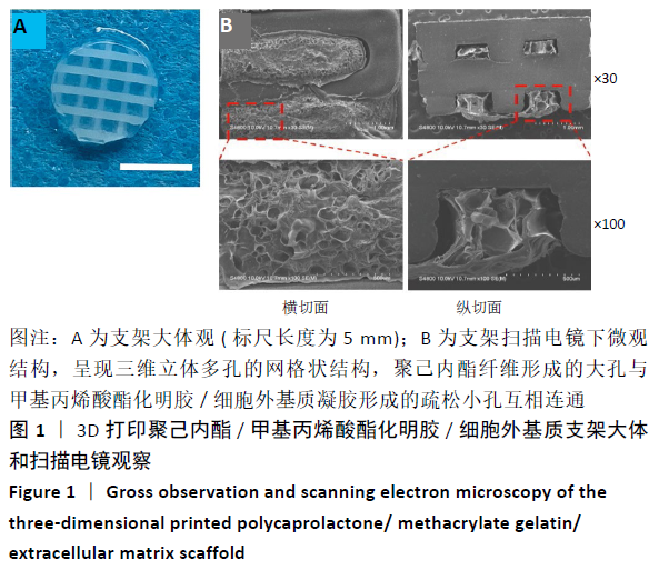
2.1 3D打印支架的大体观和微观结构观察 大体观可见PCL纤维呈现十字交叉网格结构,纤维排列整齐均匀,GelMA/ECM生物墨水材料均匀填充在PCL纤维之间,见图1A所示。通过扫描电镜观察支架的横切面和纵切面的内部结构可以发现,3D打印PCL/GelMA/ECM支架呈现三维立体多孔的网格状结构,PCL纤维形成的大孔与GelMA/ECM凝胶形成的疏松的小孔互相连通,孔径尺寸分布100-200 μm之间,见图1B所示。"
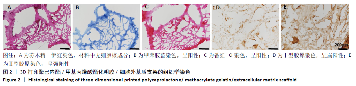
2.2 3D打印支架的组织学检测 苏木精-伊红染色可以看出GelMA/ECM生物墨水材料中无细胞核成分,提示细胞等免疫原性成分去除比较彻底(图2A)。甲苯胺蓝染色和番红染色呈阳性(图2B,C),提示PCL/GelMA/ECM支架中含有糖胺聚糖成分;Ⅰ型胶原免疫组织化学染色呈弱阳性(图2D),提示PCL/GelMA/ECM支架含有少量的Ⅰ型胶原;Ⅱ型胶原免疫组织化学染色呈强阳性(图2E),说明该支架含有大量的Ⅱ型胶原蛋白。"

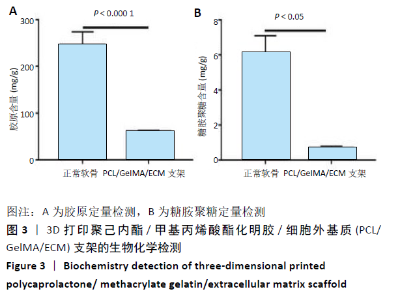
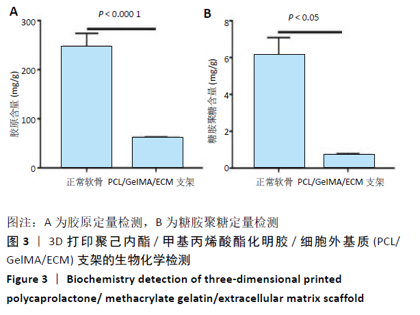
2.3 3D打印支架的生物化学检测 正常软骨的胶原含量为(246.21±27.8) mg/g,3D打印PCL/GelMA/ECM支架为(61.63± 1.27) mg/g;正常软骨的糖胺聚糖含量为(5.94±1.16) mg/g,3D打印PCL/GelMA/ECM支架仅为(0.71±0.072) mg/g,见图3。3D打印PCL/GelMA/ECM支架含有一定量的胶原和糖胺聚糖,但与正常软骨相比含量较低。"
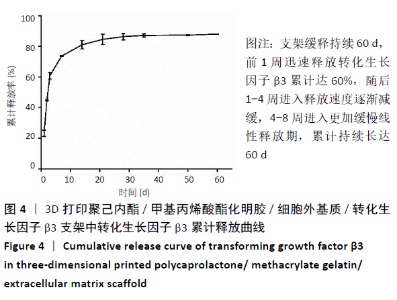
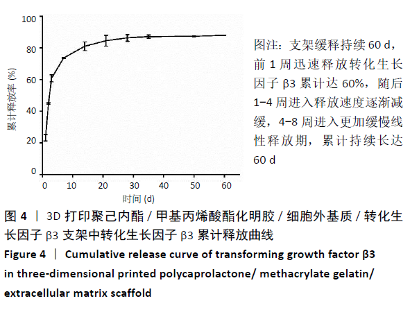
2.4 3D打印支架生物力学检测 3D生物打印PCL/GelMA/ECM支架的压缩模量为(14.24±2.44) MPa,高于PCL支架的(10.41±1.13) MPa(P < 0.05)和人膝关节正常软骨压缩模量(4.3-13.0 MPa)[28],具有良好的生物力学性能,可以在软骨再生的时间窗口内提供力学支撑。 2.5 3D打印支架缓释性检测 负载于GelMA/ECM生物墨水中的TGF-β3累计释放曲线见图4。在缓释检测的60 d内,前1周内释放较快,因子累计释放率已达约60%,呈现短期内的突释现象,随后的1-4周释放速度逐渐减缓,而4-8周的释放规律类似于零级释放动力学,60 d累计释放率约达80%,并且仍有缓慢释放的趋势。"
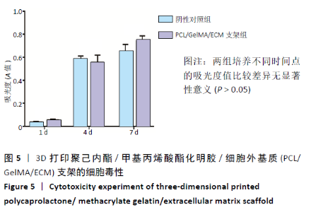
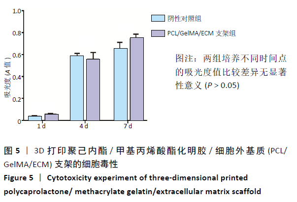
2.6 3D打印支架的细胞毒性实验 CCK-8细胞毒性实验结果显示,支架浸提液与脂肪间充质干细胞共培养1,4,7 d后,实验组与阴性对照组细胞数量明显增多,并且二者在不同时间点的吸光度值比较差异无显著性意义(P > 0.05),见图5,提示该支架无明显细胞毒性,具有良好的细胞相容性,为植入动物体内提供了有利的理论支撑。"
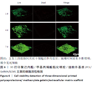
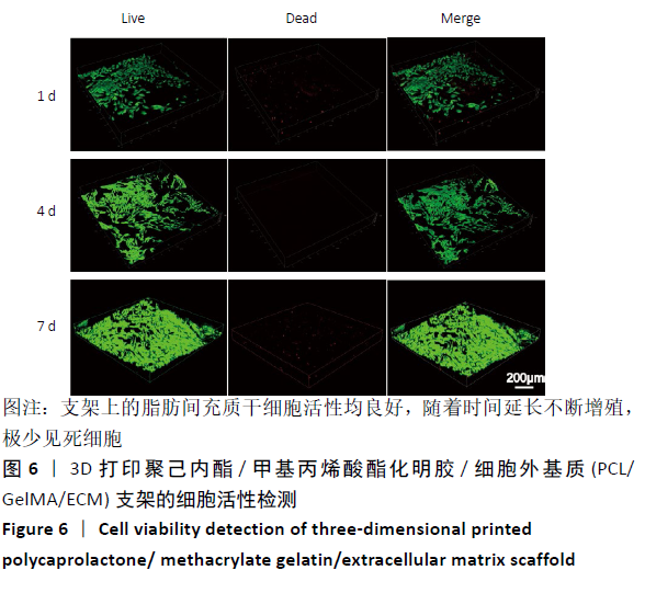
2.7 3D打印支架的细胞活性检测 兔脂肪间充质干细胞在支架上培养1,4,7 d后的死活染色结果,见图6所示,活细胞(绿色荧光)数量随着时间延长数量逐渐增加,而死细胞(红色荧光)则极少,说明细胞在支架上的活力良好,该支架有利于细胞生长增殖。"
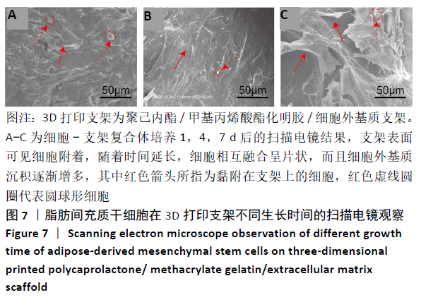
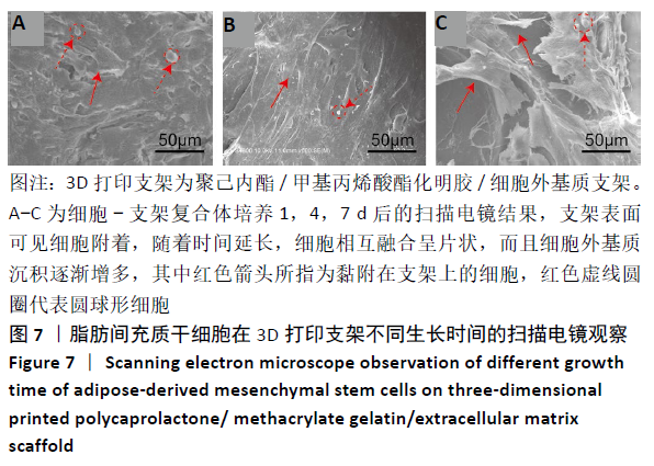
2.8 3D打印支架上的细胞黏附和形态观察 兔脂肪间充质干细胞在3D打印PCL/GelMA/ECM支架上培养1,4,7 d后的扫描电镜结果,见图7所示,可见大量片状细胞均附着于支架表面生长,形态多样且产生大量ECM,仅存在少量圆球形细胞;随着时间延长,可见细胞明显紧密贴附、相互融合成片状,并生长入微孔内,说明该支架适合细胞生长,并且促进细胞分泌大量ECM。"
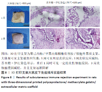
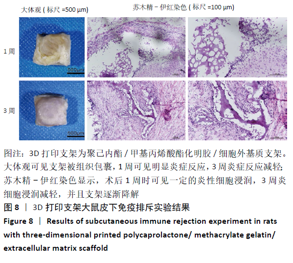
2.9 3D打印支架的免疫排斥和降解反应 将支架埋植大鼠背部皮下后,第1周出现急性炎症反应,呈现白色炎症组织块,且炎症区域大于支架;第3周可见炎症反应减轻,支架与周围组织边界不清,见图8。为了进一步观察支架植入后的组织学特点对其行苏木精-伊红染色,第1周可见炎性细胞浸润在支架内部和支架与组织交界处,而第3周炎性细胞浸润减轻,见图8,表明3D打印PCL/GelMA/ECM支架具有良好的生物相容性;在第1周时,支架GelMA/ECM成分少量降解,到第3周时该成分降解增多,反映出GelMA/ECM成分具有良好的可降解性。"
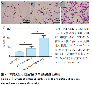
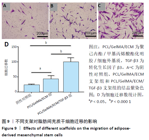
2.10 3D打印支架募集效应检测 细胞迁移结晶紫染色结果及定量分析结果见图9所示,与阴性对照组相比, PCL/GelMA/ECM支架组、PCL/GelMA/ECM/TGF-β3支架组的脂肪间充质干细胞迁移数增多(P < 0.05,P < 0.000 1);与PCL/GelMA/ECM支架相比,PCL/GelMA/ECM/TGF-β3支架组的细胞迁移数目更多(P < 0.000 1)。"
| [1]SOPHIA FOX AJ, BEDI A, RODEO SA. The basic science of articular cartilage: structure, composition, and function. Sports Health. 2009;1(6):461-468. [2] CORREA D, LIETMAN SA. Articular cartilage repair: Current needs, methods and research directions. Semin Cell Dev Biol. 2017;62:67-77. [3] VAN OSCH GJVM, BRITTBERG M, DENNIS JE, et al. Cartilage repair: past and future--lessons for regenerative medicine. J Cell Mol Med. 2009;13(5):792-810. [4] DORAN PM. Cartilage Tissue Engineering: What Have We Learned in Practice? Methods Mol Biol. 2015;1340:3-21. [5] CLAVÉ A, POTEL JF, SERVIEN E, et al. Third-generation autologous chondrocyte implantation versus mosaicplasty for knee cartilage injury: 2-year randomized trial. J Orthop Res. 2016;34(4):658-665. [6] VANDEN BERG-FOELS WS. In Situ Tissue Regeneration: Chemoattractants for Endogenous Stem Cell Recruitment. Tissue Eng Part B Rev. 2014;20(1):28-39. [7] GRANDE DA, SGAGLIONE NA. Regenerative medicine: Self-directed articular resurfacing: a new paradigm? Nat Rev Rheumatol. 2010;6(12):677-678. [8] SUN X, YIN H, WANG Y, et al. In Situ Articular Cartilage Regeneration through Endogenous Reparative Cell Homing Using a Functional Bone Marrow-Specific Scaffolding System. ACS Appl Mater Interfaces. 2018;10(45):38715-38728. [9] YANG Z, LI H, YUAN Z, et al. Endogenous cell recruitment strategy for articular cartilage regeneration. Acta Biomater. 2020;114:31-52. [10] KO IK, LEE SJ, ATALA A, et al. In situ tissue regeneration through host stem cell recruitment. Exp Mol Med. 2013;45(11):e57. [11] JING H, GAO B, GAO M, et al. Restoring tracheal defects in a rabbit model with tissue engineered patches based on TGF-beta3-encapsulating electrospun poly(l-lactic acid-co-epsilon-caprolactone)/collagen scaffolds. Artif Cells Nanomed Biotechnol. 2018;46(sup1):985-995. [12] YANG Q, TENG BH, WANG LN, et al. Silk fibroin/cartilage extracellular matrix scaffolds with sequential delivery of TGF-β3 for chondrogenic differentiation of adipose-derived stem cells. Int J Nanomedicine. 2017;12: 6721-6733. [13] LEE CH, COOK JL, MENDELSON A, et al. Regeneration of the articular surface of the rabbit synovial joint by cell homing: a proof of concept study. Lancet. 2010;376(9739):440-448. [14] LI Y, LIU Y, XUN X, et al. Three-Dimensional Porous Scaffolds with Biomimetic Microarchitecture and Bioactivity for Cartilage Tissue Engineering. ACS Appl Mater Interfaces. 2019;11(40):36359-36370. [15] YANG Q, PENG J, GUO Q, et al. A cartilage ECM-derived 3-D porous acellular matrix scaffold for in vivo cartilage tissue engineering with PKH26-labeled chondrogenic bone marrow-derived mesenchymal stem cells. Biomaterials. 2008;29(15):2378-2387. [16] KANG H, PENG J, LU S, et al. In vivo cartilage repair using adipose-derived stem cell-loaded decellularized cartilage ECM scaffolds. J Tissue Eng Regen Med. 2014;8(6):442-453. [17] 周建,田壮,田沁玉,等.不同交联密度甲基丙烯酰酯明胶/脱细胞半月板细胞外基质复合水凝胶的性能[J].中国组织工程研究,2020,24(16): 2493-2499. [18] XING F, LI L, ZHOU C, et al. Regulation and Directing Stem Cell Fate by Tissue Engineering Functional Microenvironments: Scaffold Physical and Chemical Cues. Stem Cells Int. 2019;2019:2180925. [19] LUO Y, WEI X, HUANG P. 3D bioprinting of hydrogel-based biomimetic microenvironments. J Biomed Mater Res B Appl Biomater. 2019;107(5): 1695-1705. [20] LUO Y, LIN X, HUANG P. 3D Bioprinting of Artificial Tissues: Construction of Biomimetic Microstructures. Macromol Biosci. 2018;18(6):e1800034. [21] CHEN M, FENG Z, GUO W, et al. PCL-MECM Based Hydrogel Hybrid Scaffolds and Meniscal Fibrochondrocytes Promote Whole Meniscus Regeneration in a Rabbit Meniscectomy Model. ACS Appl Mater Interfaces. 2019;41:626-639. [22] ZHOU C, YANG K, WANG K, et al. Combination of fused deposition modeling and gas foaming technique to fabricated hierarchical macro/microporous polymer scaffolds. Mater Des. 2016;109:415-424. [23] 张彬,沈师,鲜海,等.3D打印制备PLGA/脱细胞软骨细胞外基质支架材料及其理化特性研究[J].中国修复重建外科杂志,2019,33(8):1011-1018. [24] 肖统光,郝春香,荆晓光,等.关节软骨细胞外基质/人脐带Wharton胶复合多孔支架的制备及评估[J].中国组织工程研究,2017,21(22):3470-3475. [25] ALMEIDA HV, ESWARAMOORTHY R, CUNNIFFE GM, et al. Fibrin Hydrogels Functionalized with Particulated Cartilage Extracellular Matrix and Incorporating Freshly Isolated Stromal Cells as an Injectable for Cartilage Regeneration. Acta Biomater. 2016;36:55-62. [26] CHENG H, ZHANG Y, ZHANG B, et al. Biocompatibility of polypropylene mesh scaffold with adipose-derived stem cells. Exp Ther Med. 2017;13(6):2922-2926. [27] 韩爽,卢世璧,刘强,等.自体脂肪间充质干细胞复合人脐带Wharton胶支架修复兔膝关节软骨缺损[J]. 中国组织工程研究,2012,16(19):88-93. [28] SHEPHERD D, SEEDHOM B. The ‘instantaneous’ compressive modulus of human articular cartilage in joints of the lower limb. Rheumatology (Oxford, England). 1999;38(2):124-132. [29] ANDREAS K, SITTINGER M, RINGE J. Toward in situ tissue engineering: chemokine-guided stem cell recruitment. Trends Biotechnol. 2014;32(9): 483-492. [30] ZHENG X, YANG F, WANG S, et al. Fabrication and cell affinity of biomimetic structured PLGA/articular cartilage ECM composite scaffold. J Mater Sci Mater Med. 2011;22(3):693-704. [31] YUE K, TRUJILLO-DE SANTIAGO G, ALVAREZ MM, et al. Synthesis, properties, and biomedical applications of gelatin methacryloyl (GelMA) hydrogels. Biomaterials. 2015;73:254-271. [32] KLOTZ BJ, GAWLITTA D, ROSENBERG AJWP, et al. Gelatin-Methacryloyl Hydrogels: Towards Biofabrication-Based Tissue Repair. Trends Biotechnol. 2016;34(5):394-407. [33] VISSCHER D, GLEADALL A, BUSKERMOLEN J, et al. Design and fabrication of a hybrid alginate hydrogel/poly(ε-caprolactone) mold for auricular cartilage reconstruction. J Biomed Mater Res B Appl Biomater. 2019;107(5):1711-1721. [34] CHEN X, FAN H, DENG X, et al. Scaffold Structural Microenvironmental Cues to Guide Tissue Regeneration in Bone Tissue Applications. Nanomaterials (Basel, Switzerland). 2018;8(11):960. [35] LUO Z, JIANG L, XU Y, et al. Mechano growth factor (MGF) and transforming growth factor (TGF)-β3 functionalized silk scaffolds enhance articular hyaline cartilage regeneration in rabbit model. Biomaterials. 2015; 52463-52475. [36] 杨振,李浩,高仓健,等.转化生长因子β3/聚乳酸-羟基乙酸微球对干细胞的调控[J].中国组织工程研究,2020,24(28):4540-4546. [37] AGRAWAL V, BROWN BN, BEATTIE AJ, et al. Evidence of innervation following extracellular matrix scaffold-mediated remodelling of muscular tissues. J Tissue Eng Regen Med. 2009;3(8):590-600. |
| [1] | Lin Qingfan, Xie Yixin, Chen Wanqing, Ye Zhenzhong, Chen Youfang. Human placenta-derived mesenchymal stem cell conditioned medium can upregulate BeWo cell viability and zonula occludens expression under hypoxia [J]. Chinese Journal of Tissue Engineering Research, 2021, 25(在线): 4970-4975. |
| [2] | Zhou Jihui, Li Xinzhi, Zhou You, Huang Wei, Chen Wenyao. Multiple problems in the selection of implants for patellar fracture [J]. Chinese Journal of Tissue Engineering Research, 2021, 25(9): 1440-1445. |
| [3] | Wang Debin, Bi Zhenggang. Related problems in anatomy mechanics, injury characteristics, fixed repair and three-dimensional technology application for olecranon fracture-dislocations [J]. Chinese Journal of Tissue Engineering Research, 2021, 25(9): 1446-1451. |
| [4] | Chen Junming, Yue Chen, He Peilin, Zhang Juntao, Sun Moyuan, Liu Youwen. Hip arthroplasty versus proximal femoral nail antirotation for intertrochanteric fractures in older adults: a meta-analysis [J]. Chinese Journal of Tissue Engineering Research, 2021, 25(9): 1452-1457. |
| [5] | Chen Jinping, Li Kui, Chen Qian, Guo Haoran, Zhang Yingbo, Wei Peng. Meta-analysis of the efficacy and safety of tranexamic acid in open spinal surgery [J]. Chinese Journal of Tissue Engineering Research, 2021, 25(9): 1458-1464. |
| [6] | Hu Kai, Qiao Xiaohong, Zhang Yonghong, Wang Dong, Qin Sihe. Treatment of displaced intra-articular calcaneal fractures with cannulated screws and plates: a meta-analysis of 15 randomized controlled trials [J]. Chinese Journal of Tissue Engineering Research, 2021, 25(9): 1465-1470. |
| [7] | Huang Dengcheng, Wang Zhike, Cao Xuewei. Comparison of the short-term efficacy of extracorporeal shock wave therapy for middle-aged and elderly knee osteoarthritis: a meta-analysis [J]. Chinese Journal of Tissue Engineering Research, 2021, 25(9): 1471-1476. |
| [8] | Xu Feng, Kang Hui, Wei Tanjun, Xi Jintao. Biomechanical analysis of different fixation methods of pedicle screws for thoracolumbar fracture [J]. Chinese Journal of Tissue Engineering Research, 2021, 25(9): 1313-1317. |
| [9] | Jiang Yong, Luo Yi, Ding Yongli, Zhou Yong, Min Li, Tang Fan, Zhang Wenli, Duan Hong, Tu Chongqi. Von Mises stress on the influence of pelvic stability by precise sacral resection and clinical validation [J]. Chinese Journal of Tissue Engineering Research, 2021, 25(9): 1318-1323. |
| [10] | Zhang Tongtong, Wang Zhonghua, Wen Jie, Song Yuxin, Liu Lin. Application of three-dimensional printing model in surgical resection and reconstruction of cervical tumor [J]. Chinese Journal of Tissue Engineering Research, 2021, 25(9): 1335-1339. |
| [11] | Zhang Yu, Tian Shaoqi, Zeng Guobo, Hu Chuan. Risk factors for myocardial infarction following primary total joint arthroplasty [J]. Chinese Journal of Tissue Engineering Research, 2021, 25(9): 1340-1345. |
| [12] | Wei Wei, Li Jian, Huang Linhai, Lan Mindong, Lu Xianwei, Huang Shaodong. Factors affecting fall fear in the first movement of elderly patients after total knee or hip arthroplasty [J]. Chinese Journal of Tissue Engineering Research, 2021, 25(9): 1351-1355. |
| [13] | Wang Jinjun, Deng Zengfa, Liu Kang, He Zhiyong, Yu Xinping, Liang Jianji, Li Chen, Guo Zhouyang. Hemostatic effect and safety of intravenous drip of tranexamic acid combined with topical application of cocktail containing tranexamic acid in total knee arthroplasty [J]. Chinese Journal of Tissue Engineering Research, 2021, 25(9): 1356-1361. |
| [14] | Xiao Guoqing, Liu Xuanze, Yan Yuhao, Zhong Xihong. Influencing factors of knee flexion limitation after total knee arthroplasty with posterior stabilized prostheses [J]. Chinese Journal of Tissue Engineering Research, 2021, 25(9): 1362-1367. |
| [15] | Huang Zexiao, Yang Mei, Lin Shiwei, He Heyu. Correlation between the level of serum n-3 polyunsaturated fatty acids and quadriceps weakness in the early stage after total knee arthroplasty [J]. Chinese Journal of Tissue Engineering Research, 2021, 25(9): 1375-1380. |
| Viewed | ||||||
|
Full text |
|
|||||
|
Abstract |
|
|||||