Chinese Journal of Tissue Engineering Research ›› 2021, Vol. 25 ›› Issue (31): 4988-4994.doi: 10.12307/2021.141
Previous Articles Next Articles
Effect of xanthohumol on proliferation, migration, tube formation and apoptosis of endothelial progenitor cells and its mechanism
Tan Xiaowu1, Yu Xiaofan1, Jiang Huijiao1, Xing Zhikun1, Zhao Xueyuan1, Gao Fengyi1, Wu Xiangwei1, Chen Xueling2
- 1Department of General Surgery, First Affiliated Hospital of Medical College of Shihezi University, Shihezi 832008, Xinjiang Uygur Autonomous Region, China; 2Department of Immunology, Medical College of Shihezi University, Shihezi 832002, Xinjiang Uygur Autonomous Region, China
-
Received:2020-08-22Revised:2020-08-26Accepted:2020-09-26Online:2021-11-08Published:2021-04-25 -
Contact:Wu Xiangwei, MD, Professor, Chief physician, Department of General Surgery, First Affiliated Hospital of Medical College of Shihezi University, Shihezi 832008, Xinjiang Uygur Autonomous Region, China -
About author:Tan Xiaowu, Master, Department of General Surgery, First Affiliated Hospital of Medical College of Shihezi University, Shihezi 832008, Xinjiang Uygur Autonomous Region, China -
Supported by:the National Natural Science Foundation of China, No. 81760570 (to WXW); the National Natural Science Foundation of China, No. 81760371 (to CXL); the Corps Young and Middle-Aged Science and Technology Innovation Leading Talent Program, No. 2018CB017 (to WXW); the Science and Technology Public Relations Project in Key Fields of the Corps, No. 2019AB031 (to CXL)
CLC Number:
Cite this article
Tan Xiaowu, Yu Xiaofan, Jiang Huijiao, Xing Zhikun, Zhao Xueyuan, Gao Fengyi, Wu Xiangwei, Chen Xueling. Effect of xanthohumol on proliferation, migration, tube formation and apoptosis of endothelial progenitor cells and its mechanism[J]. Chinese Journal of Tissue Engineering Research, 2021, 25(31): 4988-4994.
share this article
Add to citation manager EndNote|Reference Manager|ProCite|BibTeX|RefWorks

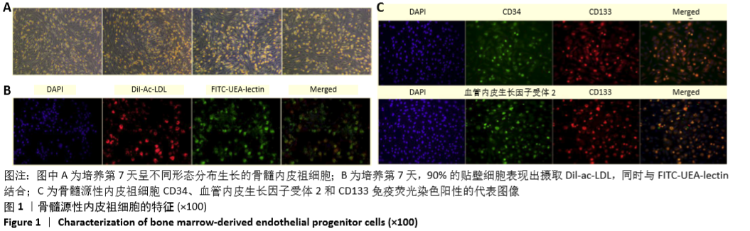
2.1 骨髓源性内皮祖细胞的特征 EGM-2MV完全培养基培养第7天获得的细胞呈梭形,为内皮细胞样形态,见图1A;90%的贴壁细胞表现出摄取Dil-ac-LDL,同时与FITC-UEA-lectin结合,见图1B,这是内皮祖细胞的重要特征[24]。此外,研究还发现这些细胞表达内皮标志物血管内皮生长因子受体2、CD34和CD133,见图1C。"
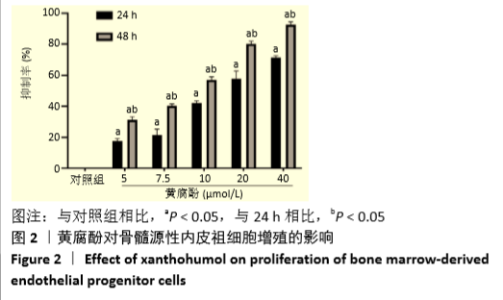
2.2 黄腐酚对骨髓源性内皮祖细胞增殖的影响 见图2。不同浓度的黄腐酚(0,5,7.5,10,20,40 μmol/L)作用于骨髓源性内皮祖细胞24,48 h后,细胞增殖抑制率明显升高,并表现出浓度依赖性和时间依赖性。"


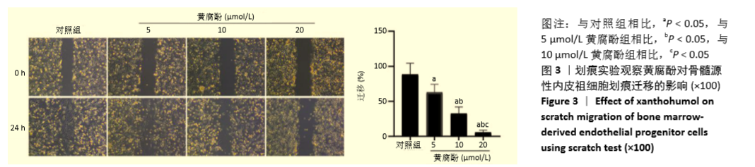
2.3 黄腐酚对骨髓源性内皮祖细胞迁移的影响 划痕实验显示,随着黄腐酚浓度的增大,骨髓源性内皮祖细胞的划痕越明显(F=62.79,两两比较均P < 0.05),且20 μmol/L黄腐酚作用最为明显,提示黄腐酚对骨髓源性内皮祖细胞具有杀伤作用,见图3。"

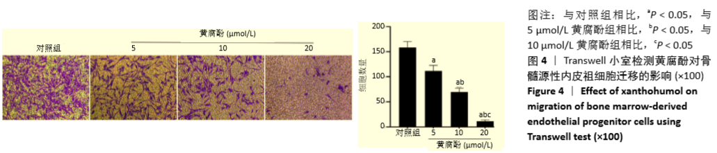
Transwell检测结果显示,与对照组相比,5,10,20 μmol/L黄腐酚组骨髓源性内皮祖细胞的迁移能力随着浓度的升高,迁移细胞数逐渐减少,差异有显著性意义(F=251.79,两两比较均P < 0.05),见图4。"

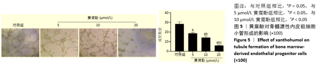
2.4 黄腐酚对骨髓源性内皮祖细胞小管形成的影响 培养4-6 h后镜下观察可见:对照组有大量小管样结构形成,而5,10 μmol/L黄腐酚组小管样结构明显减少,20 μmol/L黄腐酚组最为明显,基本上无小管样结构形成,与对照组相比,差异有显著性意义(F=198.87,两两比较均P < 0.05),见图5。"

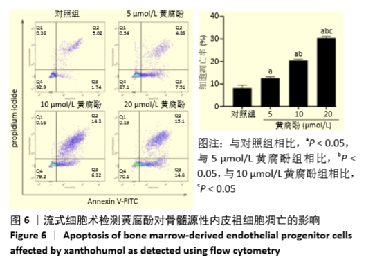
2.5 流式细胞术检测黄腐酚对骨髓源性内皮祖细胞凋亡的影响 用不同浓度黄腐酚(0,5,10,20 μmol/L)作用于骨髓源性内皮祖细胞48 h后,凋亡率分别为(8.25±1.31)%,(12.63±0.62)%,(20.47±0.57)%,(30.43±0.66)%,细胞凋亡率随着黄腐酚浓度升高而上升,具有浓度依赖性,且黄腐酚各组凋亡率均高于对照组,差异有显著性意义(F=393.45,两两比较均P < 0.05),见图6。"
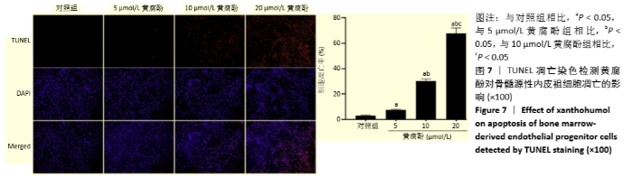
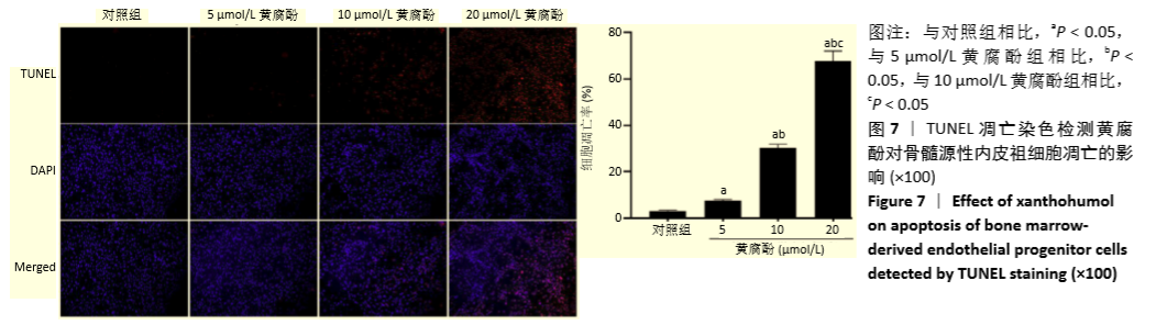
2.6 TUNEL凋亡染色检测黄腐酚对骨髓源性内皮祖细胞凋亡的影响 使用TUNEL凋亡染色法,并通过显微镜高倍视野下计算凋亡指数,如图7所示,对照组与各浓度(5,10,20 μmol/L)黄腐酚组细胞凋亡指数分别为(2.99±0.46)%,(7.56±0.50)%,(30.28±1.57)%,(67.79±4.14)%,20 μmol/L黄腐酚组凋亡率最高,有浓度依赖性,且黄腐酚各组凋亡率均高于对照组,差异有显著性意义(F=521.64,两两比较均P < 0.05)。"
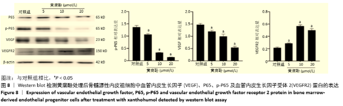
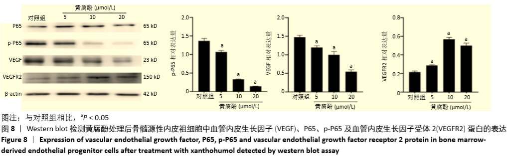
2.7 Western blot 检测黄腐酚对骨髓源性内皮祖细胞中血管内皮生长因子、P65、p-P65及血管内皮生长因子受体2蛋白表达的影响 黄腐酚处理后与对照组相比,血管内皮生长因子、p-P65蛋白表达逐渐下降,血管内皮生长因子受体2蛋白表达逐渐增高(均P < 0.05),具有浓度依赖性(均P < 0.05),见图8。"
| [1] 郑荣寿,孙可欣,张思维,等.2015年中国恶性肿瘤流行情况分析[J].中华肿瘤杂志,2019,41(1):19-28. [2] FOLKMAN J. Seminars in Medicine of the Beth Israel Hospital, Boston. Clinical applications of research on angiogenesis. N Engl J Med. 1995; 333(26):1757-1763. [3] TEPPER OM, CAPLA JM, GALIANO RD, et al. Adult vasculogenesis occurs through in situ recruitment, proliferation, and tubulization of circulating bone marrow-derived cells. Blood. 2005;105(3):1068-1077. [4] FOLKINS C, SHAKED Y, MAN S, et al. Glioma tumor stem-like cells promote tumor angiogenesis and vasculogenesis via vascular endothelial growth factor and stromal-derived factor 1. Cancer Res. 2009;69(18):7243-7251. [5] CHEN X, FANG J, WANG S, et al. A new mosaic pattern in glioma vascularization: exogenous endothelial progenitor cells integrating into the vessels containing tumor-derived endothelial cells. Oncotarget. 2014;5(7):1955-1968. [6] RUSSELL JS, BROWN JM. Circulating mouse Flk1+/c-Kit+/CD45- cells function as endothelial progenitors cells (EPCs) and stimulate the growth of human tumor xenografts. Mol Cancer. 2014;13:177. [7] XIANG Y, GUO Z, ZHU P, et al. Traditional Chinese medicine as a cancer treatment: Modern perspectives of ancient but advanced science. Cancer Med. 2019;8(5):1958-1975. [8] ZOU G, ZHANG X, WANG L, et al. Herb-sourced emodin inhibits angiogenesis of breast cancer by targeting VEGFA transcription. Theranostics. 2020;10(15):6839-6853. [9] SŁAWIŃSKA-BRYCH A, ZDZISIŃSKA B, CZERWONKA A, et al. Xanthohumol exhibits anti-myeloma activity in vitro through inhibition of cell proliferation, induction of apoptosis via the ERK and JNK-dependent mechanism, and suppression of sIL-6R and VEGF production. Biochim Biophys Acta Gen Subj. 2019;1863(11):129408. [10] LIU W, LI W, LIU H, et al. Xanthohumol inhibits colorectal cancer cells via downregulation of Hexokinases II-mediated glycolysis. Int J Biol Sci. 2019;15(11):2497-2508. [11] SAITO K, MATSUO Y, IMAFUJI H, et al. Xanthohumol inhibits angiogenesis by suppressing nuclear factor-κB activation in pancreatic cancer. Cancer Sci. 2018;109(1):132-140. [12] AGGARWAL BB. Nuclear factor-κB: The enemy within. Cancer Cell. 2004;6(3):203-208. [13] 冯文磊,张猛,印双红,等.改良差时贴壁法分离培养鉴定小鼠骨髓间充质干细胞和内皮前体细胞[J].解剖学报,2015,46(2):282-288. [14] 冯文磊,张猛,徐芳洁,等.共培养体系间充质干细胞对同源内皮祖细胞增殖和血管形成的影响[J].医学研究生学报,2015,28(3):229-235. [15] 张猛,冯文磊,王艳杰,等.全骨髓贴壁法分离培养小鼠骨髓间充质干细胞[J].石河子大学学报(自然科学版),2015,33(2):164-167. [16] 张猛,张宏伟,冯文磊,等.血管内皮前体细胞通过旁分泌功能促进间充质细胞成骨分化[J].中华医学杂志,2015,95(16):1253-1257. [17] WANG X, ZHAO Z, ZHANG H, et al. Simultaneous isolation of mesenchymal stem cells and endothelial progenitor cells derived from murine bone marrow. Exp Ther Med. 2018;16(6):5171-5177. [18] TANG B, GONG JP, SUN JM, et al. Construction of a plasmid for expression of rat platelet-derived growth factor C and its effects on proliferation, migration and adhesion of endothelial progenitor cells. Plasmid. 2013;69(3):195-201. [19] YE Y, LI X, ZHANG Y, et al. Androgen Modulates Functions of Endothelial Progenitor Cells through Activated Egr1 Signaling. Stem Cells Int. 2016; 2016:7057894. [20] ZHANG J, ZHANG H, CHEN Y, et al. Platelet‑derived growth factor D promotes the angiogenic capacity of endothelial progenitor cells. Mol Med Rep. 2019;19(1):125-132. [21] ZHOU DM, SUN LL, ZHU J, et al. MiR-9 promotes angiogenesis of endothelial progenitor cell to facilitate thrombi recanalization via targeting TRPM7 through PI3K/Akt/autophagy pathway. J Cell Mol Med. 2020;24(8):4624-4632. [22] LI Z, ZHU Y, LI C, et al. Anti-VEGFR2-interferon-α2 regulates the tumor microenvironment and exhibits potent antitumor efficacy against colorectal cancer. Oncoimmunology. 2017;6(3):e1290038. [23] CHEN Y, WANG D, PENG H, et al. Epigenetically upregulated oncoprotein PLCE1 drives esophageal carcinoma angiogenesis and proliferation via activating the PI-PLCε-NF-κB signaling pathway and VEGF-C/ Bcl-2 expression. Mol Cancer. 2019;18(1):1. [24] NIE Z, XU L, LI C, et al. Association of endothelial progenitor cells and peptic ulcer treatment in patients with type 2 diabetes mellitus. Exp Ther Med. 2016;11(5):1581-1586. [25] YODER MC, MEAD LE, PRATER D, et al. Redefining endothelial progenitor cells via clonal analysis and hematopoietic stem/progenitor cell principals. Blood. 2007;109(5):1801-1809. [26] LUGANO R, RAMACHANDRAN M, DIMBERG A. Tumor angiogenesis: causes, consequences, challenges and opportunities. Cell Mol Life Sci. 2020;77(9):1745-1770. [27] CARMELIET P, JAIN RK. Principles and mechanisms of vessel normalization for cancer and other angiogenic diseases. Nat Rev Drug Discov. 2011;10(6):417-427. [28] ALBINI A, DELL’EVA R, VENÉ R, et al. Mechanisms of the antiangiogenic activity by the hop flavonoid xanthohumol: NF-kappaB and Akt as targets. FASEB J. 2006;20(3):527-529. [29] NORTH S, MOENNER M, BIKFALVI A. Recent developments in the regulation of the angiogenic switch by cellular stress factors in tumors. Cancer Lett. 2005;218(1):1-14. [30] WONG AH, SHIN EM, TERGAONKAR V, et al. Targeting NF-κB Signaling for Multiple Myeloma. Cancers (Basel). 2020;12(8):2203. [31] HUANG F, YAO Y, WU J, et al. Curcumin inhibits gastric cancer-derived mesenchymal stem cells mediated angiogenesis by regulating NF-κB/VEGF signaling. Am J Transl Res. 2017;9(12):5538-5547. [32] BABYKUTTY S, S PP, J NR, et al. Nimbolide retards tumor cell migration, invasion, and angiogenesis by downregulating MMP-2/9 expression via inhibiting ERK1/2 and reducing DNA-binding activity of NF-κB in colon cancer cells. Mol Carcinog. 2012;51(6):475-490. [33] FERRARA N. Role of vascular endothelial growth factor in regulation of physiological angiogenesis. Am J Physiol Cell Physiol. 2001;280(6): C1358-1366. [34] CHENG WX, HUANG H, CHEN JH, et al. Genistein inhibits angiogenesis developed during rheumatoid arthritis through the IL-6/JAK2/STAT3/VEGF signalling pathway. J Orthop Translat. 2019;22:92-100. [35] SKURIKHIN EG, KRUPIN VA, PERSHINA OV, et al. Blockade of Dopamine D2 Receptors as a Novel Approach to Stimulation of Notch1+ Endothelial Progenitor Cells and Angiogenesis in C57BL/6 Mice with Pulmonary Emphysema Induced by Proteases and Deficiency of α1-Antitrypsin. Bull Exp Biol Med. 2020;168(6):718-723. |
| [1] | Yang Yufang, Yang Zhishan, Duan Mianmian, Liu Yiheng, Tang Zhenglong, Wang Yu. Application and prospects of erythropoietin in bone tissue engineering [J]. Chinese Journal of Tissue Engineering Research, 2024, 28(9): 1443-1449. |
| [2] | Yang Yifeng, Ye Nan, Wang Lin, Guo Shuaicheng, Huang Jian. Signaling pathway of dexmedetomidine against ischemia-reperfusion injury [J]. Chinese Journal of Tissue Engineering Research, 2024, 28(9): 1464-1469. |
| [3] | Yue Yun, Wang Peipei, Yuan Zhaohe, He Shengcun, Jia Xusheng, Liu Qian, Li Zhantao, Fu Huiling, Song Fei, Jia Menghui. Effects of croton cream on JNK/p38 MAPK signaling pathway and neuronal apoptosis in cerebral ischemia-reperfusion injury rats [J]. Chinese Journal of Tissue Engineering Research, 2024, 28(8): 1186-1192. |
| [4] | Liu Xin, Hu Man, Zhao Wenjie, Zhang Yu, Meng Bo, Yang Sheng, Peng Qing, Zhang Liang, Wang Jingcheng. Cadmium promotes senescence of annulus fibrosus cells via activation of PI3K/Akt signaling pathway [J]. Chinese Journal of Tissue Engineering Research, 2024, 28(8): 1217-1222. |
| [5] | Wei Juan, Li Ting, Huan Mengting, Xie Ying, Xie Zhouyu, Wei Qingbo, Wu Yunchuan. Mechanism by which static exercise improves insulin resistance in skeletal muscle of type 2 diabetes [J]. Chinese Journal of Tissue Engineering Research, 2024, 28(8): 1271-1276. |
| [6] | Zhang Kefan, Shi Hui. Research status and application prospect of cytokine therapy for osteoarthritis [J]. Chinese Journal of Tissue Engineering Research, 2024, 28(6): 961-967. |
| [7] | Zhang Ya, Mu Qiuju, Wang Zilin, Liu Hongjie, Zhu Lili. Hydrogel loaded with platelet-rich plasma promotes wound healing in diabetic rats [J]. Chinese Journal of Tissue Engineering Research, 2024, 28(5): 690-696. |
| [8] | Wang Yeyuan, Du Yilang, Yu Dehao, Ning Fengting, Bai Bing. Effect of micro-arc oxidation treatment on biological activity of medical metals [J]. Chinese Journal of Tissue Engineering Research, 2024, 28(5): 771-776. |
| [9] | Wang Jiani, Chen Junyu. Angiogenesis mechanism of metal ions and their application in bone tissue engineering [J]. Chinese Journal of Tissue Engineering Research, 2024, 28(5): 804-812. |
| [10] | Wei Yuanxun, Chen Feng, Lin Zonghan, Zhang Chi, Pan Chengzhen, Wei Zongbo. The mechanism of Notch signaling pathway in osteoporosis and its prevention and treatment with traditional Chinese medicine [J]. Chinese Journal of Tissue Engineering Research, 2024, 28(4): 587-593. |
| [11] | Yang Yuqing, Chen Zhiyu. Role and application of early transient presence of M1 macrophages in bone tissue engineering [J]. Chinese Journal of Tissue Engineering Research, 2024, 28(4): 594-601. |
| [12] | Lin Feng, Cheng Ling, Gao Yong, Zhou Jianye, Shang Qingqing. Hyaluronic acid hydrogel-encapsulated bone marrow mesenchymal stem cells promote cardiac function in myocardial infarction rats (III) [J]. Chinese Journal of Tissue Engineering Research, 2024, 28(3): 355-359. |
| [13] | Bi Yujie, Ma Dujun, Peng Liping, Zhou Ziqiong, Zhao Jing, Zhu Houjun, Zhong Qiuhui, Yang Yuxin. Strategy and significance of Chinese medicine combined with medical hydrogel for disease treatment [J]. Chinese Journal of Tissue Engineering Research, 2024, 28(3): 419-425. |
| [14] | Li Chengming, Xue Dongling, Yang Xinyu, Xiao Chi, Cui Daping. Mechanism of Chinese medicine for promoting blood circulation and removing blood stasis combined with platelet-rich plasma to improve steroid-induced necrosis of the femoral head [J]. Chinese Journal of Tissue Engineering Research, 2024, 28(2): 288-294. |
| [15] | Liu Baofang, Xu Bin, Chen Lei. Pueraria decoction in the treatment of osteoarthritis: network pharmacology analysis and animal model validation [J]. Chinese Journal of Tissue Engineering Research, 2024, 28(2): 193-199. |
| Viewed | ||||||
|
Full text |
|
|||||
|
Abstract |
|
|||||