中国组织工程研究 ›› 2017, Vol. 21 ›› Issue (17): 2766-2775.doi: 10.3969/j.issn.2095-4344.2017.17.023
• 干细胞综述 stem cell review • 上一篇 下一篇
间充质干细胞治疗心肌梗死:离临床应用还有多远?
赵启明,盛 开,张选奋
- 兰州大学第二医院,甘肃省兰州市 730030
-
修回日期:2017-04-11出版日期:2017-06-18发布日期:2017-06-29 -
作者简介:赵启明,男,1972年生,甘肃省会宁县人,汉族,2007年兰州大学毕业,博士,副教授,副主任医师,主要从事心胸血管外科工作。 -
基金资助:2012年甘肃省卫生行业科研计划资助项目(GSWST2012-02)
Mesenchymal stem cells in the treatment of myocardial infarction: how long is the way from bench to bedside?
Zhao Qi-ming, Sheng Kai, Zhang Xuan-fen
- The Second Hospital of Lanzhou University, Lanzhou 730030, Gansu Province, China
-
Revised:2017-04-11Online:2017-06-18Published:2017-06-29 -
About author:Zhao Qi-ming, M.D., Associate professor, Associate chief physician, the Second Hospital of Lanzhou University, Lanzhou 730030, Gansu Province, China -
Supported by:the Health Industry Scientific Research Plan of Guansu Province in 2012, No. GSWST2012-02
摘要:
文章快速阅读:
.jpg)
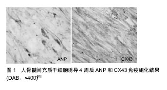
文题释义: 间充质干细胞的免疫调节作用:间充质干细胞对心肌梗死的治疗得到了大量临床证实,不仅是其自我更新和多向分化潜能分化为新生血管、心肌细胞以外,更重要的是其强大的免疫调节作用。间充质干细胞不仅能促进抗炎因子白细胞介素4和白细胞介素10、转化生长因子β等表达和抑制促炎因子白细胞介素1β、白细胞介素6、肿瘤坏死因子α和干扰素γ等表达,也抑制各类炎症反应性细胞的增殖、活化能力,还可以诱导调节性T细胞(Treg)和M2型巨噬细胞的分化。免疫调节机制对心肌梗死的治疗有重要意义。研究者可以通过基因修饰和药物诱导等方式扩大间充质干细胞在治疗心肌梗死中的免疫调节和细胞分化双重作用,这将增加间充质干细胞在心肌梗死中的治疗价值。 基因转染技术:就是将纯化的含有靶基因的质粒DNA送入细胞内,并在细胞内表达。基因转染已广泛用于基因的结构和功能分析、基因表达与调控、基因治疗与转基因动物等研究。其运载系统包括非病毒方法和病毒方法。利用基因转染技术,不仅增加了间充质干细胞移植后的存活率,还增加了其分化能力和血管再生能力。
摘要 背景:间充质干细胞作为心肌损伤后细胞移植治疗的热门种子细胞,成为治疗缺血性心脏病研究的热点。 目的:总结和分析间充质干细胞移植治疗缺血性心脏病的研究新进展。 方法:计算机检索Medline、Ovid Embase、PubMed Central、Cochrane Library、中国期刊全文数据库(CNKI)、万方数据库、中国生物医学文献数据库(CBM)等文献数据库,语种限为中文和英文,检索时间为1985至2015年。纳入标准:①与间充质干细胞治疗缺血性心脏病相关的随机对照实验或临床研究;②方法规范、思路严谨的综述论文。排除标准:观察性研究、个案报道、无标准合理设计方案的研究、无规范严谨的纳入排除标准的综述论文、交叉试验研究、整群随机试验、与论述主题无关的研究。 结果与结论:通过检索文献,总结了间充质干细胞来源、间充质干细胞的安全性和有效性、作用机制、注射方式,间充质干细胞移植的优劣势等方面的研究进展。骨髓间充质干细胞移植治疗心肌梗死给临床带来了全新的方法,但仍存在许多需要去认识、解决的问题,如在细胞质量、细胞剂量、移植方法、移植时机和患者选择等各个方面仍未得到确切的答案,故局限了其在临床上的应用。
中图分类号:
引用本文
赵启明,盛 开,张选奋. 间充质干细胞治疗心肌梗死:离临床应用还有多远?[J]. 中国组织工程研究, 2017, 21(17): 2766-2775.
Zhao Qi-ming, Sheng Kai, Zhang Xuan-fen. Mesenchymal stem cells in the treatment of myocardial infarction: how long is the way from bench to bedside?[J]. Chinese Journal of Tissue Engineering Research, 2017, 21(17): 2766-2775.
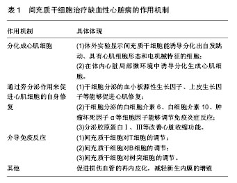
间充质干细胞治疗缺血性心脏病的作用机制见表1。
| [1] Reinhardt D, Sigusch HH, Hensse J, et al. Cardiac remodelling in end stage heart failure: upregulation of matrix metalloproteinase (MMP) irrespective of the underlying disease, and evidence for a direct inhibitory effect of ACE inhibitors on MMP. Heart. 2002;88(5):525-530.[2] Beltrami AP, Urbanek K, Kajstura J, et al. Evidence that human cardiac myocytes divide after myocardial infarction. N Engl J Med. 2001;344(23):1750-1757.[3] Herlitz J, Dellborg M, Karlson BW, et al. Prognosis after acute myocardial infarction continues to improve in the reperfusion era in the community of Göteborg. Am Heart J. 2002;144(1):89-94.[4] Henning RJ. Stem cells in cardiac repair. Future Cardiol. 2011; 7(1):99-117.[5] Bhaskar B, Mekala NK, Baadhe RR, et al. Role of signaling pathways in mesenchymal stem cell differentiation. Curr Stem Cell Res Ther. 2014;9(6):508-512.[6] 马莎. 骨髓间充质干细胞治疗缺血性心脏病的研究进展[J].医学综述, 2012, 18(16):2557-2559.[7] Zhu X, Du J, Liu G. The comparison of multilineage differentiation of bone marrow and adipose-derived mesenchymal stem cells. Clin Lab. 2012;58(9-10):897-903.[8] 唐海沁,马健,翟志敏,等.人骨髓间充质干细胞体外定向分化过程中心肌细胞特征表达及膜电位变化[J]. 临床心血管病杂志, 2007, 23(6):437-441.[9] 汤天军,杨波,杨汉东,等.人脐带血间充质干细胞体外诱导分化为心肌细胞的实验研究[J].实用医学杂志,2005,21(21):2412- 2414.[10] Wen Z, Zheng S, Zhou C, et al. Repair mechanisms of bone marrow mesenchymal stem cells in myocardial infarction. J Cell Mol Med. 2011;15(5):1032-1043.[11] Lin X, Peng P, Cheng L, et al. A natural compound induced cardiogenic differentiation of endogenous MSCs for repair of infarcted heart. Differentiation. 2012;83(1):1-9.[12] Rota M, Padin-Iruegas ME, Misao Y, et al. Local activation or implantation of cardiac progenitor cells rescues scarred infarcted myocardium improving cardiac function. Circ Res. 2008;103(1):107-116.[13] Zhao JJ, Liu XC, Kong F, et al. Bone marrow mesenchymal stem cells improve myocardial function in a swine model of acute myocardial infarction. Mol Med Rep. 2014;10(3): 1448-1454.[14] Rahbarghazi R, Nassiri SM, Ahmadi SH, et al. Dynamic induction of pro-angiogenic milieu after transplantation of marrow-derived mesenchymal stem cells in experimental myocardial infarction. Int J Cardiol. 2014;173(3):453-466.[15] Won YW, Patel AN, Bull DA. Cell surface engineering to enhance mesenchymal stem cell migration toward an SDF-1 gradient. Biomaterials. 2014;35(21):5627-5635.[16] Ma N, Ladilov Y, Moebius JM, et al. Intramyocardial delivery of human CD133+ cells in a SCID mouse cryoinjury model: Bone marrow vs. cord blood-derived cells. Cardiovasc Res. 2006;71(1):158-169.[17] Ma N, Stamm C, Kaminski A, et al. Human cord blood cells induce angiogenesis following myocardial infarction in NOD/scid-mice. Cardiovasc Res. 2005;66(1):45-54.[18] Lee OK, Kuo TK, Chen WM, et al. Isolation of multipotent mesenchymal stem cells from umbilical cord blood. Blood. 2004;103(5):1669-1675.[19] Li T, Ma Q, Ning M, et al. Cotransplantation of human umbilical cord-derived mesenchymal stem cells and umbilical cord blood-derived CD34? cells in a rabbit model of myocardial infarction. Mol Cell Biochem. 2014;387(1-2): 91-100.[20] Mareschi K, Biasin E, Piacibello W, et al. Isolation of human mesenchymal stem cells: bone marrow versus umbilical cord blood. Haematologica. 2001;86(10):1099-1100.[21] Nagamura-Inoue T, He H. Umbilical cord-derived mesenchymal stem cells: Their advantages and potential clinical utility. World J Stem Cells. 2014;6(2):195-202.[22] Fong CY, Chak LL, Biswas A, et al. Human Wharton's jelly stem cells have unique transcriptome profiles compared to human embryonic stem cells and other mesenchymal stem cells. Stem Cell Rev. 2011;7(1):1-16.[23] Musialek P, Mazurek A, Jarocha D, et al. Myocardial regeneration strategy using Wharton's jelly mesenchymal stem cells as an off-the-shelf 'unlimited' therapeutic agent: results from the Acute Myocardial Infarction First-in-Man Study. Postepy Kardiol Interwencyjnej. 2015;11(2):100-107.[24] Zhang W, Liu XC, Yang L, et al. Wharton's jelly-derived mesenchymal stem cells promote myocardial regeneration and cardiac repair after miniswine acute myocardial infarction. Coron Artery Dis. 2013;24(7):549-558.[25] Gao LR, Zhang NK, Ding QA, et al. Common expression of stemness molecular markers and early cardiac transcription factors in human Wharton's jelly-derived mesenchymal stem cells and embryonic stem cells. Cell Transplant. 2013;22(10): 1883-1900.[26] Gao LR, Chen Y, Zhang NK, et al. Intracoronary infusion of Wharton's jelly-derived mesenchymal stem cells in acute myocardial infarction: double-blind, randomized controlled trial. BMC Med. 2015;13:162.[27] Covas DT, Piccinato CE, Orellana MD, et al. Mesenchymal stem cells can be obtained from the human saphena vein. Exp Cell Res. 2005;309(2):340-344.[28] Imanishi Y, Miyagawa S, Kitagawa-Sakakida S, et al. Impact of synovial membrane-derived stem cell transplantation in a rat model of myocardial infarction. J Artif Organs. 2009;12(3): 187-193.[29] Fazel SS, Angoulvant D, Butany J, et al. Mesenchymal stem cells engineered to overexpress stem cell factor improve cardiac function but have malignant potential. J Thorac Cardiovasc Surg. 2008;136(5):1388-1389.[30] Furlani D, Li W, Pittermann E, et al. A transformed cell population derived from cultured mesenchymal stem cells has no functional effect after transplantation into the injured heart. Cell Transplant. 2009;18(3):319-331.[31] Khan M, Mohsin S, Khan SN, et al. Repair of senescent myocardium by mesenchymal stem cells is dependent on the age of donor mice. J Cell Mol Med. 2011;15(7):1515-1527.[32] 许丹,尚小明,姜玉如.经冠状动脉自体骨髓单个核细胞移植治疗急性前壁心肌梗死的研究[J].中国心血管杂志,2006,11(4): 261-265.[33] 王伟民,孙宁玲,刘健,等.经冠状动脉注入自体骨髓单个核细胞的临床研究[J].中华心血管病杂志,2006,34(2):103-106.[34] 栾云,刘晓程,张兆华,等.骨髓间充质干细胞移植治疗小型猪急性心肌梗死[J].基础医学与临床,2011,31(10):1156-1158.[35] 国海东,谭玉珍, 王海杰. 细胞因子在干细胞移植存活与分化中的作用[J]. 国际心血管病杂志,2007,34(1):23-26.[36] Tang J, Xie Q, Pan G, et al. Mesenchymal stem cells participate in angiogenesis and improve heart function in rat model of myocardial ischemia with reperfusion. Eur J Cardiothorac Surg. 2006;30(2):353-361.[37] Wu Y, Chen L, Scott PG, et al. Mesenchymal stem cells enhance wound healing through differentiation and angiogenesis. Stem Cells. 2007;25(10):2648-2659.[38] Mirotsou M, Jayawardena TM, Schmeckpeper J, et al. Paracrine mechanisms of stem cell reparative and regenerative actions in the heart. J Mol Cell Cardiol. 2011;50(2):280-289.[39] Chen L, Tredget EE, Wu PY, et al. Paracrine factors of mesenchymal stem cells recruit macrophages and endothelial lineage cells and enhance wound healing. PLoS One. 2008; 3(4):e1886.[40] Hocking AM, Gibran NS. Mesenchymal stem cells: paracrine signaling and differentiation during cutaneous wound repair. Exp Cell Res. 2010;316(14):2213-2219.[41] Uemura R, Xu M, Ahmad N, et al. Bone marrow stem cells prevent left ventricular remodeling of ischemic heart through paracrine signaling. Circ Res. 2006;98(11):1414-1421.[42] Li Z, Guo J, Chang Q, et al. Paracrine role for mesenchymal stem cells in acute myocardial infarction. Biol Pharm Bull. 2009;32(8):1343-1346.[43] Ma XH, Gao CQ, Li BJ, et al. The empirical study in transplantation of bone marrow mesenchymal stem cells for acute myocardial infarction. Zhonghua Yi Xue Za Zhi. 2009; 89(15):1067-1070.[44] Deuse T, Peter C, Fedak PW, et al. Hepatocyte growth factor or vascular endothelial growth factor gene transfer maximizes mesenchymal stem cell-based myocardial salvage after acute myocardial infarction. Circulation. 2009;120(11 Suppl): S247-254.[45] Gnecchi M, He H, Melo LG, et al. Early beneficial effects of bone marrow-derived mesenchymal stem cells overexpressing Akt on cardiac metabolism after myocardial infarction. Stem Cells. 2009;27(4):971-979.[46] 乔建晶.骨髓间充质干细胞移植调节心肌梗死后炎症反应的研究现状[J].心血管病学进展,2010,31(5):729-732.[47] 杜优优,姚瑞,蒲实,等.骨髓间充质干细胞治疗增加心脏梗死区肌纤维母细胞聚集的观察[J].中华心血管病杂志, 2012, 40(12): 1045-1050.[48] Bader AM, Brodarac A, Klose K, et al. Mechanisms of paracrine cardioprotection by cord blood mesenchymal stromal cells. Eur J Cardiothorac Surg. 2014;45(6):983- 992.[49] Li XH, Fu YH, Liu ZY, et al. Efficacy comparison between transplanting microenvironmental induced and non-induced bone marrow mesenchymal stem cells in ischemic rat hearts. Zhonghua Xin Xue Guan Bing Za Zhi. 2009;37(8):680-684.[50] Ben-Mordechai T, Holbova R, Landa-Rouben N, et al. Macrophage subpopulations are essential for infarct repair with and without stem cell therapy. J Am Coll Cardiol. 2013; 62(20):1890-1901.[51] Yan X, Shichita T, Katsumata Y, et al. Deleterious effect of the IL-23/IL-17A axis and γδT cells on left ventricular remodeling after myocardial infarction. J Am Heart Assoc. 2012;1(5): e004408.[52] Mándi Y, Vécsei L. The kynurenine system and immunoregulation. J Neural Transm (Vienna). 2012;119(2): 197-209.[53] François M, Romieu-Mourez R, Li M, et al. Human MSC suppression correlates with cytokine induction of indoleamine 2,3-dioxygenase and bystander M2 macrophage differentiation. Mol Ther. 2012;20(1):187-195.[54] Pradier A, Passweg J, Villard J, et al. Human bone marrow stromal cells and skin fibroblasts inhibit natural killer cell proliferation and cytotoxic activity. Cell Transplant. 2011; 20(5):681-691.[55] Abumaree M, Al Jumah M, Pace RA, et al. Immunosuppressive properties of mesenchymal stem cells. Stem Cell Rev. 2012;8(2):375-392.[56] Spaggiari GM, Capobianco A, Abdelrazik H, et al. Mesenchymal stem cells inhibit natural killer-cell proliferation, cytotoxicity, and cytokine production: role of indoleamine 2,3-dioxygenase and prostaglandin E2. Blood. 2008;111(3): 1327-1333.[57] 石蓓,刘志江,赵然尊,等.骨髓间充质干细胞移植对心肌梗死后心脏功能及损伤血管再狭窄的影响[J].中华医学杂志,2011,91(32): 2269-2273.[58] Assis AC, Carvalho JL, Jacoby BA, et al. Time-dependent migration of systemically delivered bone marrow mesenchymal stem cells to the infarcted heart. Cell Transplant. 2010;19(2):219-230.[59] Gyöngyösi M, Hemetsberger R, Posa A, et al. Hypoxia-inducible factor 1-alpha release after intracoronary versus intramyocardial stem cell therapy in myocardial infarction. J Cardiovasc Transl Res. 2010;3(2):114-121.[60] Dai W, Hale SL, Kay GL, et al. Delivering stem cells to the heart in a collagen matrix reduces relocation of cells to other organs as assessed by nanoparticle technology. Regen Med. 2009;4(3):387-395.[61] Tay CY, Yu H, Pal M, et al. Micropatterned matrix directs differentiation of human mesenchymal stem cells towards myocardial lineage. Exp Cell Res. 2010;316(7):1159-1168.[62] Yang Z, Zhang F, Ma W, et al. A novel approach to transplanting bone marrow stem cells to repair human myocardial infarction: delivery via a noninfarct-relative artery. Cardiovasc Ther. 2010;28(6):380-385.[63] Vulliet PR, Greeley M, Halloran SM, et al. Intra-coronary arterial injection of mesenchymal stromal cells and microinfarction in dogs. Lancet. 2004;363(9411):783-784.[64] Grieve SM, Bhindi R, Seow J, et al. Microvascular obstruction by intracoronary delivery of mesenchymal stem cells and quantification of resulting myocardial infarction by cardiac magnetic resonance. Circ Heart Fail. 2010;3(3):e5-6.[65] Gyöngyösi M, Hemetsberger R, Wolbank S, et al. Delayed recovery of myocardial blood flow after intracoronary stem cell administration. Stem Cell Rev. 2011;7(3):616-623.[66] Llano R, Epstein S, Zhou R, et al. Intracoronary delivery of mesenchymal stem cells at high flow rates after myocardial infarction improves distal coronary blood flow and decreases mortality in pigs. Catheter Cardiovasc Interv. 2009;73(2): 251-257.[67] Schmuck EG, Mulligan JD, Ertel RL, et al. Cardiac fibroblast-derived 3D extracellular matrix seeded with mesenchymal stem cells as a novel device to transfer cells to the ischemic myocardium. Cardiovasc Eng Technol. 2014; 5(1):119-131.[68] Ghanem A, Steingen C, Brenig F, et al. Focused ultrasound- induced stimulation of microbubbles augments site-targeted engraftment of mesenchymal stem cells after acute myocardial infarction. J Mol Cell Cardiol. 2009;47(3):411-418.[69] Li L, Wu S, Liu Z, et al. Ultrasound-Targeted Microbubble Destruction Improves the Migration and Homing of Mesenchymal Stem Cells after Myocardial Infarction by Upregulating SDF-1/CXCR4: A Pilot Study. Stem Cells Int. 2015;2015:691310.[70] Tano N, Narita T, Kaneko M, et al. Epicardial placement of mesenchymal stromal cell-sheets for the treatment of ischemic cardiomyopathy; in vivo proof-of-concept study. Mol Ther. 2014;22(10):1864-1871.[71] Hare JM, Traverse JH, Henry TD, et al. A randomized, double-blind, placebo-controlled, dose-escalation study of intravenous adult human mesenchymal stem cells (prochymal) after acute myocardial infarction. J Am Coll Cardiol. 2009; 54(24):2277-2286.[72] Kan CD, Lee HL, Yang YJ. Cell transplantation for myocardial injury: a preliminary comparative study. Cytotherapy. 2010; 12(5): 692-700.[73] Mathieu M, Bartunek J, El Oumeiri B, et al. Cell therapy with autologous bone marrow mononuclear stem cells is associated with superior cardiac recovery compared with use of nonmodified mesenchymal stem cells in a canine model of chronic myocardial infarction. J Thorac Cardiovasc Surg. 2009; 138(3):646-653.[74] Mazo M, Gavira JJ, Abizanda G, et al. Transplantation of mesenchymal stem cells exerts a greater long-term effect than bone marrow mononuclear cells in a chronic myocardial infarction model in rat. Cell Transplant. 2010;19(3):313-328.[75] Armiñán A, Gandía C, García-Verdugo JM, et al. Mesenchymal stem cells provide better results than hematopoietic precursors for the treatment of myocardial infarction. J Am Coll Cardiol. 2010;55(20):2244-2253.[76] Li Q, Turdi S, Thomas DP, et al. Intra-myocardial delivery of mesenchymal stem cells ameliorates left ventricular and cardiomyocyte contractile dysfunction following myocardial infarction. Toxicol Lett. 2010;195(2-3):119-126.[77] 费宇行,裘毅钢,李晶.骨髓干细胞移植改善心肌梗死患者左室重构的Meta分析[J].中国循证心血管医学杂志, 2011,3(4):255-259.[78] 王德国,曹克将.干细胞治疗与室性心律失常[J].国际心血管病杂志,2008,35(3):129-132.[79] Costa AR, Panda NC, Yong S, et al. Optical mapping of cryoinjured rat myocardium grafted with mesenchymal stem cells. Am J Physiol Heart Circ Physiol. 2012;302(1):H270-277.[80] Amado LC, Saliaris AP, Schuleri KH, et al. Cardiac repair with intramyocardial injection of allogeneic mesenchymal stem cells after myocardial infarction. Proc Natl Acad Sci U S A. 2005;102(32):11474-11479.[81] 伍徐娴,何志旭,周中军.骨髓间充质干细胞移植治疗大鼠实验性心肌梗死[J].贵阳医学院学报,2010,35(4):344-348.[82] 宋晓蓉,翟志敏,章仁品,等.兔骨髓单个核细胞自体移植对缺血心肌修复重建及心功能的改善[J].中国组织工程研究,2006, 10(37):27-29.[83] Fan L, Lin C, Zhuo S, et al. Transplantation with survivin-engineered mesenchymal stem cells results in better prognosis in a rat model of myocardial infarction. Eur J Heart Fail. 2009;11(11):1023-1030.[84] Chang W, Song BW, Lim S, et al. Mesenchymal stem cells pretreated with delivered Hph-1-Hsp70 protein are protected from hypoxia-mediated cell death and rescue heart functions from myocardial injury. Stem Cells. 2009;27(9):2283-2292.[85] Huang J, Zhang Z, Guo J, et al. Genetic modification of mesenchymal stem cells overexpressing CCR1 increases cell viability, migration, engraftment, and capillary density in the injured myocardium. Circ Res. 2010;106(11):1753-1762.[86] Alfaro MP, Vincent A, Saraswati S, et al. sFRP2 suppression of bone morphogenic protein (BMP) and Wnt signaling mediates mesenchymal stem cell (MSC) self-renewal promoting engraftment and myocardial repair. J Biol Chem. 2010;285(46):35645-35653.[87] Eun LY, Song BW, Cha MJ, et al. Overexpression of phosphoinositide-3-kinase class II alpha enhances mesenchymal stem cell survival in infarcted myocardium. Biochem Biophys Res Commun. 2010;402(2):272-279.[88] Kim HW, Haider HK, Jiang S, et al. Ischemic preconditioning augments survival of stem cells via miR-210 expression by targeting caspase-8-associated protein 2. J Biol Chem. 2009; 284(48):33161-33168.[89] Krausgrill B, Vantler M, Burst V, et al. Influence of cell treatment with PDGF-BB and reperfusion on cardiac persistence of mononuclear and mesenchymal bone marrow cells after transplantation into acute myocardial infarction in rats. Cell Transplant. 2009;18(8):847-853.[90] Cui X, Wang H, Guo H, et al. Transplantation of mesenchymal stem cells preconditioned with diazoxide, a mitochondrial ATP-sensitive potassium channel opener, promotes repair of myocardial infarction in rats. Tohoku J Exp Med. 2010;220(2): 139-147.[91] Numasawa Y, Kimura T, Miyoshi S, et al. Treatment of human mesenchymal stem cells with angiotensin receptor blocker improved efficiency of cardiomyogenic transdifferentiation and improved cardiac function via angiogenesis. Stem Cells. 2011;29(9):1405-1414.[92] Ikhapoh IA, Pelham CJ, Agrawal DK. Synergistic effect of angiotensin II on vascular endothelial growth factor-A-mediated differentiation of bone marrow-derived mesenchymal stem cells into endothelial cells. Stem Cell Res Ther. 2015;6:4.[93] Akomolafe SF, Oboh G, Akindahunsi AA, et al. Tetracarpidium conophorum (Mull.Arg) Hutch & Dalziel inhibits FeSO4-induced lipid peroxidation in rat's genitals. BMC Complement Altern Med. 2015;15:57.[94] Khan M, Meduru S, Mohan IK, et al. Hyperbaric oxygenation enhances transplanted cell graft and functional recovery in the infarct heart. J Mol Cell Cardiol. 2009;47(2):275-287.[95] Enoki C, Otani H, Sato D, et al. Enhanced mesenchymal cell engraftment by IGF-1 improves left ventricular function in rats undergoing myocardial infarction. Int J Cardiol. 2010;138(1): 9-18.[96] Eun LY, Song H, Choi E, et al. Implanted bone marrow-derived mesenchymal stem cells fail to metabolically stabilize or recover electromechanical function in infarcted hearts. Tissue Cell. 2011;43(4):238-245.[97] Fang J, Chen L, Fan L, et al. Enhanced therapeutic effects of mesenchymal stem cells on myocardial infarction by ischemic postconditioning through paracrine mechanisms in rats. J Mol Cell Cardiol. 2011;51(5):839-847.[98] Mias C, Lairez O, Trouche E, et al. Mesenchymal stem cells promote matrix metalloproteinase secretion by cardiac fibroblasts and reduce cardiac ventricular fibrosis after myocardial infarction. Stem Cells. 2009;27(11):2734-2743.[99] Chen JJ, Zhou SH. Mesenchymal stem cells overexpressing MiR-126 enhance ischemic angiogenesis via the AKT/ERK-related pathway. Cardiol J. 2011;18(6):675-681.[100] Cho J, Zhai P, Maejima Y, et al. Myocardial injection with GSK-3β-overexpressing bone marrow-derived mesenchymal stem cells attenuates cardiac dysfunction after myocardial infarction. Circ Res. 2011;108(4):478-489.[101] Gao XR, Tan YZ, Wang HJ. Overexpression of Csx/Nkx2.5 and GATA-4 enhances the efficacy of mesenchymal stem cell transplantation after myocardial infarction. Circ J. 2011;75(11): 2683-2691.[102] Jin J, Jeong SI, Shin YM, et al. Transplantation of mesenchymal stem cells within a poly(lactide-co-epsilon-caprolactone) scaffold improves cardiac function in a rat myocardial infarction model. Eur J Heart Fail. 2009;11(2):147-153.[103] Cui XJ, Xie H, Wang HJ, et al. Transplantation of mesenchymal stem cells with self-assembling polypeptide scaffolds is conducive to treating myocardial infarction in rats. Tohoku J Exp Med. 2010;222(4):281-289.[104] Wang Y, Liu XC, Zhang GW, et al. A new transmyocardial degradable stent combined with growth factor, heparin, and stem cells in acute myocardial infarction. Cardiovasc Res. 2009;84(3):461-469.[105] Luan Y, Liu XC, Zhang GW, et al. Mid-term effect of stem cells combined with transmyocardial degradable stent on swine model of acute myocardial infarction. Coron Artery Dis. 2010; 21(4):233-243.[106] Afanas'ev SA, Tsapko LP, Rogovskaya YV, et al. Radiofrequency ablation as a possible method for preparing pathologically altered myocardium for intramyocardial cell transplantation. Bull Exp Biol Med. 2012;152(4):513-515. |
| [1] | 项鑫健, 柳 芳, 吴亮亮, 贾大平, 陶 越, 赵政男, 赵 宇. 高剂量维生素C促进大鼠自体脂肪移植成活[J]. 中国组织工程研究, 2022, 26(8): 1242-1246. |
| [2] | 王 景, 熊 山, 曹 金, 冯林伟, 王 信. 白细胞介素3在骨代谢中的作用及机制[J]. 中国组织工程研究, 2022, 26(8): 1260-1265. |
| [3] | 肖 豪, 刘 静, 周 君. 脉冲电磁场治疗绝经后骨质疏松症的研究进展[J]. 中国组织工程研究, 2022, 26(8): 1266-1271. |
| [4] | 田 川, 朱向情, 杨再玲, 鄢东海, 李 晔, 王严影, 杨育坤, 何 洁, 吕冠柯, 蔡学敏, 舒丽萍, 何志旭, 潘兴华. 骨髓间充质干细胞调控猕猴卵巢的衰老[J]. 中国组织工程研究, 2022, 26(7): 985-991. |
| [5] | 胡 伟, 谢兴奇, 屠冠军. 骨髓间充质干细胞来源外泌体改善脊髓损伤后血脊髓屏障的完整性[J]. 中国组织工程研究, 2022, 26(7): 992-998. |
| [6] | 侯婧瑛, 郭天柱, 于萌蕾, 龙会宝, 吴 浩. 缺氧预处理通过激活MALAT1靶向抑制miR-195促进骨髓间充质干细胞的生存和血管形成[J]. 中国组织工程研究, 2022, 26(7): 1005-1011. |
| [7] | 周 颖, 张 幻, 廖 松, 胡凡琦, 易 静, 刘玉斌, 靳继德. 去铁胺联合干扰素γ预处理对人牙髓干细胞的免疫调节作用[J]. 中国组织工程研究, 2022, 26(7): 1012-1019. |
| [8] | 梁学振, 杨 曦, 李嘉程, 骆 帝, 许 波, 李 刚. 补肾活血胶囊介导Hedgehog信号通路调控大鼠骨髓间充质干细胞成骨成脂分化[J]. 中国组织工程研究, 2022, 26(7): 1020-1026. |
| [9] | 王继芳, 鲍 祯, 乔亚红. miR-206调控小细胞肺癌干细胞EVI1基因表达及细胞生物学行为[J]. 中国组织工程研究, 2022, 26(7): 1027-1031. |
| [10] | 刘 峰, 彭宇环, 罗良平, 吴本清. 植物源性碱性成纤维细胞生长因子维持人胚胎干细胞的生长与分化[J]. 中国组织工程研究, 2022, 26(7): 1032-1037. |
| [11] | 闻丹丹, 李 强, 沈才齐, 纪 哲, 金培生. 外用红色诺卡氏菌细胞壁骨架提高脂肪间充质干细胞活性修复糖尿病创面[J]. 中国组织工程研究, 2022, 26(7): 1038-1044. |
| [12] | 朱兵兵, 邓江华, 陈晶晶, 慕晓玲. 白细胞介素8受体可提高脐带间充质干细胞迁移和向损伤内皮的黏附[J]. 中国组织工程研究, 2022, 26(7): 1045-1050. |
| [13] | 罗小玲, 张 丽, 杨茂桦, 徐 洁, 徐晓梅. 柚皮素干预人牙周膜干细胞的成骨分化能力[J]. 中国组织工程研究, 2022, 26(7): 1051-1056. |
| [14] | 崔 兴, 孙小琪, 郑 伟, 马德欣. 黄芩汤调控自噬干预小鼠肠道急性移植物抗宿主病的机制[J]. 中国组织工程研究, 2022, 26(7): 1057-1062. |
| [15] | 熊挺淋, 应梦慧, 张丽莎, 张晓刚, 杨 燕. 诱导多能干细胞分化的心肌细胞电生理特点[J]. 中国组织工程研究, 2022, 26(7): 1063-1067. |
1.2.7 检索偏离的描述、原因及对结果的影响 因一些文献未按主题词标注或所标注关键词不规范,可能导致检索时未能完全检索主题词的近义词或扩展词,而使得部分相关文献可能被遗漏,部分文献数据因尚未发表完全或作者无法联系而被排除,导致检索结果的偏倚。
.jpg)
文题释义: 间充质干细胞的免疫调节作用:间充质干细胞对心肌梗死的治疗得到了大量临床证实,不仅是其自我更新和多向分化潜能分化为新生血管、心肌细胞以外,更重要的是其强大的免疫调节作用。间充质干细胞不仅能促进抗炎因子白细胞介素4和白细胞介素10、转化生长因子β等表达和抑制促炎因子白细胞介素1β、白细胞介素6、肿瘤坏死因子α和干扰素γ等表达,也抑制各类炎症反应性细胞的增殖、活化能力,还可以诱导调节性T细胞(Treg)和M2型巨噬细胞的分化。免疫调节机制对心肌梗死的治疗有重要意义。研究者可以通过基因修饰和药物诱导等方式扩大间充质干细胞在治疗心肌梗死中的免疫调节和细胞分化双重作用,这将增加间充质干细胞在心肌梗死中的治疗价值。 基因转染技术:就是将纯化的含有靶基因的质粒DNA送入细胞内,并在细胞内表达。基因转染已广泛用于基因的结构和功能分析、基因表达与调控、基因治疗与转基因动物等研究。其运载系统包括非病毒方法和病毒方法。利用基因转染技术,不仅增加了间充质干细胞移植后的存活率,还增加了其分化能力和血管再生能力。
骨髓间充质干细胞移植治疗心肌梗死给临床带来了全新的方法,但仍存在许多需要去认识、解决的问题,如在细胞质量、细胞剂量、移植方法、移植时机和患者选择等各个方面仍未得到确切的答案,故局限了其在临床上的应用。随着实验技术、研究方法和相关理论知识的完善,相信骨髓间充质干细胞移植必将为心肌梗死患者的治疗带来希望。
| 阅读次数 | ||||||
|
全文 |
|
|||||
|
摘要 |
|
|||||