中国组织工程研究 ›› 2016, Vol. 20 ›› Issue (33): 4890-4898.doi: 10.3969/j.issn.2095-4344.2016.33.004
• 皮肤粘膜组织构建 skin and mucosal tissue construction • 上一篇 下一篇
生肌玉红膏对增生性瘢痕的抑制作用
孙桂芳1,张晓芬1,李红昌1,潘丽芸1,陈亚峰1,徐 可2,奉典旭1
- 上海市普陀区中心医院,1普外科, 2中心实验室,上海市 200062
Shengjiyuhong ointment inhibits hypertrophic scar formation
Sun Gui-fang1, Zhang Xiao-fen1, Li Hong-chang1, Pan Li-yun1, Chen Ya-feng1, Xu Ke2, Feng Dian-xu1
- 1Department of General Surgery, 2Central Laboratory, Shanghai Putuo District Central Hospital, Shanghai 200062, China
摘要:
文章快速阅读:
.jpg) 文题释义:
增生性瘢痕:临床上增生性瘢痕几乎均继发于深达真皮网状层的皮肤损伤,如皮肤外伤或Ⅱ-Ⅲ度烧伤。病理特征主要表现为肌成纤维细胞的过度增殖及细胞外基质尤其是胶原的过度沉积,致使愈合后的组织过度挛缩,从而导致增生性瘢痕形成。不仅影响美观,还影响器官和组织功能,给患者带来巨大的身心痛苦。
α-平滑肌肌动蛋白:组织受损后,创面边缘的成纤维细胞被激活并分化成为表达α-平滑肌肌动蛋白的肌成纤维细胞,α-平滑肌肌动蛋白显著增加了肌成纤维细胞和胶原晶格的收缩力,可加速创面愈合,促进创面重塑,该过程主要受转化生长因子β1/Smads信号通路调节。
文题释义:
增生性瘢痕:临床上增生性瘢痕几乎均继发于深达真皮网状层的皮肤损伤,如皮肤外伤或Ⅱ-Ⅲ度烧伤。病理特征主要表现为肌成纤维细胞的过度增殖及细胞外基质尤其是胶原的过度沉积,致使愈合后的组织过度挛缩,从而导致增生性瘢痕形成。不仅影响美观,还影响器官和组织功能,给患者带来巨大的身心痛苦。
α-平滑肌肌动蛋白:组织受损后,创面边缘的成纤维细胞被激活并分化成为表达α-平滑肌肌动蛋白的肌成纤维细胞,α-平滑肌肌动蛋白显著增加了肌成纤维细胞和胶原晶格的收缩力,可加速创面愈合,促进创面重塑,该过程主要受转化生长因子β1/Smads信号通路调节。
文题释义:
增生性瘢痕:临床上增生性瘢痕几乎均继发于深达真皮网状层的皮肤损伤,如皮肤外伤或Ⅱ-Ⅲ度烧伤。病理特征主要表现为肌成纤维细胞的过度增殖及细胞外基质尤其是胶原的过度沉积,致使愈合后的组织过度挛缩,从而导致增生性瘢痕形成。不仅影响美观,还影响器官和组织功能,给患者带来巨大的身心痛苦。
α-平滑肌肌动蛋白:组织受损后,创面边缘的成纤维细胞被激活并分化成为表达α-平滑肌肌动蛋白的肌成纤维细胞,α-平滑肌肌动蛋白显著增加了肌成纤维细胞和胶原晶格的收缩力,可加速创面愈合,促进创面重塑,该过程主要受转化生长因子β1/Smads信号通路调节。摘要
背景:文献研究和临床使用中均发现生肌玉红膏具有减轻瘢痕的作用。
目的:进一步验证生肌玉红膏对增生性瘢痕的作用。
方法:36只日本大耳白兔,每只兔耳造4个直径1 cm圆形全层皮肤和软骨膜缺损创面,共288个创面随机分为6组,分别为模型组,基质组(不含生药),生肌玉红膏低浓度组(生药浓度为8.39%),中浓度组(生药浓度为25.18%),高浓度组(生药浓度为75.54%)和重组牛碱性成纤维细胞生长因子外用溶液阳性对照组。造模后除模型组不予处理外,其余分别给予相应药物涂抹或喷于整个创面,2次/d,直至创面愈合。造模后20,30 d根据温哥华瘢痕量表进行评分;瘢痕标本在显微镜40倍下测量并计算瘢痕增生指数,荧光定量PCR检测Ⅰ、Ⅲ型胶原、结缔组织生长因子、纤连蛋白和α-平滑肌肌动蛋白的mRNA表达,Western bloting检测Ⅰ、Ⅲ型胶原和α-平滑肌肌动蛋白的蛋白表达,免疫荧光染色检测α-平滑肌肌动蛋白的蛋白表达。
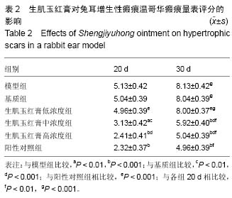
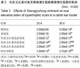
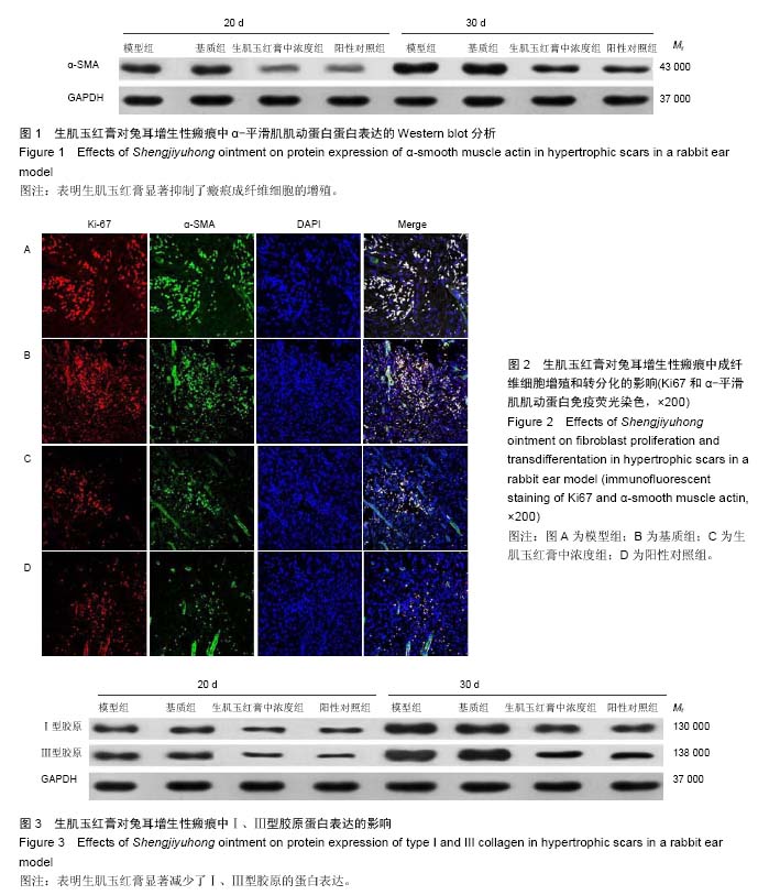
结果与结论:①各组的温哥华瘢痕量表评分和瘢痕增生指数均于30 d显著上升(P < 0.05)。与模型组比较,中、高浓度组和阳性对照组温哥华瘢痕量表评分和瘢痕增生指数显著改善(P < 0.05,P < 0.01),并显著低于基质组和低浓度组(P < 0.05,P < 0.01);②中浓度组和阳性对照组均显著抑制α-平滑肌肌动蛋白和Ⅰ、Ⅲ型胶原、结缔组织生长因子和纤连蛋白的mRNA表达,及α-平滑肌肌动蛋白和Ⅰ、Ⅲ型胶原的蛋白表达(P < 0.01);③结果表明,生肌玉红膏可抑制肌成纤维细胞增殖,减少胶原沉积,从而减轻瘢痕增生。
中国组织工程研究杂志出版内容重点:组织构建;骨细胞;软骨细胞;细胞培养;成纤维细胞;血管内皮细胞;骨质疏松;组织工程
ORCID: 0000-0001-6385-6285(孙桂芳)
中图分类号:


.jpg) 文题释义:
增生性瘢痕:临床上增生性瘢痕几乎均继发于深达真皮网状层的皮肤损伤,如皮肤外伤或Ⅱ-Ⅲ度烧伤。病理特征主要表现为肌成纤维细胞的过度增殖及细胞外基质尤其是胶原的过度沉积,致使愈合后的组织过度挛缩,从而导致增生性瘢痕形成。不仅影响美观,还影响器官和组织功能,给患者带来巨大的身心痛苦。
α-平滑肌肌动蛋白:组织受损后,创面边缘的成纤维细胞被激活并分化成为表达α-平滑肌肌动蛋白的肌成纤维细胞,α-平滑肌肌动蛋白显著增加了肌成纤维细胞和胶原晶格的收缩力,可加速创面愈合,促进创面重塑,该过程主要受转化生长因子β1/Smads信号通路调节。
文题释义:
增生性瘢痕:临床上增生性瘢痕几乎均继发于深达真皮网状层的皮肤损伤,如皮肤外伤或Ⅱ-Ⅲ度烧伤。病理特征主要表现为肌成纤维细胞的过度增殖及细胞外基质尤其是胶原的过度沉积,致使愈合后的组织过度挛缩,从而导致增生性瘢痕形成。不仅影响美观,还影响器官和组织功能,给患者带来巨大的身心痛苦。
α-平滑肌肌动蛋白:组织受损后,创面边缘的成纤维细胞被激活并分化成为表达α-平滑肌肌动蛋白的肌成纤维细胞,α-平滑肌肌动蛋白显著增加了肌成纤维细胞和胶原晶格的收缩力,可加速创面愈合,促进创面重塑,该过程主要受转化生长因子β1/Smads信号通路调节。