中国组织工程研究 ›› 2016, Vol. 20 ›› Issue (13): 1925-1931.doi: 10.3969/j.issn.2095-4344.2016.13.014

• 骨与关节生物力学 bone and joint biomechanics • 上一篇    下一篇

三维有限元分析椎体联合后部结构损伤胸椎转移瘤的生物力学特性

姜维浩,苏秀云,刘耀升,王 铖,刘蜀彬   

  1. 解放军第307医院骨科,北京市 100071
  • 收稿日期:2016-02-22 出版日期:2016-03-25 发布日期:2016-03-25
  • 通讯作者: 刘蜀彬,解放军第307医院骨科,北京市 100071
  • 作者简介:姜维浩,解放军第307医院骨科,北京市 100071
  • 基金资助:
    首都卫生发展科研专项项目(首发2011-5006-03),课题名称“个性化有限元分析预测椎体转移瘤脊髓压迫及病理骨折风险的临床应用研究”

Biomechanical properties of thoracic spine with various locations of metastatic defects: three-dimensional finite element analysis

Jiang Wei-hao, Su Xiu-yun, Liu Yao-sheng, Wang Cheng, Liu Shu-bin   

  1. Department of Orthopedics, the 307th Hospital of Chinese People’s Liberation Army, Beijing 100071, China
  • Received:2016-02-22 Online:2016-03-25 Published:2016-03-25
  • Contact: Liu Shu-bin, Department of Orthopedics, the 307th Hospital of Chinese People’s Liberation Army, Beijing 100071, China
  • About author:Jiang Wei-hao, Department of Orthopedics, the 307th Hospital of Chinese People’s Liberation Army, Beijing 100071, China
  • Supported by:

    the Capital Health Development and Scientific Research Project, No. 2011-5006-03

摘要:

文章快速阅读:

文题释义:

椎体转移瘤的生物力学特性:其生物力学特性改变与多种因素相关。整体骨强度的决定因素包括骨的几何形态、骨密度、骨的胶原特性和显微损伤等。椎体转移瘤局部生物力学特性的有限元分析及实验研究加深了对椎体转移瘤生物力学特性的认识,也证明了有限元分析方法可以模拟预测椎体转移瘤的骨折风险。
Von mises应力:是一种当量应力,它是根据第四强度理论得到的当量应力。von Mises stress 是综合的概念,考虑了第一、第二、第三主应力,可以用来对疲劳,破坏等的评价。


背景:转移瘤最常累及脊柱,以往的生物力学实验多限于模拟转移瘤累及椎体部分或椎体后部结构,而转移瘤同时累及胸椎椎体和后部不同结构时的生物力学改变还有待进一步明确。
目的:建立三维有限元模型分析转移瘤同时累及胸椎椎体及不同后部结构时的生物力学特性。
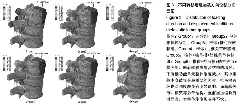
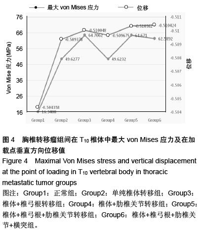
方法:基于CT数据,利用Mimics软件三维重建包括椎间盘、韧带、肋骨的胸椎(T9-T11)几何模型。模拟胸椎转移瘤累及T9椎体和不同后部结构的三维模型,分别为解剖结构完整的对照组,T10椎体右侧半椎体缺损组,椎体缺损基础上破坏同侧椎弓根组,椎体缺损基础上破坏同侧肋椎关节组,椎体缺损基础上破坏同侧椎弓根和肋椎关节组,椎体缺损基础上破坏同侧椎弓根、肋椎关节、横突组。在Abaqus软件中建立相应的三维有限元模型,分析前屈压缩负荷下的位移和Von Mises应力分布情况。
结果与结论:在前屈压缩负荷时,随着胸椎转移瘤累及后部结构的增多,整体刚度减少,其中椎体、椎弓根破坏有明显影响,而胸肋关节、横突等后部结构破坏影响并不大。椎体、椎弓根破坏时最大Von Mises应力值明显增加,而胸肋关节破坏导致应力重新分布,最大Von Mises应力值反而有所减少。 

ORCID: 0000-0001-8256-4704 (苏秀云)

关键词: 骨科植入物, 数字化骨科, 胸椎, 转移瘤, 生物力学, 有限元分析, Von Mises应力, 位移

Abstract:

BACKGROUND: The spinal column is the most common site of cancer metastases. Most of the previous biomechanical experiments utilized models with defects only in the vertebral body or posterior elements, but the biomechanical changes of the thoracic vertebrae and posterior part with various locations of metastasis deserve further research.
OBJECTIVE: To set up the three-dimensional (3D) finite element model to investigate biomechanical effects by simulating combined destruction of vertebral body and other posterior components.
METHODS: Based on CT data, we constructed the 3D geometric models of the thoracic vertebrae (T9-11), including intervertebral discs, ligaments and ribs using the Mimics software. The 3D models of T9 vertebra and different parts of the posterior thoracic vertebrae related with the metastasis could be simulated, including the control group with the intact vertebrae, the group of the T10 vertebrae with the right defective hemi-vertebrae, the group of the defective hemi-vertebrae with the defective ipsilateral pedicle, the group of the defective hemi-vertebrae with the defective ipsilateral costovertebral joint, the group of the defective hemi-vertebrae with the defective ipsilateral pedicle and costovertebral joint, the group of the defective hemi-vertebrae with the defective ipsilateral pedicle, costovertebral joint and transverse process. The corresponding 3D finite element models were established using the Abaqus software. The displacement and Von Mises stress distribution of the models were analyzed when the anterior compressive flexure load was applied.
RESULTS AND CONCLUSION: When the anterior compressive flexure load was applied, the entire stiffness was proportionally decreased when the more posterior parts destroyed, especially destruction of vertebral body and pedicle significantly decreased. The destruction of posterior structures such as the thoracic rib joints and transverse processes was not great. However, the maximal Von Mises stress increased significantly when the vertebral body and pedicle were destructed, but additional costovertebral joint destruction slightly decreased the maximal Von Mises stress because of the stress was re-distributed.