| [1] Seo BM, Miura M, Gronthos S, et al. Inrestigution of multipotent postnatal stem cells from human periodintal ligament. Lancet. 2004;364(9429): 149-155.
[2] Humphrey LL, Fu R, Buckley DI, et al. Periodotal disease and coronary heart disease incidence: A systematic review and meta-analyisis. J Gen Intern Med. 2008;23:2079-2086.
[3] Kao DW, Fiorellini JP. Regenerative periodontal therapy. Front Oral Biol. 2012;15:149-159.
[4] Mudda JA, Bajaj M. Stem cell therapy: a challenge to periodontist. Indian J Dent Res. 2011;22:132-139.
[5] Wang HL, Cooke J. Periodontal regeneration techniques for treatment of periodontal diseases. Dent Clin Nerth Am. 2005;49(3):637-659.
[6] 萨日娜,吴岩.干细胞在口腔医学中的研究与应用特点[J].中国组织工程研究与临床康复,2010,14(14): 2635-2637.
[7] 钟志华,周先略,贺国权,等.人牙周膜干细胞的多向分化潜能实验[J].临床口腔医学杂志,2012,28(4):211-214.
[8] Du L, Yang P, Ge S. Stromal cell-derived factor-1 significantly induces proliferation, migration, and collagen type I expression in a human periodontal ligament stem cell subpopulation. J Periodontol. 2012; 83(3):379-388.
[9] Monje ML, Todah, Palmer TD. Inflammatory blockade restores adult hippocampal neurogenesis. Science. 2003;302(5651):1760-1765.
[10] Pluchino S, Muzio L, Imitola J, et al. Persistent inflammation alters the function of the endogenous brain stem cell compartment. Brain. 2008;131(pt10): 2564-2578.
[11] Heitz-Mayfiele LJ, Trombelli, Heitz F, et al. A systematic review of the effect of surgical debridement vs non-surgical debridement for the treatment of chronic periodontitis. J Clin Periodontol. 2002;29 Suppl 3: 92-102; discussion 160-102.
[12] Angaji M, Gelskey S, Nogueira-Filho G, et al. A systematic review of clinical efficacy of adujunctive antibiotics in the treatment of smokers with periodontitis. J Periodontol. 2010;81(11):1518-1528.
[13] Yoshimura A, Naka T, Kubo M. SOCS proteins, cytokine signalling and immune regulation. Nat Rve Immunol. 2007;7(6):454-465.
[14] Baud V, Karin M. Signal transduction by tumor necrosis factor and its relatives. Trend cell Biol. 2001;11:372-377.
[15] Suffredini AF, Reda D, Banks SM, et al. Effects of recombinant dimeric TNF receptor on human inflammatory responses following intravenous endotoxin administration. J Immunol. 1995;155(10):5038-5045.
[16] Takahashi K, Takashiba S, Nagai A, et al. Assesement of interleukin-6 in the pathogenesis of periodontal disease. J Periodontol. 1994;65(2):147-153.
[17] Montes AH, Asensi V, Alvarez V, et al. The Toll-like receptor 4 (Asp299Gly) polymorphism is a risk factor for gram-negative and haematogenous osteomyelitis. Clin Exp Immunol. 2006;143:404-413.
[18] Itoh K, Udagawa N, Kobayashi K, et al. Lipopolysaccha- ride promotes the survival of osteoclasts via Toll-like receptor4. J Immunol. 2003;170:3688-3695.
[19] Lynn WA, Golenbock DT. Lipopolysaccharide antago-nists. Immunol Today. 1992;13:271-276.
[20] Armitage GC, Wu Y, Wang HY, et al. Low prevalence of a periodontitiss associated interleukin-1 composite genotype in individuals of Chinese heritage. J Periodontol. 2000;71(2):164-171.
[21] Ding J, Ghali O, Lencel P, et al. TNF-alpha and IL-1 bqta inhibit RUNX2 and collagen expression but increase allealine phosphatase activity and mineralization in human mesenchymal stem cells. Life Sci. 2009;84 (15-16): 499-504.
[22] Palacios D, Mozzelta C, Consalvis, et al. TNF/p38/polycomb signaling to epigenetic control of muscle regeneration. Cell Stem Cell. 2010;7(4): 455-469.
[23] Slots J, Genco RJ Black-pigmented Bacteroides species, Capnocytophaga species, and Actinobacillus actinomycetemitans in human periodontal disease: virulence factors in colonization, survival, and tissue destruction. J Dent Res. 1984;63:412-421.
[24] Lindemann RA, Economou JS, Rothermel H. Production of interleukin-1 and tumor necrsis factor by human peripheral monocytes activated by periodontal bacteria and extracted lipopolysaccharides. J Dent Res. 1988;67:1131-1135.
[25] Jang CH, Choi JH, Byun MS, et al. Chloroquine inhibits production of TNF-alpha, IL-1bate and IL-6 from lipopolysaccharide-stimulated human monocyte by different modes. Rheumatology. 2006;45: 703-710.
[26] Manjunath BC, Praveen K, Chandrashekar BR, et al. Periodontal infections: a risk factor for various systemic diseases. Natl Med J India. 2011;24:214-219.
[27] Lakschevitz F, Aboodi G, Tenenbaum H, et al. Diabetes and periodontal diseases: interplay and links. Curr Diabetes Rev. 2011;7:433-439.
[28] Ulevitch RJ, Tobias PS. Receptor-dependent mechanisms of cell stimulation by bacterial endotoxin. Annu Rev Immunol. 1995;13:437-457.
[29] Hla T, Lee MJ, Ancellin N, et al. Lyso-phospholipids-receptor revelations. Science 2001;294:1875-1878.
[30] Pober JS, Sessa WC. Evolving functions of endothelial cells in inflammation. Nat Rev Immunol. 2007;7: 803-815.
[31] Wei S, Kitaura H, Zhou P, et al. IL-1 mediates TNF-induced osteoclastogenesis. J Clin Invest. 2005;115:282-290.
[32] Redlich K, Hayer S, Ricci R, et al. Osteoclasts are essential for TNF-a-mediated joint destruction. J Clin Invest. 2002;110:1419-1427.
[33] Lipsky PE, van der Heijde DM, St Clair EW, et al. Anti-Tumor Necrosis Factor Trial in Rheumatoid Arthritis with Concomitant Therapy Study Group. N Eng1 J Med. 2000;343:1594-1602.
[34] Weinblatt ME, Kremer JM, Bankhurst AD, et al. A trial of etanercept, a recombinant tumor necrosis factor receptor: Fc fusion protein, in patients with rheumatoid arthritis receiving methotrrxate. N Engl J Med. 1999; 340: 253-259.
[35] Gilbert LC, Rubin J, Nanes MS. The p55 TNF receptor mediates TNF inhibition of osteoblast differentiation inde-pendently of apoptosis. Am J Physiol Endocrinol Metab. 2005;288:E1011-E1018.
[36] Zhou FH, Foster BK, Zhou XF. et al. TNF-alpha mediatcs p38 MAP kinase activation and negatively regulates bone formation at the injured growth plate in rats. J Bone Miner Res. 2006;21:1075-1088.
[37] Kitaura H, Sands MS, Aya K, et al. Marrow stromal cells and osteoclast precursors differentially contribute to TNF-a-induced osteoclastogenesis in vivo. J Immunol. 2004;173:4838-4846.
[38] Nanes MS Tumor necrosis factor: Molecular and cel-lular mechanisms in skeletal pathology. Gene. 2003;321:1-15.
[39] Hess K, Ushmorov A, Fiedler J, et al. TNF-alpha primotes osteogenic differentiation of human mesenchymal stem cells by triggering the NF-kappaB signaling pathway. Bone. 2009;45:367-376. |
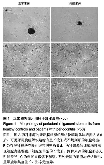
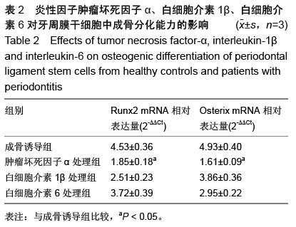
.jpg)
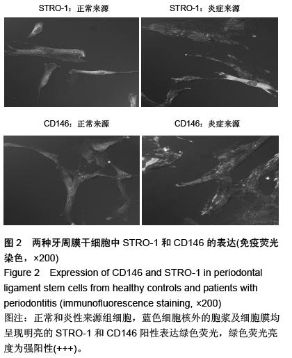
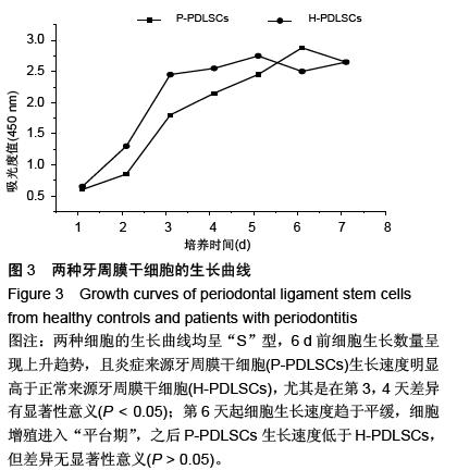
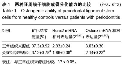
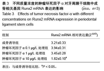
.jpg)