中国组织工程研究 ›› 2016, Vol. 20 ›› Issue (5): 635-639.doi: 10.3969/j.issn.2095-4344.2016.05.005

• 周围神经损伤动物模型 Animal models of peripheral nerve injury • 上一篇    下一篇

新型脊髓完全横断缺损模型大鼠的建立

贺 丰1,俞 兴2,穆晓红2,赵 赫1,李少刚1,杨永栋1,李朋安1,元小红1,朱陵群3,付玲玲1,徐 林2   

  1. 1北京中医药大学,北京市100029;2北京中医药大学东直门医院,北京市 100700;3北京中医药大学东直门医院中医内科学教育部重点实验室和北京市重点实验室,北京市 100700
  • 收稿日期:2015-11-20 出版日期:2016-01-29 发布日期:2016-01-29
  • 通讯作者: 徐林,博士,教授,主任医师,北京中医药大学东直门医院,北京市 100700
  • 作者简介:贺丰,男,1986年生,内蒙古自治区人,鄂伦春族,北京中医药大学在读博士,主要从事脊柱外科方面的研究。
  • 基金资助:
    教育部2012年度“新世纪优秀人才支持计划”(NCET-12-0805)

Establishment of a new rat model of complete spinal cord transection and defect

He Feng1, Yu Xing2, Mu Xiao-hong2, Zhao He1, Li Shao-gang1, Yang Yong-dong1, Li Peng-an1, Yuan Xiao-hong1, Zhu Ling-qun3, Fu Ling-ling1, Xu Lin2   

  1. 1Beijing University of Chinese Medicine, Beijing 100029, China; 2Dongzhimen Hospital, Beijing University of Chinese Medicine, Beijing 100700, China; 3Key Laboratory of the Ministry of Education and the Key Laboratory of Beijing City, Department of Internal Medicine of Traditional Chinese Medicine, Dongzhimen Hospital, Beijing University of Chinese Medicine, Beijing 100700, China
  • Received:2015-11-20 Online:2016-01-29 Published:2016-01-29
  • Contact: Xu Lin, M.D., Professor, Chief physician, Dongzhimen Hospital, Beijing University of Chinese Medicine, Beijing 100700, China
  • About author:He Feng, Studying for doctorate, Beijing University of Chinese Medicine, Beijing 100029, China
  • Supported by:

    the New Century Excellent Talents Support Plan of Ministry of Education in 2012, No. NCET-12-0805

摘要:

文章快速阅读:


文题释义:
脊髓完全横断缺损:在脊髓完全横断的基础上造成适当缺损,以保证断端组织彻底分离。

脊髓损伤:是指由于外界直接或间接因素导致脊髓损伤,从而出现各种运动、感觉和括约肌功能障碍,肌张力异常及病理反射等相应改变。脊髓损伤可分为原发性脊髓损伤和继发性脊髓损伤两种,前者属机械性打击造成脊髓组织的结构破坏,为不可逆过程。后者为伴随原发损伤而产生的一系列有害的体内生物化学反应,包括出血、水中、炎性反应等病理变化。


背景:大鼠脊髓完全性横断模型是研究神经组织工程的常用模型,使用既往造模方法横断脊髓后,无法保证断端间隙长度的相对统一,以致无法客观评价各种治疗方法或组织工程材料的效果。
目的:采用自制的双刃显微剪刀建立大鼠脊髓完全横断缺损模型,并通过与常规造模方法比较,探索这种新模型的可行性。
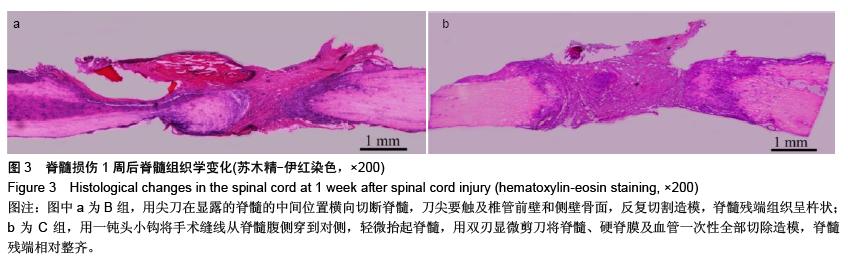
方法:成年雌性SD大鼠42只,随机分为A组(n=6)、B 组(n=18)和C组(n=18),A 组仅行椎板切除;B组用尖刀在已显露脊髓的中间位置横向切断脊髓,刀尖要触及椎管前壁和侧壁骨面,反复切割,制备脊髓完全横断模型;C 组采用自制双刃显微剪刀制备脊髓完全横断模型,用一钝头小钩将手术缝线从已显露脊髓的腹侧穿到对侧,轻微抬起脊髓,用双刃显微剪刀将脊髓、硬脊膜及血管一次性全部切除。
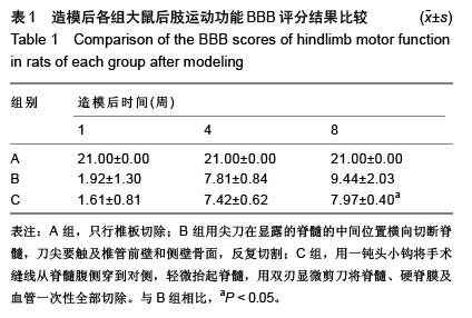
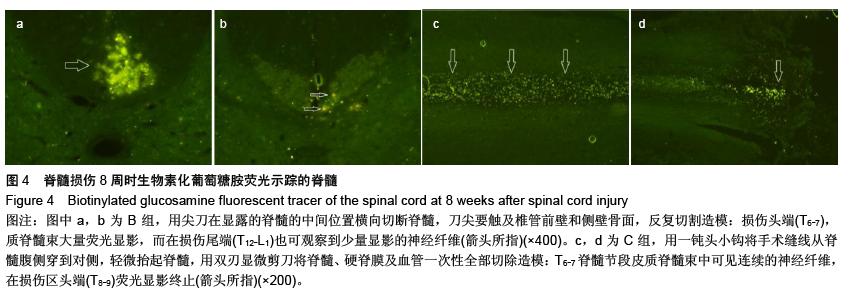
结果与结论:①B和C组大鼠造模后1周,后肢完全性瘫痪,BBB评分接近,但脊髓残端间距离有显著差异。②造模后4周,两组大鼠后肢功能有不同程度恢复,但BBB评分差异无显著性意义。③造模后8周,两组大鼠后肢运动功能评分差异有显著性意义;生物素化葡萄糖胺示踪显示:B组大鼠损伤尾侧可观察到少量被标记的轴突纤维,而C组大鼠尾侧无被标记的神经纤维,说明脊髓横断完全。④以上结果提示:使用自制的双刃显微剪刀造模方法可有效消除个体差异,有利于治疗效果的量化分析和研究对比。 
ORCID: 0000-0001-7412-7520(贺丰)

关键词: 实验动物, 神经损伤与修复动物模型, 脊髓损伤, 模型, 外科器械, 完全横断, 大鼠, 生物素葡聚糖胺, 神经示踪, 皮质脊髓束

Abstract:

BACKGROUND: Rat models of complete spinal cord transection are common models for neural tissue engineering. After transecting the spinal cord by the previous methods, gap length of broken end cannot keep relatively uniform, so we cannot objectively evaluate effects of various treatments or tissue engineering materials.
OBJECTIVE: The spinal cord transection models were established by using double edged micro scissors, and the feasibility of this new model was explored by comparing with the conventional method.
METHODS: A total of 42 adult female Sprague-Dawley rats were divided randomly into group A (n=6), group B (n=18) and group C (n=18). Group A only received laminectomy. In the group B, the spinal cord was transected with a sharp-pointed knife. Knife point should touch anterior wall of spinal canal and sidewall bone surface. Complete spinal cord transection models were prepared by repeated cutting. In group C, complete spinal cord transection models were established by using self-made double edged micro scissors.
RESULTS AND CONCLUSION: (1) At 1 week after model establishment, in the groups B and C, complete paralysis of the hind limbs was found, and BBB scores were similar. However, significant differences in the spacing of broken end were detected. (2) At 4 weeks after model establishment, hind limb functions could restore to different degrees in both groups, but no significant difference in BBB scores was found. (3) At 8 weeks after model establishment, significant differences in hindlimb motor function scores were detectable between both groups. Biotin glucosamine tracer display: In group B, a few labeled axon fibers were observed at the caudal side of the injured spinal cord. In group C, spinal cord was completely transected, and labeled axon fibers cannot be found at the caudal side. (4) Results suggested that the modeling method of self-made double edged micro scissors could effectively eliminate individual differences, contribute to quantitative analysis and comparative study of therapeutic effects.