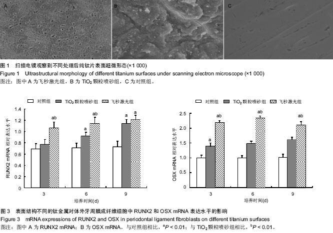
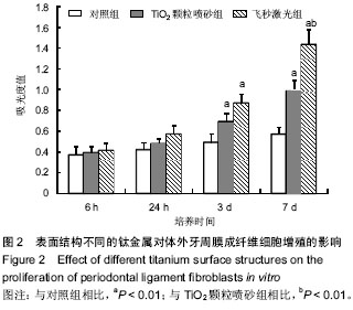
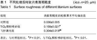
| [1] Lindhe J, Meyle J; Group D of European Workshop on Periodontology. Peri-implant diseases: Consensus Report of the Sixth European Workshop on Periodontology. J Clin Periodontol. 2008;35(8 Suppl):282-285.
[2] 张丁华,孙皎.牙种植体材料的研究进展[J].口腔材料器械杂志,2006,15(1):33-36.
[3] 赵晶妹.口腔纯钛种植体表面改性及其生物性能研究[D].上海:上海交通大学,2013
[4] 李春艳.纯钛种植体表面飞秒激光改性及性能评价[D].天津:天津医科大学,2012.
[5] 王怀柳.间隙杂质对工业纯钛性能的影响[J].特钢技术,2006,3(1):25-28.
[6] 张锋,韩莉,徐媛媛,等.纯钛钛片表面胶原/透明质酸聚电解质复合膜涂层的构建[J].口腔医学,2015,2(35):96-100.
[7] 曲露露,李美华,罗云纲.纯钛种植体表面处理技术促进骨整合研究进展[J].中国老年学杂志,2015(12):3474-3476.
[8] Ironside JG, Swain MV. Ceramic in dental restorations-a review and critical issues. J Aust Ceram Soc. 1998;34(2):78-91.
[9] Lemons JE. Dental implant biomaterials. J Am Dent Assoc. 1990;121(6):716-719.
[10] 陈静.纯钛表面粗糙度对人牙龈成纤维细胞影响的体外实验研究[D].成都:四川大学,2004.
[11] 廖湘凌,李声伟.生物活性种植牙的研究发展[J].中国口腔种植学杂志,1999,4(2):90-93.
[12] Tsukamoto M, Asuka K, Nakano H, et al. Periodic microstructures produced by femtosecond laser irradiation on titanium plate. Vacuum. 2006;80(11-12):1346-1350.
[13] Liang JH, Kang J, Pan YL, et al. Ex vivo evaluation of femtosecond pulse laser incision of urinary tract tissue in a liquid environment: implications for endoscopic treatment of benign ureteral strictures. Lasers Surg Med. 2011; 43(6): 516-521.
[14] Kara V, Kizil H. Titanium micromachining by femtosecond laser. Opt Lasers Eng. 2012;50(2):140-147.
[15] Vorobyev AY, Guo C. Femtosecond laser structuring of titanium implants. Appl Surf Sci. 2007;253(17):7272-7280.
[16] Stamp R, Fox P, O'Neill W, et al. The development of a scanning strategy for the manufacture of porous biomaterials by selective laser melting. J Mater Sci Mater Med. 2009;20(9): 1839-1848.
[17] Wieland M, Chehroudi B, Textor M, et al. Use of Ti-coated replicas to investigate the effects on fibroblast shape of surfaces with varying roughness and constant chemical composition. J Biomed Mater Res. 2002;60(3):434-444.
[18] 刘潇.纯钛表面不同处理对人牙龈成纤维细胞增殖的影响[D].石家庄:河北医科大学,2011.
[19] Lowenberg BF, Pilliar RM, Aubin JE, et al. Migration, attachment, and orientation of human gingival fibroblasts to root slices, naked and porous-surfaced titanium alloy discs, and Zircalloy 2 discs in vitro. J Dent Res. 1987;66(5): 1000-1005.
[20] Kim SY, Oh N, Lee MH, et al. Surface microgrooves and acid etching on titanium substrata alter various cell behaviors of cultured human gingival fibroblasts. Clin Oral Implants Res. 2009;20(3):262-272.
[21] Pi SH, Lee SK, Hwang YS, et al. Differential expression of periodontal ligament-specific markers and osteogenic differentiation in human papilloma virus 16-immortalized human gingival fibroblasts and periodontal ligament cells. J Periodontal Res. 2007;42(2):104-113.
[22] Yamaguchi A, Komori T, Suda T. Regulation of osteoblast differentiation mediated by bone morphogenetic proteins, hedgehogs, and Cbfa1. Endocr Rev. 2000;21(4):393-411.
[23] Liu CJ, Chang E, Yu J, et al. The interferon-inducible p204 protein acts as a transcriptional coactivator of Cbfa1 and enhances osteoblast differentiation. J Biol Chem. 2005; 280(4):2788-2796.
[24] Gu K, Zhang L, Jin T, et al. Identification of potential modifiers of Runx2/Cbfa1 activity in C2C12 cells in response to bone morphogenetic protein-7. Cells Tissues Organs. 2004;176 (1-3): 28-40.
[25] Lee MH, Javed A, Kim HJ, et al. Transient upregulation of CBFA1 in response to bone morphogenetic protein-2 and transforming growth factor beta1 in C2C12 myogenic cells coincides with suppression of the myogenic phenotype but is not sufficient for osteoblast differentiation. J Cell Biochem. 1999;73(1):114-125.
[26] Nishimura R, Hata K, Harris SE, et al. Core-binding factor alpha 1 (Cbfa1) induces osteoblastic differentiation of C2C12 cells without interactions with Smad1 and Smad5. Bone. 2002;31(2):303-312.
[27] Banerjee C, Javed A, Choi JY, et al. Differential regulation of the two principal Runx2/Cbfa1 n-terminal isoforms in response to bone morphogenetic protein-2 during development of the osteoblast phenotype. Endocrinology. 2001;142(9):4026-4039.
[28] Zhao M, Qiao M, Oyajobi BO, et al. E3 ubiquitin ligase Smurf1 mediates core-binding factor alpha1/Runx2 degradation and plays a specific role in osteoblast differentiation. J Biol Chem. 2003;278(30):27939-27944.
[29] Ohyama Y, Nifuji A, Maeda Y, et al. Spaciotemporal association and bone morphogenetic protein regulation of sclerostin and osterix expression during embryonic osteogenesis. Endocrinology. 2004;145(10):4685-4692. Epub 2004 Jun 24.
[30] Balemans W, Van Hul W. Identification of the disease-causing gene in sclerosteosis--discovery of a novel bone anabolic target? J Musculoskelet Neuronal Interact. 2004;4(2):139- 142.
[31] Nifuji A, Miura N, Kato N, et al. Bone morphogenetic protein regulation of forkhead/winged helix transcription factor Foxc2 (Mfh1) in a murine mesodermal cell line C1 and in skeletal precursor cells. J Bone Miner Res. 2001;16(10):1765-1771.
[32] Celil AB, Campbell PG. BMP-2 and insulin-like growth factor-I mediate Osterix (Osx) expression in human mesenchymal stem cells via the MAPK and protein kinase D signaling pathways. J Biol Chem. 2005;280(36):31353-31359.
[33] Takagi M, Kamiya N, Takahashi T, et al. Effects of bone morphogenetic protein-2 and transforming growth factor beta1 on gene expression of transcription factors, AJ18 and Runx2 in cultured osteoblastic cells. J Mol Histol. 2004;35(1): 81-90.
[34] Li IW, Cheifetz S, McCulloch CA, et al. Effects of osteogenic protein-1 (OP-1, BMP-7) on bone matrix protein expression by fetal rat calvarial cells are differentiation stage specific. J Cell Physiol. 1996;169(1):115-125.
[35] Abe E, Yamamoto M, Taguchi Y, et al. Essential requirement of BMPs-2/4 for both osteoblast and osteoclast formation in murine bone marrow cultures from adult mice: antagonism by noggin. J Bone Miner Res. 2000;15(4):663-673. |