中国组织工程研究 ›› 2015, Vol. 19 ›› Issue (33): 5338-5343.doi: 10.3969/j.issn.2095-4344.2015.33.018

• 组织构建与生物活性因子 tissue construction and bioactive factors • 上一篇    下一篇

巨噬细胞移动抑制因子在瘢痕疙瘩中的表达

李桂锋1,王春梅2,周 显3,闫 伦2,项晓飞2,徐 伟2,杨思奋2,任家骠2   

  1. 1广州中医药大学第二临床医学院,广东省广州市 510502;东莞康华医院,2烧伤整形科,3病理科,广东省东莞市 523000
  • 出版日期:2015-08-13 发布日期:2015-08-13
  • 通讯作者: 王春梅,博士,教授,主任医师,东莞康华医院烧伤整形科,广东省东莞市 523000
  • 作者简介:李桂锋,男,1987年生,河南省信阳市人,汉族,广州中医药大学第二临床医学院在读硕士,主要从事病理性瘢痕方面的基础研究。

Expression of macrophage migration inhibitory factor in keloid 

Li Gui-feng1, Wang Chun-mei2, Zhou Xian3, Yan Lun2, Xiang Xiao-fei2, Xu Wei2, Yang Si-fen2, Ren Jia-biao2   

  1. 1The Second Clinical Medical College of Guangzhou Traditional Chinese Medicine, Guangzhou 510502, Guangdong Province, China; 2Department of Burn and Plastic Surgery, 3Department of Pathology, Kanghua Hospital of Dongguan, Dongguan 523000, Guangdong Province, China
  • Online:2015-08-13 Published:2015-08-13
  • Contact: Wang Chun-mei, M.D., Professor, Chief physician, Department of Burn and Plastic Surgery, Kanghua Hospital of Dongguan, Dongguan 523000, Guangdong Province, China
  • About author:Li Gui-feng, Studying for master’s degree, the Second Clinical Medical College of Guangzhou Traditional Chinese Medicine, Guangzhou 510502, Guangdong Province, China

摘要:

背景:巨噬细胞移动抑制因子参与了多种疾病的发生、发展过程,它在肿瘤、自身免疫性疾病、炎症反应、血管新生、纤维化等方面发挥重要作用,这些生物学特征与瘢痕疙瘩较为相似。

目的:检测巨噬细胞移动抑制因子(MIF)在瘢痕疙瘩、增生性瘢痕、正常皮肤组织中的表达情况,说明巨噬细胞移动抑制因子分布位置和数量的差异。

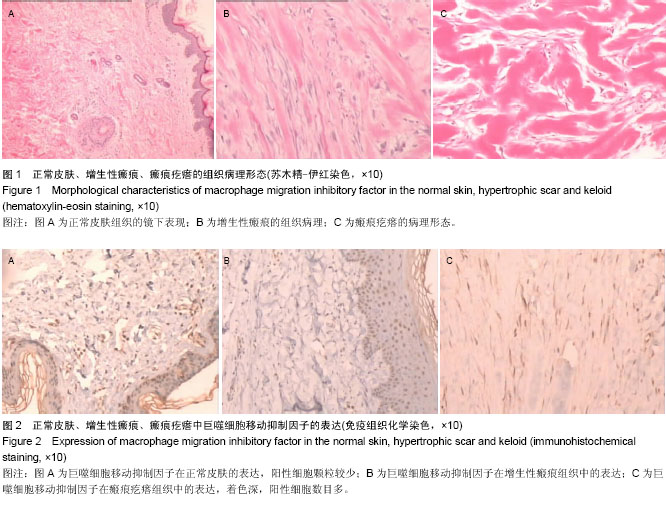
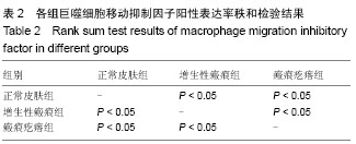
方法:收集临床病理性瘢痕切除后标本共40例,其中瘢痕疙瘩组20例,增生性瘢痕组20例,另取正常皮肤标本10例作为对照。分别做苏木精-伊红染色和免疫组织化学染色观察巨噬细胞移动抑制因子在病理性瘢痕及正常皮肤中的表达情况。

结果与结论:巨噬细胞移动抑制因子在正常皮肤、增生性瘢痕、瘢痕疙瘩中均有表达,但在瘢痕疙瘩中表达明显强于增生性瘢痕和正常皮肤(P < 0.01)。说明巨噬细胞移动抑制因子在瘢痕疙瘩中的异常浸润可能与瘢痕疙瘩的形成有关。

中国组织工程研究杂志出版内容重点:组织构建;骨细胞;软骨细胞;细胞培养;成纤维细胞;血管内皮细胞;骨质疏松组织工程

关键词: 组织构建, 组织工程, 巨噬细胞移动抑制因子, 瘢痕疙瘩, 增生性瘢痕, 免疫组织化学

Abstract:

BACKGROUND: Macrophage migration inhibitory factor is involved in the process of a variety of diseases, and plays a very important role in the tumor, autoimmune diseases, inflammation, angiogenesis, fibrotic diseases and so on. These biological characteristics are similar to keloids.

OBJECTIVE: To compare the distribution and number of macrophage migration inhibitory factor in normal skin, hypertrophic scar and keloid.

METHODS: We collected 40 clinical pathological scar specimens after surgery, including 20 hypertrophic scars and 20 keloids. Another 10 samples of the normal skin were used as control group. Hematoxylin-eosin staining and immunohistochemistry staining were performed to test the expression of macrophage migration inhibitory factor in pathological scars and normal skin.

RESULTS AND CONCLUSION: Macrophage migration inhibitory factor was positively expressed in the normal skin, hypertrophic scar and keloid, and the expression of macrophage migration inhibitory factor in keloid was significantly higher than that in hypertrophic scar and normal skin (P < 0.01). It means that the abnormal infiltration of macrophage migration inhibitory factor may be associated with the formation of keloid.

中国组织工程研究杂志出版内容重点:组织构建;骨细胞;软骨细胞;细胞培养;成纤维细胞;血管内皮细胞;骨质疏松组织工程

Key words: Neoplasms, Immunity, Inflammation, Fibrosis

中图分类号: