中国组织工程研究 ›› 2015, Vol. 19 ›› Issue (27): 4304-4308.doi: 10.3969/j.issn.2095-4344.2015.27.008

• 骨髓干细胞 bone marrow stem cells • 上一篇    下一篇

以线栓法构建脑梗死模型大鼠的微血管新生及电针干预效应

金哲峰,石  磊,杜元灏   

  1. 天津中医药大学第一附属医院,天津市  300193
  • 出版日期:2015-06-30 发布日期:2015-06-30
  • 通讯作者: 石磊,博士,副主任医师,天津中医药大学第一附属医院,天津市 300193
  • 作者简介:金哲峰,男,1979年生,内蒙古自治区通辽市人,朝鲜族,2008年中国中医科学院毕业,硕士,主治医师,主要从事骨科生物力学及细胞分子生物学的研究。
  • 基金资助:

    国家自然科学基金资助项目(81303021)“电针干预脑梗塞后神经血管单元构型调整的分子机制及远期效应研究”

Effects of electroacupuncture intervention on angiogenesis in rat models of cerebral infraction constructed by suture method

Jin Zhe-feng, Shi Lei, Du Yuan-hao   

  1. First Teaching Hospital of Tianjin University of Traditional Chinese Medicine, Tianjin 300193, China
  • Online:2015-06-30 Published:2015-06-30
  • Contact: Shi Lei, M.D., Associate chief physician, First Teaching Hospital of Tianjin University of Traditional Chinese Medicine, Tianjin 300193, China
  • About author:Jin Zhe-feng, Master, Attending physician, First Teaching Hospital of Tianjin University of Traditional Chinese Medicine, Tianjin 300193, China
  • Supported by:

    the National Natural Science Foundation of China, No. 81303021

摘要:

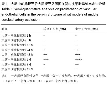
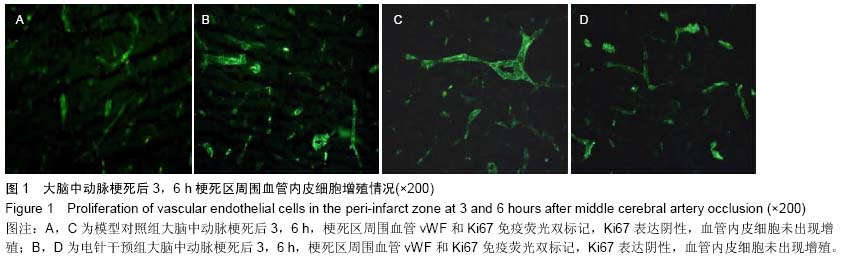
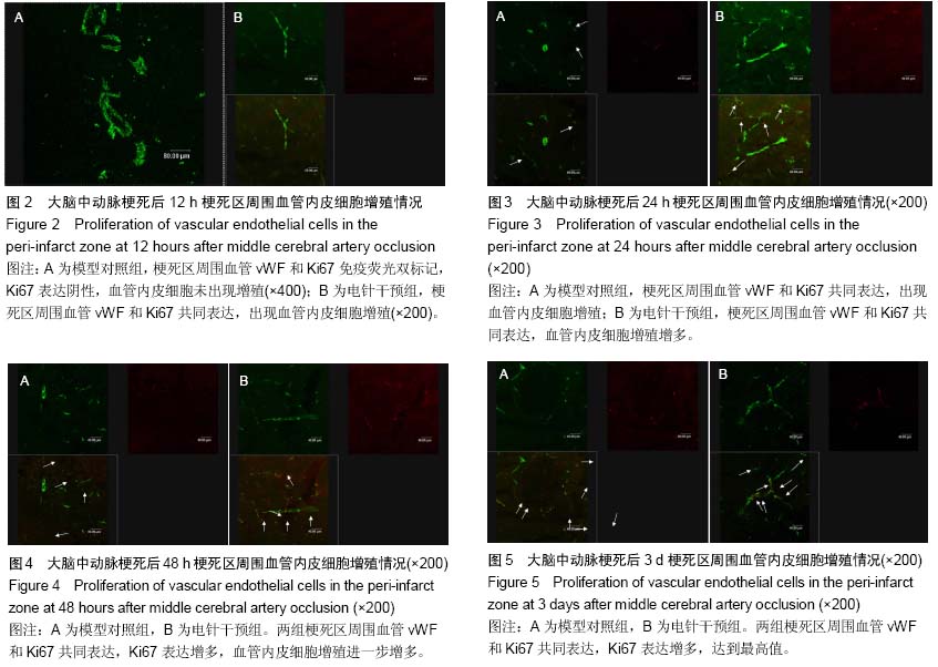
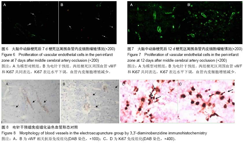
背景:脑梗死发生后缺血半暗带存在的状态决定了最终梗死灶的体积,在这一区域出现的选择性基因表达、蛋白合成减少、乳酸中毒和细胞毒性水肿,对神经细胞的抢救性治疗以及对神经血管单元的持续调节,成为后期神经功能恢复的结构和生理基础。
目的:以脑梗死后微血管新生过程为研究切入点,探索微血管新生随时间延续呈现的基本规律以及电针干预对局部血管增殖产生的影响。
方法:96只Wistar大鼠随机分为模型对照组和电针干预组,以线栓法复制大鼠大脑中动脉梗死模型,电针干预组在造模后即刻针刺人中穴,给予15 Hz,1 mA的电刺激,持续20 min。模型对照组同样抓取固定,但不进行任何治疗。在脑梗死后不同时间(3,6,12,24,48 h以及3,7,12 d)采用vWF、Ki67免疫荧光双标记检测血管的新生情况。
结果与结论:在大脑中动脉梗死后3,6,12 h,模型对照组梗死区周围无血管内皮细胞增殖标记,24 h出现血管内皮细胞增殖,48 h血管内皮细胞增殖增加,3 d时达到高峰,7 d时血管内皮细胞增殖水平下降,12 d时无血管内皮细胞增殖标记;在大脑中动脉梗死后3,6 h,电针干预组无血管内皮细胞增殖标记,12 h出现血管内皮细胞增殖,24,48 h血管内皮细胞增殖进一步增加,3 d时达到高峰,7 d时血管内皮细胞增殖水平下降,12 d时无血管内皮细胞增殖标记。与模型对照组比较,电针干预组血管内皮细胞增殖出现时间提前,血管内皮细胞增殖数量多于同时相模型对照组,而两组所有时相梗死区和梗死灶对侧半球均无血管内皮细胞增殖标记。结果表明电针干预可促进大脑中动脉梗死模型大鼠梗死区周围脑血管内皮细胞增殖,并将血管内皮细胞增殖出现时间提前,由此可改善脑梗死的预后。

中国组织工程研究杂志出版内容重点:肾移植肝移植移植;心脏移植;组织移植;皮肤移植;皮瓣移植;血管移植;器官移植组织工程

关键词: 实验动物, 脑及脊髓损伤动物模型, 脑梗死, 血管新生, vWF, Ki67, 电针, 国家自然科学基金

Abstract:

BACKGROUND: The state of ischemic penumbra after cerebral infraction determines the final infarct volume. Decreased selective gene expression and protein synthesis, lactic acidosis and cytotoxic edema, salvage therapy of nerve cells, and continous regulation of neurovascular unit in the ischemic penumbra are areas of interest for recovery of neurological function.
OBJECTIVE: Taking the neoangiogenesis process of post-infarction as the research point, to investigate the basic law of neovascularization with time and the effect of electroacupuncture intervention on proliferation of local blood vessels.
METHODS: Ninety-six Wistar rats were randomly and evenly divided into model and electroacupuncture groups. 
The rat models of middle cerebral artery infarction were established by suture method. Immediately after modeling, the rats in the electroacupuncture group were given electrical stimulation (15 Hz, 1mA, 20 minutes) at the Renzhong (Du26) acupoint. The rats in the model group received similar procedures with the exception of no treatment. The angiogenesis was observed using double-immunofluorescence labeling for von Willebrand Factor (vWF) and Ki-67 antibodies at 3, 6, 12, 24, 48 hours and 3, 7, 12 days after middle cerebral artery occlusion.
RESULTS AND CONCLUSION: In the model group, vascular endothelial cell proliferation in the peri-infarct region was not observed at 3, 6 and 12 hours, occurred at 24 hours, increased at 48 hours, reached a maximum at 3 days, began to decrease at 7 days, and disappeared at 12 days after middle cerebral artery occlusion. In the electroacupuncture group, vascular endothelial cell proliferation in the peri-infarct region was not observed at 3 and 6 hours, occurred at 12 hours, increased at 24 and 48 hours, peaked at 3 days, began to decerase at 7 days, and disappeared at 12 days after middle cerebral artery occlusion. Compared with the model group, the time at which vascular endothelial cell proliferation started was earlier, and the number of proliferated vascular endothelial cells was greater in the electroacupuncture group. Vascular endothelial cell proliferation in the infarcted regions and contralateral hemisphere was not observed in either group at all designated time points. These results demonstrate that electroacupuncture intervention can improve the prognosis of cerebral infarction by promoting the proliferation of vascular endothelial cells in the peri-infarct region and advancing the time at which vascular endothelial cell proliferation started in rat models of middle cerebral artery occlusion. 

中国组织工程研究杂志出版内容重点:肾移植肝移植移植;心脏移植;组织移植;皮肤移植;皮瓣移植;血管移植;器官移植组织工程

Key words: Infarction, Middle Cerebral Artery, Ki-67 Antigen, Electroacupuncture

中图分类号: