中国组织工程研究 ›› 2015, Vol. 19 ›› Issue (25): 3990-3995.doi: 10.3969/j.issn.2095-4344.2015.25.011

• 膜生物材料 membrane biomaterials • 上一篇    下一篇

静脉注射羧基化单壁碳纳米管在大鼠腋窝淋巴结的积聚

李苏宁1,秦  悦1,成晓静2,傅宣皓1,冯建海3,赖泽锋1,刘华钢1   

  1. 广西医科大学,1药学院,
    2医学实验中心,广西壮族自治区南宁市  530021;
    3广西中医药大学药学院,广西壮族自治区南宁市  530200
  • 出版日期:2015-06-18 发布日期:2015-06-18
  • 通讯作者: 赖泽锋,博士,讲师,广西医科大学药学院,广西壮族自治区南宁市 530021
  • 作者简介:李苏宁,男,1987年生,壮族,广西医科大学在读硕士,主要从事纳米药物载体和纳米毒理学方面的研究。
  • 基金资助:

    国家自然科学基金项目(21367006);广西自然科学基金项目(2012GXNSFBA053112);广西教育厅科研项目(200103YB024)

Accumulation of intravenously injected carboxylated single-walled carbon nanotubes in rat axillary lymph nodes 

Li Su-ning1, Qin Yue1, Cheng Xiao-jing2, Fu Xuan-hao1, Feng Jian-hai3, Lai Ze-feng1, Liu Hua-gang1   

  1. 1School of Pharmacy, Guangxi Medical University, Nanning 530021, Guangxi Zhuang Autonomous Region, China; 
    2Medical Research Center, Guangxi Medical University, Nanning 530021, Guangxi Zhuang Autonomous Region, China;
    3School of Pharmacy, Guangxi University of Chinese Medicine, Nanning 530200, Guangxi Zhuang Autonomous Region, China
  • Online:2015-06-18 Published:2015-06-18
  • Contact: Lai Ze-feng, Ph D., Lecturer, School of Pharmacy, Guangxi Medical University, Nanning 530021, Guangxi Zhuang Autonomous Region, China
  • About author:Li Su-ning, Studying for master’s degree, School of Pharmacy, Guangxi Medical University, Nanning 530021, Guangxi Zhuang Autonomous Region, China
  • Supported by:

    the National Natural Science Foundation of China, No. 21367006; Guangxi Natural Science Fund, No. 2012GXNSFBA053112; Research Program of Education Department of Guangxi, No. 200103YB024

摘要:

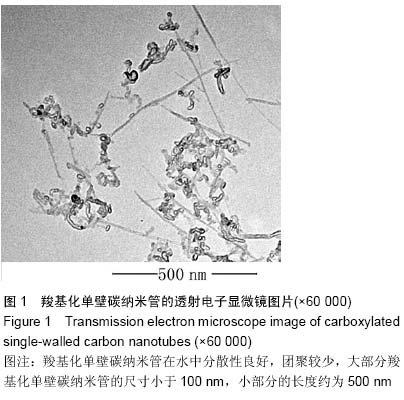
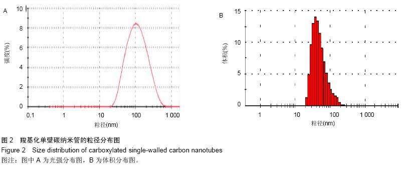
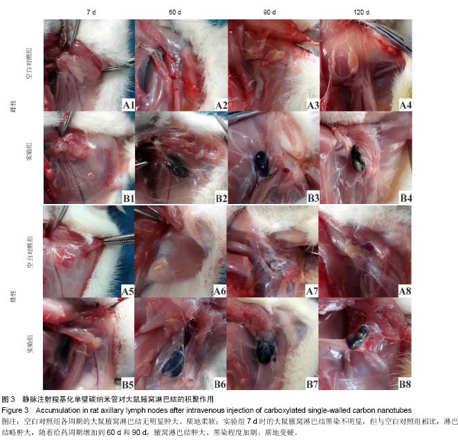
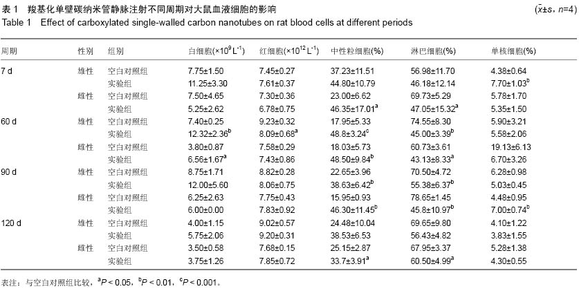
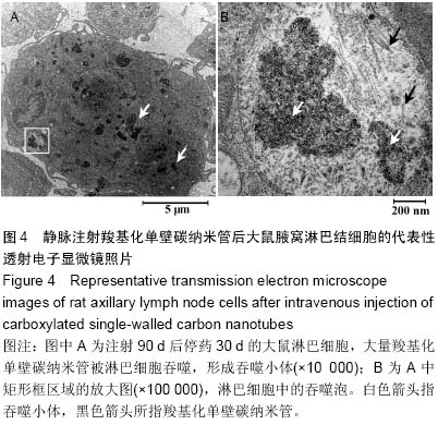
背景:基于碳纳米管的淋巴靶向示踪和治疗是肿瘤靶向诊疗的研究热点之一,评价碳纳米管对腋窝淋巴结的聚集作用,能够为开发淋巴特异性更高、生物兼容性更好的纳米示踪剂和药物载体提供实验依据。
目的:观察静脉注射羧基化单壁碳纳米管在SD大鼠腋窝淋巴结的积聚作用,并评价其对血液细胞的影响。
方法:将64只SD大鼠随机均分为两组,实验组尾静脉注射羧基化单壁碳纳米管2 mg/kg,空白对照组尾静脉注射5%葡萄糖溶液1 mL/kg,3次/周,设定7,60,90,120 d(120 d为给药90 d后停药30 d)共4个周期,每个周期结束时每组随机抽取8只大鼠,采集腹主动脉血液进行血常规检测,观察腋窝淋巴结变化,采集实验组120 d淋巴结标本进行透射电子显微镜观测。
结果与结论:与空白对照组相比,实验组7 d的腋窝淋巴结无明显黑染;随着给药周期的增加,实验组大鼠淋巴结肿大、质地变硬、黑染加深,伴随血液中性粒细胞百分数显著升高(P < 0.01或P < 0.001);停药30 d后,腋窝淋巴结缩小、黑染局部褪去,中性粒细胞百分数下降。停药30 d后,透射电子显微镜观测证实实验组大量羧基化单壁碳纳米管被淋巴细胞吞噬,形成大量吞噬泡。表明尾静脉注射羧基化单壁碳纳米管对SD大鼠腋窝淋巴结的短期靶向示踪作用较弱,长期静脉注射会逐渐在大鼠腋窝淋巴结形成积聚,并引起中性粒细胞增多;停止给药后,羧基化单壁碳纳米管能够被淋巴结缓慢清除。

中国组织工程研究杂志出版内容重点:生物材料;骨生物材料; 口腔生物材料; 纳米材料; 缓释材料; 材料相容性;组织工程

关键词: 生物材料, 材料相容性, 碳纳米管, 淋巴结, 中性粒细胞, 积聚, 静脉注射, 示踪剂, 国家自然科学基金

Abstract:

BACKGROUND: Lymph-targeted tracing and therapy based on carbon nanotubes have been one of the hottest researches on targeting tumor diagnosis and treatment. To evaluate the accumulation of carbon nanotubes in axillary lymph node can provide experimental evidences for developing nano-tracers and drug carriers which are more lymph-specific and more biocompatible.       
OBJECTIVE: To study the accumulation of the intravenously injected carboxylated single-walled carbon nanotubes in axillary lymph nodes of Sprague-Dawley rats, and to evaluate their effect on blood cells.
METHODS: Sixty-four Sprague-Dawley rats were randomly divided into two groups. Rats in testing group were injected with carboxylated single-walled carbon nanotubes suspension (2 mg/kg), while those in control group were injected with 5% glucose solution (1 mL/kg), both through the tail vein, three times per week. Four periods of 7, 60, 90 and 120 days were set (the 120-day period referred to 90 days of administration followed by 30 days of 
drug withdrawal). At the end of each period, eight rats from each group were randomly picked out, to collect blood samples via the abdominal aorta for blood routine test. Finally the axillary lymph nodes were observed, and the lymph node samples of rats in the testing group were collected and analyzed at 120 days by transmission electron microscope.
RESULTS AND CONCLUSION: Compared with the control group, black staining of axillary lymph nodes of rats in testing group was not obvious at the end of the 7-day period. However, with the increase of the dosing periods, the lymph nodes of the rats in the testing group became enlarged, firm and black stained, coupled with a significant rising in the percentage of blood neutrophils. After 30 days of drug withdrawal, the size of the rat axillary lymph nodes was reduced and black staining partly faded, with the decreasing of blood neutrophil percentage. Under the transmission electron microscope, abundant carboxylated single-walled carbon nanotubes were uptaken by lymphocytes to form a large number of phagocytic vacuoles after drug withdrawal for 30 days. It indicates that the short-term tracing of rat axillary lymph nodes by carboxylated single-walled carbon nanotubes injected through the tail vein is relatively weak, while the long-term intravenous injection can cause their accumulation in rat axillary lymph nodes, coupled with the increase of neutrophils; after drug withdrawal, the carboxylated single-walled carbon nanotubes can be slowly cleared by the lymph nodes.

中国组织工程研究杂志出版内容重点:生物材料;骨生物材料; 口腔生物材料; 纳米材料; 缓释材料; 材料相容性;组织工程

Key words: Nanotubes, Carbon, Lymph Nodes, Lymphocytes

中图分类号: