| [1] 耿沁,董强刚,姚明,等.肺癌干细胞的球体形成与致瘤性分析[J].肿瘤,2008,28(9):751-754.
[2] 徐瑜.耐药肺癌干细胞的分离、鉴定及多梳家族成员Bmi-1对其更新和增殖的调控[D].第三军医大学,2011.
[3] 梁冬,马秀梅.人肺癌干细胞分离培养及鉴定的实验研究[J].医学信息(中旬刊),2011,24(5):1718-1719.
[4] Gottschling S,Granzow M,Kuner R,et al.Mesenchymal stem cells in non-small cell lung cancer-Different from others? Insights from comparative molecular and functional analyses. Lung Cancer.2013;80(1):19-29.
[5] Yi BR,Kim SU,Kim YB,et al.Antitumor effects of genetically engineered stem cells expressing yeast cytosine deaminase in lung cancer brain metastases via their tumor-tropic properties.Oncol Rep.2012;27(6):1823-1828.
[6] 郑必强.人肺腺癌干细胞的实验及临床初步研究[D].上海交通大学,2009.
[7] 熊吕平.人肺腺癌A549干细胞球和A549细胞系线粒体融合蛋白表达的研究[D].南昌大学,2013.
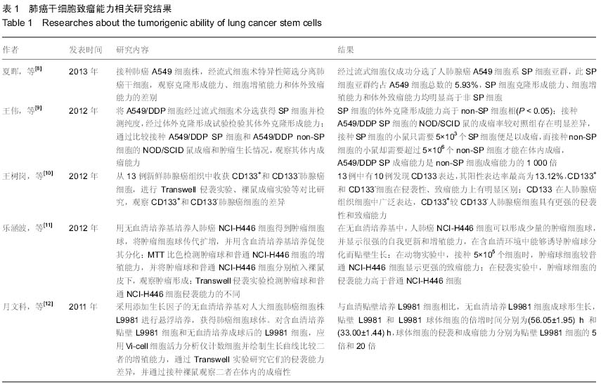
[8] 夏晖,于长海,张文,等.人肺癌A549细胞系肿瘤干细胞的筛选和鉴定[J].中国肺癌杂志,2013,16(8):400-404
[9] 王伟,李勇,牛旗.从耐顺铂肺腺癌A549/DDP细胞系分离和鉴定肺癌干细胞的研究[J].中华临床医师杂志(电子版),2012,6(22): 7229-7232.
[10] 王树岗,曾志勇,杨胜生,等.CD133+肺癌细胞的干细胞特性研究[J].临床肺科杂志,2012,17(5):871-872.
[11] 乐涵波,刘晓光,曾芳.人肺癌NCI-H446细胞生成肿瘤干细胞球[J].解剖学报,2012,43(4):500-505.
[12] 月文科,焦锋,刘彬,等.人大细胞肺癌干细胞样细胞的富集及其功能研究[J].中国肺癌杂志,2011,14(6):484-491.
[13] 月文科.肺癌干细胞的表面筛选标志分子和富集方法的研究[D].天津医科大学,2011.
[14] 易恒仲,龙灵芝,周源,等.LY294002抑制小细胞肺癌干细胞样细胞自我更新[J].湖南师范大学学报:医学版,2013,3(1):15-18.
[15] 王光辉.反义肽核酸封闭线粒体DNA转录启动子对肺癌细胞/肺癌干细胞生物学特性影响的初步研究[D].第三军医大学,2009.
[16] 刘攀,周向东.大细胞肺癌H460细胞系肿瘤干细胞样细胞生物学特性[J].南方医科大学学报,2014,34(4):453-457,462.
[17] 蔡华荣,江跃全.多肽修饰载紫杉醇脂质体靶向A549肺癌干细胞的研究[J].中国生化药物杂志,2014,34(4):12-14,18.
[18] 高全立,汪萍.小细胞肺癌干细胞样细胞的分离鉴定[J].中国癌症杂志,2010,20(6):421-426.
[19] 刘芃芃,于文文,程亚楠,等.基于3D细胞培养系统的肺癌类干细胞的分离和鉴定[J].中国肿瘤临床,2014,41(16):1013-1016.
[20] 付军科,吴齐飞,张勇,等.Id1在人肺癌细胞系A549肿瘤细胞球中的表达[J].现代肿瘤医学,2012,20(12):2483-2485.
[21] 邹爱梅,吴爱兵,黄维甄,等.肺癌A549细胞株中ALDH1+细胞的生物学功能研究[J].中国肿瘤临床,2011,38(14):817-821.
[22] 辛艳泓.转录因子POU5F1在肺癌侵袭转移中的机制研究及胶质瘤肿瘤干细胞免疫逃逸的初步探讨[D].第三军医大学,2013.
[23] 江爱桂.人肺腺癌干细胞的分离、培养与鉴定及PI3K/AKT信号传导通路对其增殖及自我更新的影响[D].苏州大学,2013.
[24] 王波,杨欢,黄玉政,等.人小细胞肺癌细胞株H446侧群细胞的生物学特征[J].癌症,2010,29(3):272-278.
[25] 闫秀萍.肺癌干细胞的分选及鉴定实验研究[D].第三军医大学, 2012.
[26] 闫秀萍,罗虎,周向东,等.人肺癌细胞系A549中肿瘤干细胞样细胞的分离及鉴定[J].第三军医大学学报,2012,34(12):1153-1157.
[27] 高全立,汪萍,Gunnar Kvalheim,等.大细胞肺癌干细胞样细胞的分离及鉴定[J].中国肿瘤生物治疗杂志,2010,17(3):274-279.
[28] Normolle DP,Donnenberg VS,Donnenberg AD.Statistical classification of multivariate flow cytometry data analyzed by manual gating: Stem, progenitor, and epithelial marker expression in nonsmall cell lung cancer and normal lung. Cytometry A.2013;83(1):150-160.
[29] Wang H,Zhang G,Zhang H,et al.Acquisition of epithelial- mesenchymal transition phenotype and cancer stem cell-like properties in cisplatin-resistant lung cancer cells through AKT/p-catenin/Snail signaling pathway.Eur J Pharmacol.2014; 723:156-166.
[30] 谢爱民.Notch1及Bmi-1在肺癌干细胞中表达的相关性及临床意义[J].实用医学杂志,2014,30(7):1104-1106,1107.
[31] 赵欣.载microRNA-34a脂质体靶向治疗肺癌干细胞靶的研究[J].中国生化药物杂志,2014,34(1):68-71.
[32] Andey TMarepally S,Patel A,et al.Cationic lipid guided short-hairpin RNA interference of annexin A2 attenuates tumor growth and metastasis in a mouse lung cancer stem cell model.J Control Release.2014;184:67-78.
[33] Corominas-Faja B,Oliveras-Ferraros C,Cuyàs E,et al.Stem cell-like ALDHbright cellular states in EGFR-mutant non-small cell lung cancer a novel mechanism of acquired resistance to erlotinib targetable with the natural polyphenol silibinin.Cell Cycle. 2013;12(21):3390-3404.
[34] 肖桦,秦旭华,赖宇,等.西黄丸含药血清经Wnt信号转导通路细胞周期蛋白D1调控人肺癌干细胞增殖的机制[J].中国实验方剂学杂志,2014,20(15):172-176.
[35] 赵雅瑞,张立凡,刘特,等.姜黄素上调caspase-3和p53表达后对人肺癌干细胞增殖与侵袭的影响[J].中国临床医学,2013,20(3): 259-262.
[36] Karimi-Busheri F,Zadorozhny V,Carrier E,et al.Molecular integrity and global gene expression of breast and lung cancer stem cells under long-term storage and recovery.Cell Tissue Bank.2013;14(2):175-186.
[37] Reddy BY,Greco SJ,Patel PS, et al.RE-1-silencing transcription factor shows tumor-suppressor functions and negatively regulates the oncogenic TAC1 in breast cancer cells.Proc Natl Acad Sci U S A.2009;106(11):4408-4413.
[38] 黄学德,熊吕平,叶小群,等.低氧对人肺腺癌A549多细胞球体上皮-间质转化促转移的影响[J].肿瘤,2012,32(3):182-188.
[39] 郑必强,周瑾,耿沁,等.人肺腺癌干细胞源自肺脏细支气管肺泡干细胞的初步研究[J].中国肺癌杂志,2008,11(6):759-764.
[40] Liu Y,Lu WL,Guo J,et al.A potential target associated with both cancer and cancer stem cells: A combination therapy for eradication of breast cancer using vinorelbine stealthy liposomes plus parthenolide stealthy liposomes.J Controll Release.2008;129(1):18-25.
[41] 李寒雪,侯文婧.肺干细胞、肺癌干细胞和肺癌[J].世界最新医学信息文摘(电子版),2013,13(7):67-68.
[42] 曾梁,王共先.肺癌干细胞在肺癌诊断和治疗中的研究进展[J].天津医药,2011,39(12):1173-1175. |