中国组织工程研究 ›› 2014, Vol. 18 ›› Issue (41): 6684-6688.doi: 10.3969/j.issn.2095-4344.2014.41.022

• 干细胞培养与分化 stem cell culture and differentiation • 上一篇    下一篇

维甲酸和睾酮单独与联合诱导脂肪源干细胞周期的变化

段富华,曾文钦,杨 春,杨会营,余美春,陶 晖,戴景兴,原 林   

  1. 南方医科大学人体解剖学教研室,广东省广州市 510515
  • 修回日期:2014-08-20 出版日期:2014-10-01 发布日期:2014-10-01
  • 通讯作者: 原林,教授,博士生导师,南方医科大学人体解剖学教研室,广东省广州市 510515
  • 作者简介:段富华,女,1986年生,河南省郑州市人,南方医科大学在读硕士,主要从事脂肪源干细胞的研究。
  • 基金资助:

    国家重点基础研究计划 (973计划)项目(2007 CB512705);国家自然科学基金青年面上项目(30801464)

Retinoic acid, testosterone or their combination affects the cell cycle of adipose-derived stem cells

Duan Fu-hua, Zeng Wen-qin, Yang Chun, Yang Hui-ying, Yu Mei-chun, Tao Hui, Dai Jing-xing, Yuan Lin   

  1. Department of Human Anatomy, Southern Medical University, Guangzhou 510515, Guangdong Province, China
  • Revised:2014-08-20 Online:2014-10-01 Published:2014-10-01
  • Contact: Yuan Lin, Professor, Doctoral supervisor, Department of Human Anatomy, Southern Medical University, Guangzhou 510515, Guangdong Province, China
  • About author:Duan Fu-hua, Studying for master’s degree, Department of Human Anatomy, Southern Medical University, Guangzhou 510515, Guangdong Province, China
  • Supported by:

    the National Key Basic Research Program (973 Planning), No. 2007CB512705; the National Natural Science Foundation of China, No. 30801464

摘要:

背景:目前关于维甲酸对干细胞增殖作用的研究较少见,对睾酮的研究主要集中于其抑制细胞衰老的作用方面。

目的:探索维甲酸和睾酮单独和联合诱导对脂肪源干细胞细胞周期的影响。 
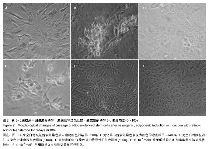
方法:分离培养2月龄SD雌性大鼠脂肪源干细胞,传至第3代进行成脂、成骨诱导和表面标记鉴定。将其分为6组:①对照组。②10-5 mol/L 维甲酸组。③10-6 mol/L 维甲酸组。④10-5 mol/L维甲酸+睾酮组。⑤10-6 mol/L维甲酸+睾酮组。⑥睾酮组。对照组应用DMEM+体积分数10%胎牛血清培养液,其余各组均在对照组的基础上加相应剂量维甲酸或睾酮或二者联合诱导剂。各组培养36 h后行流式细胞仪检测各细胞分期的变化。
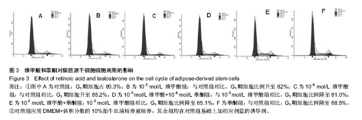
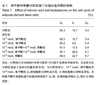
结果与结论:与对照组相比,10-5 mol/L 维甲酸组和10-6 mol/L 维甲酸组G1期细胞数比例明显升高,S期细胞数比例明显降低。与对照组相比,睾酮组G1期细胞数比例明显下降,S期细胞数比例为明显上升。10-5 mol/L维甲酸+睾酮组与10-5 mol/L 维甲酸组比较,10-6 mol/L维甲酸+睾酮组与10-6 mol/L 维甲酸组比较,G1期细胞数比例有所下降,S期细胞数比例为明显上升。结果表明,维甲酸可将脂肪源干细胞周期阻滞于G1期,延缓细胞周期由G1期向S期的进程;睾酮可加速脂肪源干细胞周期由G1期向S期的进程;联合诱导可加速脂肪源干细胞的细胞周期由G1期向S期的进程。

中国组织工程研究杂志出版内容重点:干细胞;骨髓干细胞;造血干细胞;脂肪干细胞;肿瘤干细胞;胚胎干细胞;脐带脐血干细胞;干细胞诱导;干细胞分化;组织工程


全文链接:

关键词: 干细胞, 脂肪干细胞, 维甲酸, 睾酮, 脂肪源干细胞, 细胞周期, 细胞增殖, 成脂诱导, 成骨诱导, 诱导剂, 大鼠, 国家自然科学基金

Abstract:

BACKGROUND: The researches about the effect of retinoic acid on the proliferation of adipose-derived stem cells are rare, and the researches on the testosterone are mainly on the inhibition of cell aging.

OBJECTIVE: To study the effects of retinoic acid and testosterone or combination on the cell cycle of adipose derived stem cells.
METHODS: Adipose derived stem cells were isolated from adult female Sprague Dawley rats with 2 months age and cultured in vitro till passage 3 adipose derived stem cells, and then the 3rd passage adipose-derived stem cells were performed with adipogenic induction, osteogenic induction and surface marker identification. The cells were divided into six groups: (1) Control group; (2) 10-5 mol/L retinoic acid group; (3) Retinoic acid group; (4)     10-5 mol/L retinoic acid+testosterone group; (5) 10-6 mol/L retinoic acid+testosterone group; (6) Testosterone group. The adipose-derived stem cells in the control group were cultured with Dulbecco’s modified Eagle’s medium+10% fetal bovine serum culture medium, and the adipose-derived stem cells in the other five groups were induced with corresponding dose of retinoic acid and testosterone on the basis of control group. After cultured for 36 hours, the flow cytometry was used to detect the changes of cell cycle.
RESULTS AND CONCLUSION: Compared with the control group, cell proportions in phase G1 of 10-5 mol/L retinoic acid group and 10-6 mol/L retinoic acid group were increased significantly, and the cell proportions in phase S were decreased. Compared with control group, the cell proportion in phase G1 of testosterone group was significantly reduced, and the cell proportion in phase S was increased. Compared with 10-5 mol/L retinoic acid group and 10-6 mol/L retinoic acid group, cell proportions in phase G1 of 10-5 mol/L retinoic acid+testosterone group and 10-6 mol/L retinoic acid+testosterone group were reduced significantly and the cell proportions in phase S were increased. Retinoic acid can inhibit the cell cycle of adipose-derived stem cells in phase G1, and delay the process of the cell cycle from phase G1 to phase S; while testosterone can promote the cell cycle of adipose-derived stem cells from phase G1 to phase S; the combination induction of retinoic acid and testosterone can accelerate the process of the cell cycle of adipose-derived stem cells from phase G1 to phase S.

中国组织工程研究杂志出版内容重点:干细胞;骨髓干细胞;造血干细胞;脂肪干细胞;肿瘤干细胞;胚胎干细胞;脐带脐血干细胞;干细胞诱导;干细胞分化;组织工程


全文链接:

Key words:  stem cells, cell cycle, tretinoin, testosterone

中图分类号: