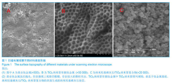
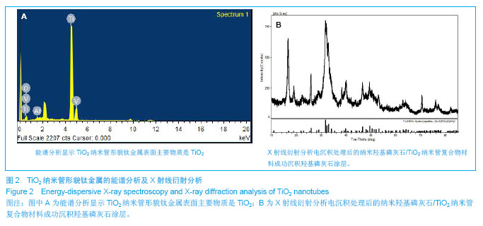
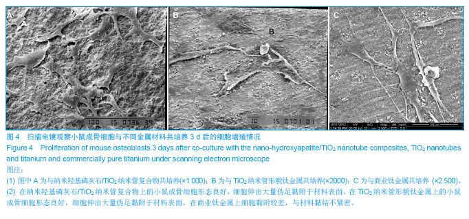
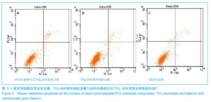
| [1]Lentino JR.Prosthetic joint infections: Bane of orthopedists, challenge for infectious disease specialists.Clin Infect Dis. 2003;36:1157-1161.[2]Darouiche RO. Treatment of infections associated with surgical implants. New Engl J Med.2004;350:1422-1429.[3]Li S,Huang B,Chen Y,et al.Hydroxyapatite-coated femoral stems in primary total hip arthroplasty: a meta-analysis of randomized controlled trials. Int J Surg.2013;11(6):477-482. [4]Oonishi H.A long term histological analysis of effect of interposed hydroxyapatite between bone and bone cement in THA and TKA.J Long Term Eff Med Implants.2012;22(2): 165-176.[5]Munzinger U,Guggi T,Kaptein B,et al.A titanium plasma-sprayed cup with and without hydroxyapatite-coating: a randomised radiostereometric study of stability and osseointegration.Hip Int.2013;23(1):33-39. [6]Cossetto DJ.Mid-term outcome of a modular, cementless, proximally hydroxyapatite-coated, anatomic femoral stem.J Orthop Surg. 2012;20(3):322-326.[7]Herrera A, Mateo J, Lobo-Escolar A et al. Long-term outcomes of a new model of anatomical hydroxyapatite-coated hip prosthesis.J Arthroplasty. 2013; 28(7):1160-1166. [8]Eschen J,Kring S,Brix M,et al.Outcome of an uncemented hydroxyapatite coated hemiarthroplasty for displaced femoral neck fractures: a clinical and radiographic 2-year follow-up study.Hip Int.2012;22(5):574-579.[9]Lazarinis S,Kärrholm J,Hailer NP.Effects of hydroxyapatite coating of cups used in hip revision arthroplasty.Acta Orthop. 2012;83(5):427-435.[10]Melton JT,Mayahi R,Baxter SE,et al.Long-term outcome in an uncemented, hydroxyapatite-coated total knee replacement: a 15- to 18-year survivorship analysis.J Bone Joint Surg Br. 2012;94(8):1067-1070.[11]Martin JY,Schwartz Z,Hummert TW,et al.Effect of titanium surface roughness on proliferation, differentiation, and protein synthesis of human osteoblast-like cells (MG63).J Biomed Mater Res.1995;29(3):389-401.[12]Del Curto B,Brunella MF,Giordano C,et al.Decreased bacterial adhesion to surface-treated titanium.Int J Artif Organs.2005;28(7):718-730.[13]Rajesh P,Muraleedharan CV,Komath M,et al.Laser surface modification of titanium substrate for pulsed laser deposition of highly adherent hydroxyapatite. J Mater Sci Mater Med. 2011;22(7):1671-1679.[14]Popat KC,Eltgroth M,Latempa TJ,et al.Decreased Staphylococcus epidermis adhesion and increased osteoblast functionality on antibiotic-loaded titania nanotubes. Biomaterials. 2007;28(32):4880-4888.[15]George EA,Chang Y,Thomas JW.Enhanced osteoblast adhesion to drug-coated anodized nanotular titanium safaces. Int J Nanomed.2008;3:257-264.[16]Peng L,Mendelsohn AD,Latempa TJ,et al.Long-term small molecule and protein elution from TiO(2) nanotubes.Nano Lett.2009;9(5):1932-1936.[17]田昂,王超,管格非,等.Nano-HA 涂层与HA/ CTS 复合涂层抑制胶质母细胞瘤细胞活力作用的比较[J].功能材料,2010,2(41): 300-303.[18]Tian A,Xue XX,Liu C,et al.Electrodeposited hydroxyapatite coatings in static magnetic field.MaterLett.2010;64(10): 1197-1199.[19]Tian A,Wang C,Xue XX,et al.Inhibitory Effect of Nano-HA Particles on Human U87 Glioblastoma Cells Viability. J Inorganic Mater.2010;25(1): 101-106.[20]Piao Z,Qiu J,Wu Y,et al.Effects of the nano-tubular anodic TiO2 buffer layer on bioactive hydroxyapatite coating.J Nanosci Nanotechnol.2011;11(1):286-290.[21]Tsuchiya H, Macak JM, Müller L,et al.Hydroxyapatite growth on anodic TiO2 nanotubes.J Biomed Mater Res A.2006; 77(3):534-541.[22]Oh SH,Finõnes RR,Daraio C,et al.Growth of nano-scale hydroxyapatite using chemically treated titanium oxide nanotubes.Biomaterials.2005;26(24):4938-4943.[23]Li W, Kabius B,Auciello O.Science and technology of biocompatible thin films for implantable biomedical devices.Conf Proc IEEE Eng Med Biol Soc. 2010;2010: 6237-6242.[24]Bose S,Roy M,Das K,et al.Surface modification of titanium for load-bearing applications.J Mater Sci Mater Med.2009;20 Suppl 1:S19-24.[25]Xie J,Luan BL.Nanometer-scale surface modification of Ti6Al4V alloy for orthopedic applications.J Biomed Mater Res A.2008;84(1):63-72.[26]Yang BC,Uchida M,Kim HM,et al.Preparation of bioactive titanium metal via anodic oxidation treatment.Biomaterials. 2004;25(6):1003-1010.[27]Cooper LF.A role for surface topography in creating and maintaining bone at titanium endosseous implants.J Prosthet Dent.2000;84(5):522-534.[28]Webster TJ,Ejiofor JU.Increased osteoblast adhesion on nanophase metals: Ti, Ti6Al4V, and CoCrMo.Biomaterials. 2004;25:4731-4739.[29]Shinto Y,Uchida A,Korkusuz F,et al.Calcium hydroxyapatite ceramic used as a delivery system for antibiotics.J Bone Joint Surg Br.1992;74(4):600-604.[30]Brammer KS,Seunghan O,Cobb CJ,et al.Improved bone-forming functionality on diameter-controlled TiO2 nanotube surface. Acta biomater.2009;5:3215-3223.[31]von Wilmowsky C,Bauer S,Lutz R,et al.In vivo evaluation of anodic TiO2 nanotubes: an experimental study in the pig.J Biomed Mater Res B. 2009;89(1):165-171.[32]Bjursten LM,Rasmusson L,Oh S,et al.Titanium dioxide nanotubes enhance bone bonding in vivo.2010;92(3): 1218-1224. |

.jpg)