中国组织工程研究 ›› 2013, Vol. 17 ›› Issue (34): 6144-6151.doi: 10.3969/j.issn.2095-4344.2013.34.013

• 材料生物相容性 material biocompatibility • 上一篇    下一篇

皮下植入生肌玉红胶原后的血管新生与瘢痕抑制

曹东阳1,姚 昶2,陈德轩2,卞卫和2,张晓清2,尹 恒3,郭檬檬1   

  1. 1南京中医药大学2011级硕士研究生,江苏省南京市 210029;2江苏省中医院,江苏省南京市 210029;3南京中医药大学2012级博士研究生,江苏省南京市 210046
  • 出版日期:2013-08-20 发布日期:2013-08-20
  • 通讯作者: 姚昶,博士,主任医师,博士生导师,江苏省中医院,江苏省南京市 210029 yaochang67@126.com
  • 作者简介:曹东阳,男,1988年生,河南省南阳市人,汉族,南京中医药大学在读硕士,主要从事中医外科与创伤修复研究。
  • 基金资助:

    国家自然科学基金资助项目(30973596);康缘中医药科技创新基金(HZ1009KY);江苏省中医药领军人才基金(LJ2009002)。

Angiogenesis and scar inhibition after subcutaneous implantation of Shengji Yuhong collagen

Cao Dong-yang1, Yao Chang2, Chen De-xuan2, Bian Wei-he2, Zhang Xiao-qing2, Yin Heng3, Guo Meng-meng1   

  1. 1Postgraduate Student of Grade 2011, Nanjing University of Chinese Medicine, Nanjing  210029, Jiangsu Province, China; 2Jiangsu Province Hospital of TCM, Nanjing 210029, Jiangsu Province, China; 3Doctoral Student of Grade 2012, Nanjing University of Chinese Medicine, Nanjing  210046, Jiangsu Province, China
  • Online:2013-08-20 Published:2013-08-20
  • Contact: Yao Chang, M.D., Chief physician, Doctoral supervisor, Jiangsu Province Hospital of TCM, Nanjing 210029, Jiangsu Province, China yaochang67@126.com
  • About author:Cao Dong-yang, Studying for master’s degree, Postgraduate Student of Grade 2011, Nanjing University of Chinese Medicine, Nanjing 210029, Jiangsu Province, China
  • Supported by:

    the National Natural Science Foundation of China, No. 30973596*; the Kangyuan TCM Technology Innovation Foundation, No. HZ1009KY*; the TCM Leading Talent Foundation of Jiangsu Province, No. LJ2009002*

摘要:

背景:前期研究显示生肌玉红胶原促进血管新生与组织愈合的疗效显著好于生肌玉红膏及单纯胶原或明胶。

目的:探察兔皮下植入生肌玉红胶原促进血管新生与修复的疗效与机制。

方法:在新西兰兔背部两侧对称制作皮下口袋模型,并对称植入生肌玉红胶原(实验组)与胶原(对照组),于术后3,7,14,28,56 d取植入标本及标本周围组织,制作病理切片观察标本周围组织修复状况,测定胶原内血红蛋白水平,免疫荧光与CD34 染色标记法观察周围组织微血管新生情况,Western Blot法检测血管内皮生长因子、血管生成素1的表达,免疫组织化学法观察周围组织Ⅰ、Ⅲ型胶原的分泌及两者的比例。

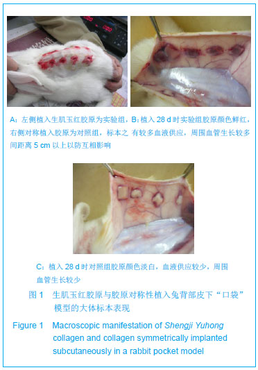
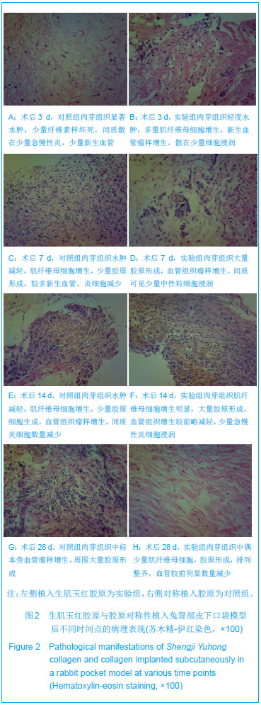
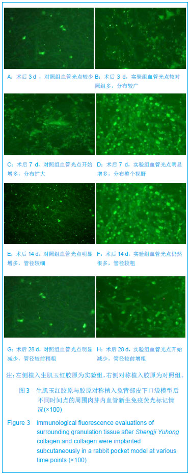
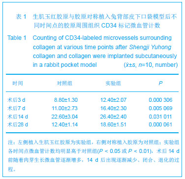
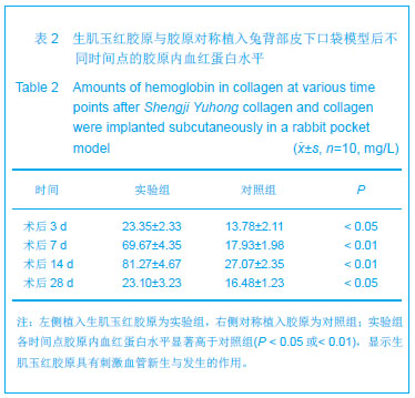
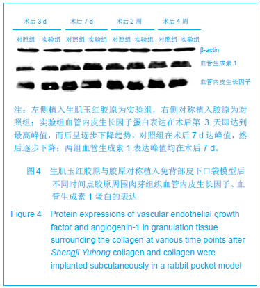
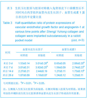
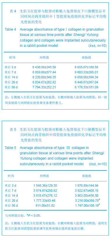
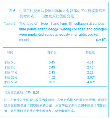
结果与结论:实验组标本周围皮下血管增多,胶原周围组织随着时间推移炎性渗出减少,肉芽组织增生,到28 d时已形成成熟的纤维结缔组织,并且促进微血管新生作用及血红蛋白水平高于对照组(P < 0.05或P < 0.01),术后3,7 d血管内皮生长因子、血管生成素1表达高于对照组(P < 0.05或P < 0.01);实验组Ⅰ型胶原分泌与对照组相当,但术后28,56 d Ⅲ型胶原的分泌高于对照组(P < 0.05),且术后28,56 dⅠ、Ⅲ型胶原的比例低于对照组(P < 0.01)。表明生肌玉红胶原可显著促进周围组织微血管新生,提高血管内皮生长因子与血管生成素1的表达,同时协同调节胶原形成及Ⅰ、Ⅲ型胶原的比例达到减少瘢痕愈合的作用。

关键词: 生物材料, 材料生物相容性, 生肌玉红胶原, 血管新生, Ⅰ型胶原, Ⅲ型胶原, 血管内皮生长因子, 血管生成素1, 国家自然科学基金

Abstract:

BACKGROUND: Shengji Yuhong collagen showed good curative effect of promoting angiogenesis and tissue healing compared with Shengji Yuhong Gao and collagen alone or gelatin alone.

OBJECTIVE: To explore the curative effect and mechanism of subcutaneous implantation of Shengji Yuhong collagen in rabbits in promoting angiogenesis and repair.

METHODS:Shengji Yuhong collagen as the experimental group and collagen as the control group was implanted inside the rabbit subcutaneous pockets of the back of New Zealand rabbits. The implanted samples and surrounding tissues were obtained at 3, 7, 14, 28 and 56 days following surgery. Pathological sections were made and the repair of surrounding tissue was observed. Hemoglobin levels in collagen were measured. Immunofluorescence and CD34 dyeing marking method were utilized to observe capillary angiogenesis. Western  blot assay was employed to examine vascular endothelial growth factor and angiogenin-1 expression. Immunohistochemistry was used to observe the secretion of typeⅠ and Ⅲ collagen on the surrounding tissues.

RESULTS AND CONCLUSION: The experimental group showed increased subcutaneous vascularization. There were reduced inflammatory exudation, granulation tissue hyperplasia, and mature fiber connective tissue at 28 days. Angiogenesis and hemoglobin contents were greater in the experimental group than in the control group (P < 0.05 or P < 0.01). At 3 and 7 days following surgery, vascular endothelial growth factor and angiogenin-1 expression was greater in the experimental group than that in the control group (P < 0.05 or P < 0.01). The secretion of type Ⅰ collagen was identical between the experimental and control groups. However, the secretion of type Ⅲ collagen was higher in the experimental group than in the control group at 28 and 56 days (P < 0.05), and the proportion of type Ⅰ and Ⅲ collagen was lower in the experimental group than in the control group at 28 and 56 days (P < 0.01). These suggested that Shengji Yuhong collagen can significantly promote angiogenesis in the surrounding tissues with the possible mechanisms of adjusting the protein expression of vascular endothelial growth factor and angiogenin-1. At the same time, it has the function of regulating collagen formation with better ratio of typeⅠ and type Ⅲ collagen to acquire higher quality of wound healing with reduced scar formation.

Key words: biomaterials, material biocompatibility, Shengji Yuhong collagen, angiogenesis, typeⅠcollagen, type Ⅲ collagen, vascular endothelial growth factor, angiogenin-1, National Natural Science Foundation of China

中图分类号: