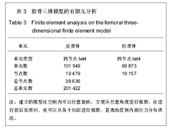
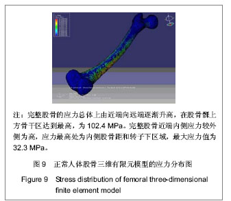
有限元方法是随计算机技术不断进步而逐渐发展起来的一种很有效的数值方法,其基本原理是源于化整为零、集零为整的基本思想。将连续的弹性体分割成有限个单元,单元之间靠结点连接,模拟而成不同几何形状的求解小区域,然后对单元进行力学分析,最后再行整体分析。利用有限元分析法可以对试验对象整体以及各个组成部分在不同的生理条件下的应力分布情况,可以设计出接近于人体反应的试验模型以预测在相似条件下可能形成的结果。随着数字技术和计算机技术的高速发展,有限元方法越来越成为骨科生物力学研究之中必不可少的重要工具之一;己成为结构优化设计、材料非线性及几何非线性分析的一种实用而有效的应力分析方法。
3.1 股骨三维有限元模型的建立
3.1.1 以往的有限元建模方法 由于人体骨骼解剖结构的复杂性及不规则性,在骨科的生物力学研究中有限元分析大部分时间都花在建模上;尤其对于解剖结构及形态功能都很复杂的股骨。在目前医学有限元分析建模的方法主要有以下几种:
磨片及切片法[6]:由于切割、破坏了模型,在断面很薄的情况下很难获得一致的断面厚度。而且需要充分的时间准备模型和将断面几何形状数字化,并且误差大己经被淘汰。
简单建模及基于解剖数据的建模[7]:简单建模将人体器官之几何形状作相应的简化,用相对规则的几何体来代替复杂的实际形状。比如说用圆形的柱体代替骨干,用较规则的球体和凹面来代替关节等。此种方法只是对真实解剖的极度简化,缺乏计算的精确性。基于解剖数据的建模是根据实际形体用样条曲线、曲面等元素进行建模,建立的模型比之用简单几何体模拟要相对精确很多,但误差也较大。
三维测量法[8]:比如激光表面扫描建模,通过激光表面扫描设备将物体表面的轮廓进行扫描,再将其数据输入到逆向软件建立实体模型;再汇入有限元前处理模块中。三维测量法成本比较高,数据处理时间比较长,只能建立表层的结构,无法反映出组织内在的材料特性。在医学研究中主要应用在一些可以不考虑结构内部特性的分析如假体模型建立、动力学分析等,目前也不常用。
基于断层图像的建模[9]:先通过成像系统(CT、MRI、PET)取得原始图像序列后,经过一系列诸如去噪、分割、增强、配准等图像处理工作之后,根据每个断层的图像信息与三维空间坐标,运用数据三维可视化的方法建立起模型。
目前最常用者是CT图像处理法,其主要流程可以概括成为:①对所研究对象进行CT扫描,得到原始数据。②将CT图片通过扫描摄像等各种方法输入到计算机,以位图格式存储及获得二维图像。③在专业图像分析软件中形成轮廓位图同时获取各图像边界的数据资料;同时将得到的数据输入有限元分析软件中进行处理从而最终得到三维有限元模型。这种办法需要人工将CT图片上的每一帧图转换为计算机可以识别的位图格式,且需要在图像处理软件中人工准确对位,对位的不准确会直接影响着所建立模型精确性;同时这种方法要花费大量的人力及物力,且在通过胶片扫描传递数据的过程中容易丢失很多信息。
3.1.2 本试验建模方法 基于DICOM数据直接建模。
DICOM(Digital Imaging and Communication of Medicine)医学数字图像通讯,是医学信息图像系统领域的核心。它主要涉及信息系统中最主要也是最困难的医学图像的存储与通信,可以直接应用到放射学信息系统和图像存档和通信系统中[10],近几年在医学信息领域蓬勃的兴起。DICOM文件提供了非常精确的组织密度信息,同时也包含对研究对象非常完整的几何信息。
利用DICOM格式文件数据直接建模不需将数据做转换,可以直接读取出数据并处理,避免反复操作而造成的数据失真或丢失,显著提高了模型的精确度。其基本过程为:①CT或MRI扫描。②DICOM数据的读取。③图像的分割。④轮廓提取并生成轮廓曲线。⑤建模,将轮廓曲线输入建模软件后再使用相应的前处理模块进行建模。
本建模方法的优点:①精确建模:以往的有限元建模方法大多是通过断层图像在每一层采一些关键点组成轮廓 后将这些点的坐标参数一一导入有限元程序之中,由此建立出三维有限元实体模型。这要处理大量繁杂的数据,且通过胶片传输数据 易丢失信息,再者对位的不准确 直接影响着建立模型的精确性。②利用薄层CT扫描技术可以获得更为准确的CT断层影像。③用Mimics软件的自动建立模型功能显著地提高建模的速度。Mimics(Matorialise's interactive medical image control system)是比利时的Materialise公司研制的交互式医学影像控制系统[11],是一个CT,MRI和超声图像的显示和分割工具;是介于医学及机械领域之间的一套逆向软件;可以将断层扫描的结果转换成为机械领域中的CAD/CAM软件可以处理的数据格式;是医学断层扫描和机械工程之间的接口软件。可以直接读取DICOM数据并直接建立三维模型并转换为ABQUS 6.8等三维有限元软件可以识别的格式,不用进行图像重新定位和二次处理自动生成三维模型。从文章建立的股骨三维有限元模型上来看,该法所建立的模型非常逼真而明显优于以往建模过程。另外在后期处理之中Mimics具有强大的断层图像处理功能,通过设定适当的阈值(Threshold)后可以有效提取每层图像的边缘轮廓数据;并且可以方便的对一层及多层图像进行精确地调整及修正(如选择性编辑、边缘性分割、补洞处理等)。④可重复性强。⑤利用Mimics软件建立的股骨三维模型可以用于精确测量股骨诸空间各点之间的距离,在空间内可以任意旋转、方便从任意角度观察。在进行前后处理之时,也可以从各个切面观察,可以直观地看到内部应力的分布状况。还可选取模型的任意部分,查看其相应计算结果。
3.2 模型生物力学特点
股骨颈力学特性:由试验结果可见股骨近端股骨颈区是主要的应力分布和集中之处,且以股骨颈的中下段为主;并以小转子上方压力骨小粱和股骨距区集中明显。这与股骨颈处解剖特征相一致,股骨距部位的骨皮质最厚而且密度最高、因而才可承受身体上部的质量在该部位形成的高应力而不致于疲劳骨折。这与既往的研究结果相互佐证,充分说明了在生理载荷下压力骨小粱和股骨距是最主要的承重结构,它具有重要的负重作用。这种应力及骨密度分布方式符合最基本的力学原理,又是WOLFF定律的又一印证,再次体现出人体内各种组织结构和功能的一致性。
试验还发现生理载荷下应力经由股骨头压力骨小粱和股骨距向股骨干传导,压力骨小粱与股骨距在传导应力上起重要的作用。由以上分析可以指导临床内固定的固定方式,内固定物的放置要循压力骨小梁方向紧贴股骨距才是符合股骨的主应力方向,有利于应力经由股骨头向股骨干的传导及减小骨折处的剪力,而有利于骨折断端的加压。这样才符合生物力学的原理。
股骨干力学特性:通过实验的结果可以看出在生理载荷下股骨干上的应力分布范围始终主要集中在下中1/3处。故可知载荷的传导主要通过压力骨小粱及股骨距由股骨颈区传倒至股骨干的中下1/3之处;此处是股骨在负重情况下的应力集中处,这与Visuri等[12]报道3名新兵股骨干应力骨折,均出现在股骨干中下1/3交界处相符,这主要与载荷下应力集中导致骨质吸收有关,也与工程力学原理符合。解剖上股骨干上段呈现圆形,向下延伸逐渐变为椭圆形状。在股骨干的中下段移行部,股骨干内外侧的骨皮质迅速变薄而骨髓腔明显增大。按照工程力学原理:在构件断面突然改变的附近区域或构件表面粗糙不平且光洁度不好的区域 会引起应力集中,从而容易发生疲劳断裂。本试验同样发现应力集中在股骨干的中下段1/3处,而且内侧主要表现为压应力外侧主要表现为拉应力,与此理论相符合。
3.3 三维有限元模型的评价 对于股骨三维有限元模型,单元的大小、网格的划分,形状及数目,股骨几何形态完整性与受力情况等均影响模型的精确性;评价有限元模型与有限元计算结果的准确性,很大程度上取决于以上因素。
因此,对所构建的股骨三维有限元模型应进行恰当的优化:①单元数量:从有限元的角度来说,单元个数越多则有限元精度越高;但是计算则越复杂,费时而费力,普通的计算机运行非常困难。然而单元个数过少后,则计算精确度太低,应力和位移值跳跃性比较大;其整体力学特征与实际情况误差也增大,本试验中采用的股骨模型单元数量比较适中。②网格划分:股骨三维有限元稀疏分布要能与受力情况相一致。即应力梯度大的部位采用比较 密集的网格分布,应力梯度小的部位则采用比较稀疏的网格分布。③模型的受力情况必须尽量接近人体的实际力学条件,这样才能保证分析结果准确可靠。然目前在动态下精确模拟人体的动态力学条件几乎不可能;因此作者提出选择占主导地位的载荷进行加载。
3.4 有限元模型的验证 目前,有限元研究方法作为生物力学的一种数学模拟方法,其结论的真实性最终仍需要实验生物力学的检验;它的优点在于可计算骨结构每一点的应力、应变,提供实验手段中不易得到的详细数据,并可改变骨结构等的参数;观察这种改变对完整结构的影响,从而可解释病理或生理过程中的各种变化机制。只有正确合理的模型才可以用于今后的研究和计算。
验证模型的方法主要包括与实验研究的结果做比较,与相似的己经验证的计算机模型做比较,或者结合以上两种方法。如果所得结果基本一致,那么说明模型可以较好地模拟真实情况,从而可以进行有效地计算和分析。通过与以往的文献比较研究发现,本模型可以较好地表现骨关节等组织应力分布的总体趋势,与以往的研究结果相近
[13],从而证实了本模型可以用于今后的有限元分析计算。


.jpg)
.jpg)
.jpg)
.jpg)
.jpg)
.jpg)