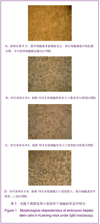
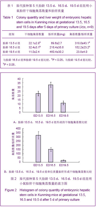
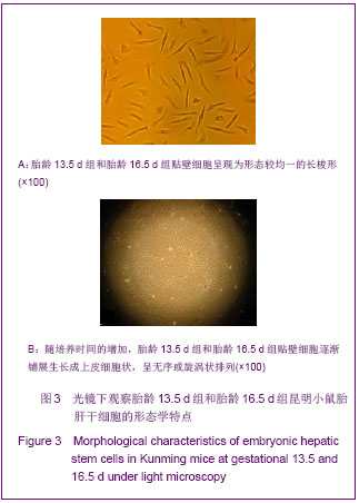
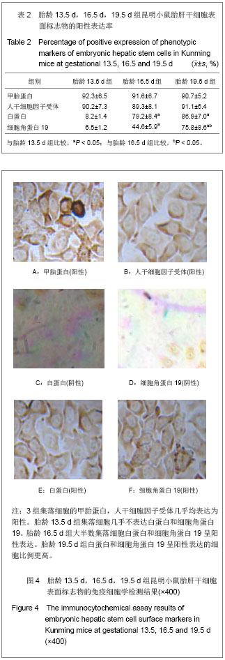
| [1]Cho J, Rameshwar P, Sadoshima J. Distinct roles of glycogen synthase kinase (GSK)-3alpha and GSK-3beta in mediating cardiomyocyte differentiation in murine bone marrow-derived mesenchymal stem cells. J Biol Chem. 2009;284(52): 36647-36658.[2]Yuan YH,Wang Y,Ding Y,et al. Xibao yu Fenzi Mianyixue Zazhi. 2010;26(11):1097-1100.袁雅红,王泳,丁妍,等.人胎盘源间充质干细胞与人脐血单个核细胞联合移植促进SCID鼠早期造血重建[J].细胞与分子免疫学杂志, 2010,26(11):1097-1100.[3]Zhang WZ,Fan Y,Chen YQ,et al. Disi Junyi Daxue Xuebao. 2008;29(8):692-695.张卫泽,樊艳,陈永清,等.血管紧张素Ⅱ对成人脂肪间充质干细胞向心肌细胞分化的影响[J].第四军医大学学报,2008,29(8):692-695.[4]Koninckx R, Hensen K, Daniëls A, et al. Human bone marrow stem cells co-cultured with neonatal rat cardiomyocytes display limited cardiomyogenic plasticity.Cytotherapy. 2009; 11(6):778-792.[5]Khurana S, Mukhopadhyay A. Hematopoietic progenitors from early murine fetal liver possess hepatic differentiation potential. Am J Pathol. 2008;173(6):1818-1827.[6]Li H, Li X, Lam KS,et al. Adeno-associated virus-mediated pancreatic and duodenal homeobox gene-1 expression enhanced differentiation of hepatic oval stem cells to insulin- producing cells in diabetic rats. J Biomed Sci. 2008; 15(4): 487-497.[7]Wang XB,Cai DH,Zhang H,et al. Guangdong Yixue. 2009; 30(3):336-338.王先宝,蔡德鸿,张桦,等.磁标记猪骨髓间充质干细胞肝脏移植示踪研究[J].广东医学, 2009,30(3):336-338.[8]Götherström C, Ringdén O, Tammik C,et al. Immunologic properties of human fetal mesenchymal stem cells. Am J Obstet Gynecol. 2004;190(1):239-245.[9]Weiss TS, Lichtenauer M, Kirchner S,et al. Hepatic progenitor cells from adult human livers for cell transplantation.Gut. 2008; 57(8):1129-1138.[10]Dan YY, Yeoh GC. Liver stem cells: a scientific and clinical perspective. J Gastroenterol Hepatol. 2008;23(5):687-698.[11]Li H, Li X, Lam KS,et al. Adeno-associated virus-mediated pancreatic and duodenal homeobox gene-1 expression enhanced differentiation of hepatic oval stem cells to insulin- producing cells in diabetic rats. J Biomed Sci. 2008;15(4): 487-497.[12]Yovchev MI, Zhang J, Neufeld DS,et al. Thymus cell antigen-1-expressing cells in the oval cell compartment. Hepatology. 2009;50(2):601-611.[13]Pichard V, Ferry N. Origin of small hepatocyte-like progenitor in retrorsine-treated rats. J Hepatol. 2008;48(2):368-369.[14]Best DH, Coleman WB. Treatment with 2-AAF blocks the small hepatocyte-like progenitor cell response in retrorsine-exposed rats. J Hepatol. 2007;46(6):1055-1063.[15]Gao ZQ,Tao KS,Li R,et al. Weichangbingxue he Ganbingxue Zazhi. 2010;19(6): 513-516.高植泉,陶开山,李韧,等.体外不同诱导条件下对大鼠胚胎肝干细胞分化影响的实验研究[J].胃肠病学和肝病学杂志, 2010,19(6): 513-516.[16]Zhao WX,Yin ZY,Yu QF,et al. Zhongguo Zuzhi Gongcheng Yanjiu yu Linchuang Kangfu. 2009;13(36):7103-7107.赵文秀,尹震宇,余强峰,等.小鼠胚胎肝干细胞体外向肝样细胞的诱导分化[J].中国组织工程研究与临床康复, 2009,13(36): 7103-7107.[17]Chang J, Cheng JB, Jia FP, et al. Transdiferentiation of fetal liver-delivered mesenchymal stem cells into cardiomyocyte- like cells. South China Journal of Cardiology.2006; 7(2):78-85. [18]Bin WT,Sun Y,An W,et al. Ganzhang. 2006;11(5):330-334.宾文婷,孙燕,安威,等.小鼠胚胎与成体肝脏中干细胞变化的探讨[J].肝脏,2006,11(5):330-334.[19]Wu BG,Zhang XG,Chang J,et al. Zhongguo Zuzhi Gongcheng Yanjiu yu Linchuang Kangfu. 2009;13(10):1877-1880.吴必刚,张晓刚,常静,等.小鼠胎肝干细胞的分离培养与鉴定[J].中国组织工程研究与临床康复,2009,13(10):1877-1880.[20]Mancino MG, Carpino G, Onori P,et al. Hepatic "stem" cells: state of the art. Ital J Anat Embryol. 2007;112(2):93-109.[21]Yovchev MI, Grozdanov PN, Zhou H,et al. Identification of adult hepatic progenitor cells capable of repopulating injured rat liver. Hepatology. 2008;47(2):636-647.[22]Herrera MB, Bruno S, Buttiglieri S,et al. Isolation and characterization of a stem cell population from adult human liver. Stem Cells. 2006;24(12):2840-2850.[23]Lowes KN, Croager EJ, Olynyk JK,et al. Oval cell-mediated liver regeneration: Role of cytokines and growth factors. J Gastroenterol Hepatol. 2003;18(1):4-12.[24]Tanimizu N, Tsujimura T, Takahide K,et al. Expression of Dlk/Pref-1 defines a subpopulation in the oval cell compartment of rat liver. Gene Expr Patterns. 2004;5(2): 209-218.[25]Sun XY, An J. Expression of nestin, an intermediate filament protein, in human fetal hepatic stem cells. Di Yi Jun Yi Da Xue Xue Bao. 2004;24(2):207-209.[26]Yang F,Wang CJ,Su XL,et al. Jichu Yixue yu Linchuang. 2007; 27(4):386-390.杨帆,王春景,苏小玲,等.人胚胎肝细胞的体外培养及表型观察[J].基础医学与临床,2007,27(4):386-390.[27]Wan DJ,Li LJ,Shi XQ,et al. Zhongguo Bijiao Yixue Zazhi. 2003; 13(2):68-71.万东君,李六金,施新猷,等.大鼠胎肝前体细胞的分离培养[J].中国比较医学杂志,2003,13(2):68-71. |
.jpg)