中国组织工程研究 ›› 2013, Vol. 17 ›› Issue (23): 4212-4215.doi: 10.3969/j.issn.2095-4344.2013.23.005

• 脐带脐血干细胞 umbilical cord blood stem cells • 上一篇    下一篇

人脐带间充质干细胞经5-氮胞苷诱导后心肌特异性肌钙蛋白I的表达

唐  欣1,王妮妮1,易海波2,王  岩3,庞天舒1   

  1. 1辽河油田中心医院循环内科,辽宁省盘锦市  124000
    2辽河油田第二职工医院内科,辽宁省盘锦市  124000
    3辽河油田中心医院放射线科,辽宁省盘锦市  124000
  • 出版日期:2013-06-04 发布日期:2013-06-04
  • 通讯作者: 王岩,辽河油田中心医院放射线科,辽宁省盘锦市 124000
  • 作者简介:唐欣,1978年生,辽宁省营口市人,汉族,2001年大连医科大学毕业,主治医师,主要从事心血管疾病诊断与治疗研究。 tangxin1978@qq.com

Cardiac troponin I expression in human umbilical cord mesenchymal stem cells after 5-azacytidine induction in vitro

Tang Xin1, Wang Ni-ni1 , Yi Hai-bo2 , Wang Yan3 , Pang Tian-shu1
  

  1. 1 Department of Cardiology, Central Hospital of Liaohe Oil Field, Panjin  124000, Liaoning Province, China
    2 Department of Internal Medicine, Second Workers Hospital of Liaohe Oil Field, Panjin  124000, Liaoning Province, China
    3 Department of Radiology, Central Hospital of Liaohe Oil Field, Panjin  124000, Liaoning Province, China
  • Online:2013-06-04 Published:2013-06-04
  • Contact: Wang Yan, Department of Radiology, Central Hospital of Liaohe Oil Field, Panjin 124000, Liaoning Province, China
  • About author:Tang Xin, Attending physician, Department of Cardiology, Central Hospital of Liaohe Oil Field, Panjin 124000, Liaoning Province, China tangxin1978@qq.com

摘要:

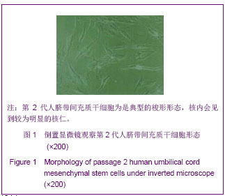
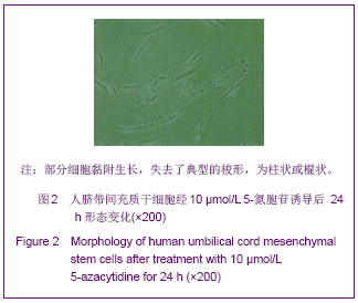
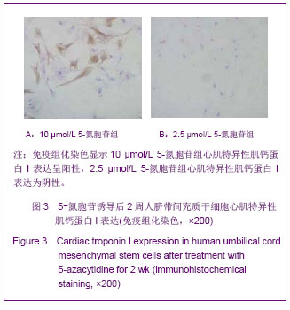
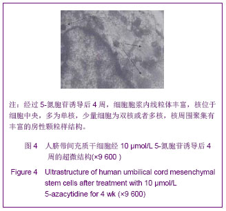
背景:人脐带间充质干细胞具有多向分化潜能,经诱导后是否可向心肌细胞分化?目前的报道尚不多。
目的:观察人脐带间充质干细胞经5-氮胞苷诱导分化后肌钙蛋白I的表达情况。
方法:取第2代人脐带间充质干细胞标本,经消化、离心、沉淀后,分为7份,制成细胞悬液,调整细胞浓度为2×10⁷ L-1,分别加入浓度为80,40,20,10,5,2.5,0 μmol/L 5-氮胞苷于同样条件继续培养,作用24 h后除去,继续培养4周。
结果与结论:①人脐带间充质干细胞经5-氮胞苷处理24 h之后,各组细胞都会出现部分死亡,失去典型的梭形形态,成为棍状或者柱状,40,80 μmol/L组细胞形态变化明显。②免疫组化染色显示5,10,20,40,80 μmol/L组诱导后2周心肌特异性肌钙蛋白I表达呈阳性,对照组与2.5 μmol/L组心肌特异性肌钙蛋白I表达为阴性。选取5个高倍视野进行细胞计数,80,40,20,10 μmol/L组间心肌样细胞的转化率差异无显著性意义,均明显高于5 μmol/L组(P < 0.05)。③超微结构观察显示经5-氮胞苷诱导后4周时部分人脐带间充质干细胞内可见典型的肌小节及心房颗粒。而未经5-氮胞苷诱导的对照组无以上改变。结果提示经5-氮胞苷诱导后人脐带间充质干细胞能够向心肌样细胞转化。

关键词: 干细胞, 干细胞培养与分化, 脐带间充质干细胞, 5-氮胞苷, 免疫组化, 心肌细胞, 肌钙蛋白I, 干细胞图片文章

Abstract:

BACKGROUND: Human umbilical cord mesenchymal stem cells possess multi-differentiation potential. There are few studies describing whether human umbilical cord mesenchymal stem cells can be induced to differentiate into cardiomyocytes .
OBJECTIVE: To investigate cardiac troponin I expression in human umbilical cord mesenchymal stem cells after 5-azacytidine induction in vitro.
METHODS: Passage 2 human umbilical cord mesenchymal stem cells were digested, centrifuged and deposited. Cell suspension was prepared and cell concentration was adjusted to be 2×10⁷/L. Then cells were cultured for 24 hours after addition of various concentrations of 5-azacytidine (80, 40, 20, 10, 5, 2.5, 0 μmol/L). After removal of 5-azacytidine, cells were cultured for another 4 weeks.
RESULTS AND CONCLUSION: After treatment with 5-azacytidine for 24 hours, some cells died in each group, and typical fusiform appearance turned into stick-like or column-like appearance, especially in the 40 and 80 μmol/L 5-azacytidine groups. Immunohistochemical staining results showed that cardiac troponin I expression was detected in human umbilical cord mesenchymal stem cells in 5, 10, 20, 40, 80 μmol/L 5-azacytidine groups after induction for 2 weeks, but it was not detected in the control and 2.5 μmol/L 5-azacytidine groups. Five high-fold fields were selected for cell counting. The number of cardiomyocytes was not significantly different between 80, 40, 20, 10 μmol/L 5-azacytidine groups, but it was significantly higher compared to that in the 5 μmol/L 5-azacytidine group. After 5-azacytidine treatment for 4 weeks, typical sarcomeres and cardiac atrium particles were observed in some human umbilical cord mesenchymal stem cells. Such changes were not observed in the control group in which 5-azacytidine induction was not used. These findings suggest that human umbilical cord mesenchymal stem cells were induced to cardiomyocyte-like cells by 5-azacytidine.

Key words: stem cells, stem cell culture and differentiation, umbilical cord mesenchymal stem cells, 5-azacytidine, immunohistochemistry, cardiomyocyte, cardiac troponin I, stem cell photographs-containing paper

中图分类号: